The Role of the FEI Receptor Kinases in the Regulation of Cell Wall Function in Arabidopsis thaliana
Blaire Janell Steinwand
A dissertation submitted to the faculty of the University of North Carolina at Chapel Hill in partial fulfillment of the requirements for the degree of Doctor of Philosophy in
the Department of Biology.
Chapel Hill 2013
Approved by:
Joseph Kieber PhD
Greg Copenhaver PhD
Jason Reed PhD
Candace Haigler PhD
© 2013
ABSTRACT
BLAIRE JANELL STEINWAND: The Role of the FEI Receptor Kinases in the Regulation of Cell Wall Function in Arabidopsis thaliana
(Under the direction of Joseph Kieber)
The plant cell wall is a staple in the human diet and provides the raw material
used to manufacture paper, textiles, and more recently, biofuel as it is the most
abundant reservoir of carbon in nature. In plants, the cell wall provides structural
support, acts as a barrier to pathogen attack, and determines both the direction and
the extent of cell expansion. The cell wall is a dynamic structure that functions
throughout plant growth and development and in response to developmental and
environmental cues. Despite the importance of the cell wall, the molecular
components and signal transduction pathways involved in regulating its function
remain largely unknown.
FEI1 and FEI2 are two leucine-rich repeat receptor-like kinases (LRR-RLKs)
that promote cell wall function in Arabidopsis thaliana. Mutations in both FEI1 and
FEI2 disrupt cell wall synthesis and this leads to a loss of cell elongation and a short,
swollen root phenotype. In order to determine how exactly the FEI proteins regulate
cell wall function, we sought to identify novel components of the FEI pathway and
have isolated suppressors of the fei1 fei2 mutant phenotype. Further
FEI proteins may act in a complex with other LRR-RLKs and ACC synthase to
ACKNOWLEDGEMENTS
The support that I received from my family, friends, and co-workers has made
this dissertation possible. First, I would like to thank my advisor, Joe Kieber for his
ongoing enthusiasm and support throughout the years. Joe has always had my best
interest in mind and has given me the space and time away from research to pursue
opportunities to teach and has never hesitated to allow me to mentor and train
undergraduates. I thank my committee (Candace Haigler, Greg Copenhaver, Jason
Reed, Mara Duncan, and Pat Gensel) for their helpful comments and suggestions
throughout the years. Candace Haigler has been kind enough to travel over to UNC
in Chapel Hill from North Carolina State in Raleigh each year and has significantly
contributed to my research. Greg Copenhaver was kind enough to join my
committee several years after it had initially formed. Without his support and
encouragement during a very crucial point in my graduate career, I am confident I
would not be writing this dissertation today. In addition to those that have supported
me in my research, I thank the lecturers in Biology for supporting me as a teacher.
Corey Johnson, Gidi Shemer, Jean DeSaix, Jennifer Coble, and the one person that
has made the biggest impact on me during my graduate career both personally and
professionally, Kelly Hogan. Kelly has mentored me in the pedagogy of teaching and
learning and taught me what it truly means to be a fantastic teacher. Kelly and I
share an enthusiasm for teaching biology that has taken us to conferences together
importantly, our love for teaching has brought us together as very close friends over
the years. Without Kelly, I would not have found the strength to finish this degree.
I am grateful for my lab-mates (present – Asia Polko, Apurva Bhargava, Carly
Sacks, Chia-Yi Cheng, Christian Burr, Gyoeng Mee Yoon, Tracy Raines, Wenjing
Zhang) and (past- Cris Argueso, Elizabeth Shin, Jason Punwani, Kaylon Patterson,
Shouling Xu, and Smadar Harpaz-Saad) who have been helpful, friendly faces
throughout the years. There is one graduate student I have always been particularly
glad to have in my life. Tracy Raines has worked across the bench from me from the
day I joined the Kieber lab and I simply could not imagine my graduate career
without her. We have always known how to make the most of everything. Together,
we never failed to make a bad better. In addition, I have been blessed with the best
undergraduate one could ever hope for. Stephanie Doctor has helped me obtain a
large fraction of the data presented here. She is an incredibly bright and motivated
student but also a fun, young spirit. What started as a mentoring relationship has
turned into a very close friendship that will only continue to grow for years to come.
Finally, I have to thank my friends and family. My friends have never failed to
fill my evenings and weekends with joy. I have to thank my best friend and neighbor,
Eliza Peterson for anything and everything from the hundreds of miles that we have
logged road biking to the many, many tears that have fallen over the years. I look
forward to a new chapter in our lives back west as our friendship continues in
Seattle. I thank my Mom for picking up the phone every day and for having the
importance of a good education and hard work, Laurie, and my brother his love and
support. For every single day of this experience, I thank my ever-supportive and
loving partner, Kyle for believing in me even when I have not believed in myself.
Last, but not least, I thank my two kitty cats Scooby Doo and Nala for always making
TABLE OF CONTENTS
LIST OF TABLES ... xi
LIST OF FIGURES ... xii
LIST OF ABBREVIATIONS ... xiv
I. BACKGROUND AND SIGNIFICANCE ... 1
II. Alterations in Auxin Homeostasis Suppress Defects in Cell Wall Function ... 21
OVERVIEW ... 21
INTRODUCTION ... 22
RESULTS ... 26
DISCUSSION ... 32
REFERENCES ... 40
III. Further Characterization of the FEI Pathway ... 54
INTRODUCTION ... 55
RESULTS ... 57
DISCUSSION ... 65
MATERIALS AND METHODS ... 68
REFERENCES ... 72
LIST OF TABLES Table
2.1 Markers used to map shou2-1 ... 44
3.2 Phenotypes of fei1 fei2 suppressors ... 74
LIST OF FIGURES Figure
2.1 Isolation of the shou2 suppressor ... 45
2.2 Positional cloning of shou2 ... 46
2.3 Mutations in the auxin biosynthesis genes WEI8 and TAR2 suppress fei1 fei2 ... 47
2.4 Additional phenotypes of fei1 fei2 iar4-5 ... 48
2.5 iar4 does not suppress defects in the seed coat mucilage of fei1 fei2 ... 49
2.6 Mutations in iar4-5 suppress other cell wall mutants ... 50
2.7 Mutations in IAR4 confer resistance to isoxaben ... 51
2.8 iar4 suppresses lignin accumulation in cell wall mutants ... 52
2.9 Model of the FEI pathway ... 53
3.1 Root elongation of fei1 fei2 suppressors ... 76
3.2 Totoal root elongation of fei1 fei2 suppressors in the presenece of 2,4D ... 77
3.3 Quantification of hypocotyl widths of fei1 fei2 seedlings ... 78
3.4 SHOU4 maps to the bottom arm of chromosome 1 ... 79
3.5 SHOU3 maps to the top arm of chromosome 1 ... 80
3.6 Whole genome sequencing of fei1 fei2 shou4 ... 81
3.7 The shou4 mutation leads to a 45 bp or 15 amino acid in frame deletion ... 82
3.8 shou4 does not suppress the seed mucilage phenotype of fei1 fei2 ... 83
3.9 FEI1 and FEI2 interact with ACS in planta ... 84
3.10 Relative abundance of CESA3 ... 85
3.12 ACS interacts with LRR-RLKs that are co-expressed or
LIST OF ABBREVIATIONS
2,4D dichlorophenoxyacetic acid
ACC 1 aminocyclopropane-1-carboxylate
ACS 1 AMINOCYCLOPROPANE-1-CARBOXYLATE SYNTHASE
AIB α-aminoisobutyric acid
AOA Aminooxy-acetic acid
BAK1 BRI1 ASSOCIATED KINASE 1
BiFC Bimolecular fluorescence complementation
BRI BRASSINOSTEROID 1
CESA Cellulose synthase
ChD Discordant chastity
COB COBRA
CSC Cellulose synthase complex
DCB 2,6-dichlorobenzonitrile
ELI1 ECTOPIC LIGNIFICATION 1
FER FERONIA
FIL FEI INTERACTING LEUCINE RICH REPEAT RECEPTOR LIKE KINASE
HERK HERCULES
IXB isoxaben
KOR KORRIGAN
MAPK MITOGEN ACTIVATED PROTEIN KINASE
PRC PROCUSTE
RLK receptor-like kinase
RSW1 ROOT SWELLING 1
SNP Single nucleotide polymorphism
SOS5 SALT OVERLY SENSITIVE 5
THE THESEUS
WAK WAL-ASSOCIATED KINASE
____________________________
1Portions of this chapter have been reproduced/amended from: Steinwand, B. J., and J.J. Kieber. 2013. The Role of
Receptor-Like Kinases in Regulating Cell Wall Function. Plant Physiol. 153, 479-484. CHAPTER 1
BACKGROUND AND SIGNIFICANCE
The plant cell wall is a rigid but highly dynamic structure that provides
mechanical support, protection against pathogen attack, and determines the direction
and extent of cell expansion (Humphrey et al., 2007). The dynamic nature of the plant
cell wall allows growing cells to expand while providing the mechanical strength
required to resist the forces of turgor pressure exerted on the cell (Cosgrove, 2000).
The properties of the cell wall are modified during growth and development as well as in
response to a wide variety of environmental stimuli. In order to maintain the integrity of
the wall and to adjust its properties to accommodate the changing needs of the cell,
plants respond to perturbations to the wall and environmental cues by remodeling
matrix polysaccharides and by regulating the cell wall biosynthetic machinery. The
components and mechanisms underlying such a signaling system remain largely
unknown, but emerging evidence has implicated several receptor-like kinases as
regulators of cell wall function.
Plant cell walls are composite structures composed primarily of cellulose and
matrix polysaccharides such as hemicelluloses and pectins (Somerville et al., 2004). In
contribution to the wall. The major load-bearing components of the cell wall are the
cellulose microfibrils which, in a longitudinally expanding cells, are deposited primarily
inan orientation perpendicular to the axis of expansion, thus constricting radial
expansion (Green, 1980; Taiz, 1984; Baskin, 2005).
Consistent with a role in differential cell expansion, cellulose-deficient mutants
and seedlings treated with inhibitors of cellulose synthesis display reduced or no growth
anisotropy, and this is generally accompanied by cell and organ swelling (Somerville,
2006). The oriented deposition of cellulose is guided by underlying cortical
microtubules, and thus cortical microtubules are thought to be key determinants of
anisotropic growth (Baskin, 2001; Paredez et al., 2006; Lucas and Shaw, 2008).
In the primary cell wall, cellulose is synthesized at the plasma membrane by a
hexameric protein complex called cellulose synthase (CESA). Each hexamer is
comprised of six CESA proteins that each synthesize a β-1,4-linked glucan chain. A
combination of expression analyses, genetic studies, and co-immunoprecipitation
experiments have defined roles for the various CESA isoforms in Arabidopsis. CESA1,
CESA3, and CESA6 interact with each other to form a class of rosettes that function in
primary cell wall biosynthesis (Desprez et al., 2007). CESA2, CESA5 and CESA9 also
likely function in primary cell wall synthesis in a manner such that they are partially
redundant with CESA6 at different stages of growth (Desprez et al., 2007; Persson et
al., 2007). CESA4, CESA7, and CESA8 comprise a distinct subset of rosettes that
function in secondary cell wall biosynthesis (Taylor et al., 2000; Taylor NG, 2003).
cellulose biosynthesis, which suggests that a functional cellulose synthase complex
requires contributions from three different CESA subunits (Desnos et al., 1996; Arioli et
al., 1998; Taylor et al., 1999; Fagard et al., 2000; Taylor et al., 2000; Cano-Delgado et
al., 2003; Taylor, 2008).
The same structure and composition that lends strength and rigidity to the wall
also serves to constrain cell expansion. While cell wall loosening is essential for
expansion, this must be balanced with polymer synthesis and wall re-strengthening to
prevent the cell wall from rupturing. Such wall remodeling is facilitated by the activity of
loosening and strengthening agents that modify cell wall polysaccharides. For example,
wall loosening is accomplished through the activities of hydroxyl radicals, expansins,
xyloglucan endoglucosylase/hydrolases, and endog-(1,4)-ß-D-glucanases,whereas the
extensins and peroxidases function in wall rigidification (Cosgrove, 2005; Humphrey et
al., 2007). Coordinating wall loosening with wall strengthening activities during cell
expansion requires the ability of the cell to monitor changes in wall integrity and to
signal back to regulate the machinery involved in the synthesis and modification of the
cell wall components.
Such a cell wall signaling system has been well characterized in the yeast
Saccharomyces cerevisiae (Levin, 2005). In this system, the WSC and MID2 cell
surface receptors function as sensors of cell wall integrity. Both WSC and MID2 contain
an extracellular domain rich in Ser/Thr residues, a single transmembrane domain, and a
small carboxy terminal cytoplasmic domain that interacts with the guanine exchange
stimulate the small GTP binding protein Rho1, which in turn initiates a variety of
processes, including changes in the synthesis of β-glucan, nucleation of actin filaments,
secretory vesicle targeting, and activation of a MAP kinase cascade that leads to
changes in gene expression related to cell wall biogenesis (Ozaki et al., 1996; Levin,
2005).
The hypothesis that plant cells have the ability to sense and respond to changes
in wall integrity is supported by the observation that genetic or chemical perturbation of
cellulose biosynthesis results in an ectopic deposition of lignin. Lignification, which
increases the rigidity of the cell wall, normally occurs in the secondary cell walls of the
vascular tissue and in response to pathogen attack. Ectopic lignin deposition has been
observed in several cellulose deficient mutants (Vance, 1980), including rsw1 (root
swelling1,) eli1 (ectopic lignification), and prc1 (procuste), which disrupt CESA1, CESA3
and CESA6 respectively, as well as the kor (korrigan) and fei mutants (Nicol et al.,
1998; Fagard et al., 2000; Williamson et al., 2001; Cano-Delgado et al., 2003; Xu et al.,
2008). Ectopic lignin deposition also occurs in seedlings treated with the cellulose
synthesis inhibitors 2,6-dichlorobenzonitrile (DCB) and isoxaben (IXB) (Caño-Delgado
et al., 2003). In addition to increased lignin deposition, disruption of cellulose synthesis
also results in other changes, including changes in gene expression, activation of
ethylene and jasmonic acid signaling pathways, and the inhibition of cell elongation
(Caño-Delgado et al., 2003; Duva and Beaudoin, 2009). These changes in cellular
function indicate that the cell not only senses changes in the wall, but that there is a
Relatively little is known about the molecular components and signal transduction
pathways involved in the regulation of plant cell wall function. Recent studies have
implicated multiple receptor like kinases (RLKs) in cell wall signaling. RLKs represent a
large (~600 in Arabidopsis), diverse family of proteins (Shiu and Bleecker, 2001) that
physically link the cell wall to the cytoplasm, making them ideal candidates for cell wall
sensors. RLKs are situated at the plasma membrane and contain an extracellular
domain, a transmembrane domain, and an intracellular serine/threonine kinase domain.
They have been implicated in various signaling pathways, including meristem function,
brassinosteroid perception, floral abscission, ovule development and embryogenesis,
plant defense, and overall plant morphology (Becraft, 2002). This review highlights the
role of RLKs in cell wall function.
The Wall Associated Kinases (WAKs)
The wall-associated kinases (WAKs) are a set of RLKs that are tightly bound to
the cell wall (He et al., 1996).There are five highly conserved WAK genes in
Arabidopsis, and an additional 26 WAK-like genes that encode proteins with divergent
extracellular domains (Verica et al., 2003). The WAK proteins consist of an extracellular
domain, a transmembrane domain, and a cytoplasmic serine/threonine protein kinase
domain. The extracellular domains of the WAKs are 40%-60% identical to each other
and contain two epidermal growth factor (EGF)-like repeats. This domain binds tightly to
pectin in a calcium-dependent fashion (Decreux and Messiaen, 2005). This association
with pectin first occurs in an endomembrane compartment, most likely the Golgi
conserved than their extracellular domains, which might reflect similar downstream
targets or, alternatively, this catalytic domain may be more evolutionarily constrained.
All five WAKs are expressed widely throughout the plant in the expanding cells of
leaves, stems, roots, and fruits, and their expression is differentially regulated by
environmental and developmental cues such as wounding, pathogen infection, and
aluminum (He et al., 1998; Wagner and Kohorn, 2001; Sivaguru et al., 2003).
WAKs are required for cell expansion during plant development. Disruption of
WAK function using inducible expression of full length WAK2 anti-sense RNA, which
likely disrupts multiple WAKs, compromised leaf cell expansion (Lally et al., 2001;
Wagner and Kohorn, 2001). Consistent with these results, root cell elongation is
impaired in wak2 loss-of-function mutants and in seedlings expressing WAK4
anti-sense RNA (Lally et al., 2001; Kohorn et al., 2006). The growth of a wak2
loss-of-function mutant was dependent on exogenous sugars, suggesting that the mutation
may alter sugar metabolism (Lally et al., 2001; Kohorn et al., 2006). This idea is
supported by the finding that wak2 mutant roots show reduced vacuolar invertase
activity, which is critical for the generation of solutes required to maintain turgor
pressure during cell expansion (Kohorn et al., 2006). Furthermore, both the expression
of INV1, which encodes an invertase enzyme, and MAPK3 activity are induced in
Arabidopsis mesophyll protoplasts treated with pectin in a WAK2-dependent manner
(Kohorn et al., 2009). Loss-of-function mapk3 mutants, which are aphenotypic,
enhanced the phenotypic effects of a WAK2 dominant negative transgene. Together,
these results suggest that WAK2 and MAPK3 may be involved in a pathway that
wall (Kohorn et al., 2009).
A combination of in vitro and in vivo studies have identified a WAK1 protein
complex that includes a glycine-rich-extracellular protein (AtGRP-3) and a
kinase-associated protein phosphatase (KAPP) (Park et al., 2001). In plants, glycine-rich
proteins are considered structural components of the cell wall (Keller, 1993), and thus,
in addition to binding pectin, the extracellular domain of WAK1 likely also binds
AtGRP-3. The KAPP protein binds to the cytoplasmic kinase domain of multiple receptor
kinases in a phosphorylation-dependent manner (Braun et al., 1997; Shah et al., 2002),
and in several cases this interaction has been demonstrated to be functionally relevant.
AtGRP-3 specifically interacts with WAK1; however, KAPP binds to the kinase domains
of both WAK1 and WAK2, as well as to the kinase domains of other RLKs. The
expression of WAK1 and AtGRP-3 were up-regulated by exogenously added AtGRP-3
protein, suggesting that they are regulated by a positive feedback loop (Park et al.,
2001). Although the biological significance of the WAK1/AtGRP-3 interaction has not
been determined, the specificity of this interaction, together with the distinct expression
patterns of the various WAK genes suggests the possibility that the WAKs may sense
different signals from the wall.
The Catharanthus roseus RLK1 Like (CrRLK1L) Family
The Catharanthus roseus RLK1 Like family is named after its founding member,
CrRLK1, which was identified from the plant Catharanthus roseus (Schulze-Muth,
1996). There are 17 members of the Arabidopsis CrRLK1L subfamily of RLKs and four
THESEUS1 (THE1), HERCULES1 (HERK1) and HERK2 (Hematy and Hofte, 2008;
Guo et al., 2009a; Guo et al., 2009b).
The FER RLK was identified by its role in pollen tube function (Huck et al., 2003).
FER-dependent signaling in the synergid cell appears to be required for pollen tube
growth arrest and the release of sperm cells in the female gametophyte during
fertilization (Huck et al., 2003; Escobar-Restrepo et al., 2007). In fer mutant ovules,
pollen tubes fail to cease growth and to rupture upon reaching the micropylar entrance
of the embryo sac; instead, they continue to grow within the embryo sac, thus failing to
fertilize the ovule (Escobar-Restrepo et al., 2007). More recent studies have
demonstrated that fer mutant seedlings display a pronounced decrease in hypocotyl
elongation, petiole length, and overall shoot growth when compared to wild-type
seedlings, suggesting that FER also regulates cell elongation in these contexts (Guo et
al., 2009a).
The THE1 RLK was identified as a suppressor of the hypocotyl elongation defect
of a loss-of-function mutation in the catalytic subunit cellulose synthase 6 (cesA6prc1).
the1 was found to also suppress the hypocotyl growth inhibition of a subset of other
mutants altered in cell wall function, including cesA3eli1 and cesArsw1, and to suppress
the ectopic lignin accumulation observed in these cellulose deficient mutants. However,
surprisingly, the1 did not suppress the defect in cellulose biosynthesis of the cesA6prc1
mutant. These results suggest that the inhibition of hypocotyl elongation and the ectopic
lignin deposition in the cesA6prc1mutant is an active response to cell wall defects that
profiling identified thirty-six genes that were altered by cesA6prc1 in a THE1-dependent
manner. The THE1-dependent genes included two transcription factors, several
proteins involved in protecting the cell against oxidative stress, potential pathogen
defense proteins, and multiple genes encoding cell wall proteins (Hematy et al., 2007).
Single loss-of-function mutations in THE1 in an otherwise wild type background
did not result in any detectable change in plant growth and development (Hematy et al.,
2007), suggesting that THE1 function is only revealed when the cell wall is perturbed.
However, recent studies have shown that THE1 is genetically redundant with other
members of the CrRLK1L gene family, as combining the1 with herk1 and/or herk2
mutations, single mutants that are also aphenotypic, resulted in strong effects on cell
expansion, including decreased petiole length and shoot growth (Guo et al., 2009a; Guo
et al., 2009b) similar to the effects of the fer mutation. The overall decreased growth in
the double the1 herk1 mutants was found to be a consequence of reduced cell
elongation, implicating these RLKs as important regulators of cell expansion within the
cell wall.
Interestingly, six of the 17 CrRLK1L genes are regulated by brassinosteroid,
including THE1, HERK1, HERK2 and FER (Guo et al., 2009a; Guo et al., 2009b).
Furthermore, the herk1 the1 mutations enhanced the dwarfed phenotype of the
loss-of-function brassinosteroid receptor mutant, bri1, and partially suppressed the excessive
cell elongation phenotype of a gain-of-function bes1-D mutant (Guo et al., 2009a).
Expression profiling of the the1 herk1 double mutant and the fer single mutant suggests
affected in these mutants are regulated by brassinosteroid. These data suggest that
while THE1, HERK1, HERK2 and FER may act in a common pathway required for cell
elongation, there is crosstalk between this pathway and and the pathway mediating
brassinosteroid-regulated cell elongation.
The reduced cell expansion observed in the the1/herk multiple mutants at first
seems at odds with the increased cell expansion brought about by the the1 mutation in
the cesA6prc1 background. One simple model to resolve this apparent discrepancy
invokes a threshold mechanism: the reduction in THE1/HERK signaling resulting from
single the1 mutations is enough to disrupt the feedback system involved in perception of
the altered cell wall function of the cesA6prc1 mutant, but is not drastic enough to
substantially alter basal cell wall synthesis. In contrast, further disruption of this class of
receptors (i.e. the the1 herk multiple mutants) decreases signaling below a threshold
necessary for proper regulation of cell wall synthesis even in basal conditions. The cell
must maintain a delicate and dynamic balance between wall rigidity and extensibility
during growth, and thus perturbing this proposed feedback system to different levels
could shift this balance with distinct outcomes.
The Leucine-Rich Repeat RLKs (LRR-RLKs)
The leucine-rich repeat (LRR) RLK family represents the largest group of RLKs
encoded by higher plant genomes. The Arabidopsis LRR-RLK family is comprised of
216 genes distributed among 13 different subfamilies (Shiu and Bleecker, 2001). In
animals, LRR proteins are important signaling components of many developmental and
cytoplasmic protein kinase domain, but instead transduce signals across the plasma
membrane by activating co-receptors, a mechanism that may be conserved in plants.
Recently, two LRR-RLKs (FEI1 and FEI2) were demonstrated to play a role in
the regulation of cell wall function. Although single fei1 and fei2 mutants showed no
obvious phenotypes, double fei1 fei2 mutants displayed conditional root anisotropic
growth and ectopic lignin deposition. These phenotypes are characteristic of cellulose
deficiency and, indeed, fei1 fei2 mutant roots displayed a significant decrease in the
synthesis of cellulose and possibly other cell wall polymers when grown in
non-permissive conditions, suggesting that the FEI receptors regulate the synthesis of cell
wall components (Xu et al., 2008). The sos5 mutant, which was isolated as a mutant
that displayed a swollen root tip in the presence of moderately high salt (Shi et al.,
2003), was found to have a similar phenotype to fei1 fei2. Genetic analysis revealed
that SOS5 and the FEIs act through the same pathway to regulate cell wall function (Xu
et al., 2008). SOS5 encodes a cell surface GPI-anchored protein with fasciclin-like
domains (Shi et al., 2003), and could act as, or may be involved in, the production or
presentation of a FEI ligand.
Both the fei1 fei2 and sos5 mutants display swollen root phenotypes only when
elevated levels of sucrose or salt are present in the media, which is also observed in
several other root swelling mutants, including cesA6prc1, weak alleles of cobra (Xu et al.,
2008), and pom1 and pom2 mutants (Hauser et al., 1995). This suggests that elevated
levels of sucrose or salt sensitize roots to perturbations in cell wall synthesis through an
phenotype of cobra and several other root swelling mutants could be linked to the
relative rate of root growth, with defects occurring only under conditions of maximal
growth rates (Hauser et al., 1995). However, fei1 fei2 (and sos5 and weak cobra alleles)
also display swollen roots on media containing moderately elevated levels of NaCl (Xu
et al., 2008), a condition that decreases the rate of root growth.
Further analysis indicated a role for 1-aminocyclopropane-1-carboxylate (ACC)
synthase, which catalyzes the rate-limiting step in ethylene biosynthesis, in FEI function.
Both FEI1 and FEI2 directly interact with ACS as shown by yeast two-hybrid assays and
inhibition of ACC function using either α-aminoisobutyric acid (AIB), a structural analog
of ACC, or aminooxy-acetic acid (AOA), which inhibits enzymes that require pyroxidal
phosphate including ACC synthase, suppressed the root swelling phenotype of both the
fei1 fei2 and the sos5 mutants. As AOA and AIB block ethylene biosynthesis by distinct
mechanisms, it is unlikely that this phenotypic reversion of fei1 fei2 is due to off-target
effects of the inhibitors. Furthermore, this is not a general effect of AIB as it did not
revert the root swelling phenotype of the cobra mutant (Xu et al., 2008). Surprisingly,
inhibition of ethylene perception via mutations or chemical inhibitors had no appreciable
effect on the root phenotype of fei1 fei2 or sos5-2 mutants (Xu et al., 2008). This
suggests that either swelling in the absence of FEI depends on a hitherto undiscovered
pathway for ethylene perception, or that ACC itself is acting as a signaling molecule.
Consistent with this hypothesis, recent data indicates that, in addition to acting as the
immediate precursor to ethylene, ACC itself may also act as an essential regulator of
plant growth and development (Tsuchisaka et al., 2009). Genetic disruption of all eight
that eliminate ethylene perception, such as etr1 or ein2, which have only relatively
modest effects on plant development. The precise role of ACC in plant development in
general and in the FEI pathway specifically, and how this potential signaling molecule is
perceived, are important questions that need to be addressed.
Conclusions
While the identification of multiple RLKs that likely play a role in regulating cell
wall function is an important begininning in our understanding of cell wall signaling, the
field is only in its infancy and many questions remain unanswered. There are over 600
RLKs in Arabidopsis, and it is likely that additional RLKs play a role in regulating wall
function. To further enhance our understanding of this signaling system, it is crucial to
identify the immediate targets of the RLKs implicated in cell wall function. One potential
target could be the cellulose synthase enzyme itself as multiple phosphorylation sites
have been identified, clustered primarily in the N-terminal domain, of several CESA
proteins (Nuhse et al., 2004; Brown et al., 2005; Persson et al., 2007), and
phosphorylation of CESA7 has been linked to its degradation via a 26S proteasome
dependant pathway (Taylor, 2007). However, an intact kinase catalytic domain is not
required for the function of FEI1/FEI2 (Xu et al., 2008), raising the possibility that, at
least for this class of RLKs, the targets may not be regulated solely by phosphorylation.
How these RLKs interact with each other and with other signaling pathways to regulate
cell wall function is unknown. Finally, while pectin has been identified as a possible
ligand for the WAKs, there are no clear candidate ligands for the other RLKs. The near
model describing the mechanisms by which cell walls perceive and respond to signals
REFERENCES
Arioli T, Peng L, Betzner A, Burn J, Wittke W, Herth W, Camilleri C, Höfte H, Plazinski J, Birch R, Cork A, Glover J, Redmond J, Williamson RE (1998) Molecular analysis of cellulose biosynthesis in Arabidopsis. Science 279: 717-720
Baskin TI (2001) On the alignment of cellulose microfibrils by cortical microtubules: A review and a model. Protoplasma 215: 150-171
Baskin TI (2005) Anisotropic expansion of the plant cell wall. Annu Rev Cell Dev Biol 21: 203-222
Becraft PW (2002) Receptor kinase signaling in plant development. Annu Rev Cell Dev Biol 18: 163-192
Braun DM, Stone JM, Walker JC (1997) Interaction of the maize and Arabidopsis kinase interaction domains with a subset of receptor-like protein kinases: implications for transmembrane signaling in plants. Plant J 12: 83-95
Brown DM, Zeef LA, Ellis J, Goodacre R, Turner SR (2005) Identification of novel genes in Arabidopsis involved in secondary cell wall formation using expression profiling and reverse genetics. Plant Cell 17: 2281-2295
Cano-Delgado A, Penfield S, Smith C, Catley M, Bevan M (2003) Reduced cellulose synthesis invokes lignification and defense responses in Arabidopsis thaliana. Plant J 34: 351-362
Cosgrove DJ (2000) Expansive growth of plant cell walls. Plant Physiol Biochem 38: 109-124
Cosgrove DJ (2005) Growth of the plant cell wall. Nat Rev Mol Cell Biol 6: 850-861 Decreux A, Messiaen J (2005) Wall-associated kinase WAK1 interacts with cell wall
pectins in a calcium-induced conformation. Plant Cell Physiol 46: 268-278
Desnos T, Orbovic V, Bellini C, Kronenberger J, Caboche M, Traas J, Höfte H (1996) Procuste1 mutants identify two distinct genetic pathways controlling hypocotyl cell elongation, respectively in dark- and light-grown Arabidopsis seedlings. Development 122: 683-693
Duva lI, Beaudoin N (2009) Transcriptional profiling in response to inhibition of cellulose synthesis by thaxtomin A and isoxaben in Arabidopsis thaliana suspension cells. Plant Cell Rep 28: 811-830
Escobar-Restrepo JM, Huck N, Kessler S, Gagliardini V, Gheyselinck J, Yang WC, Grossniklaus U (2007) The FERONIA receptor-like kinase mediates male-female interactions during pollen tube reception. Science 317: 656-660
Fagard M, Desnos T, Desprez T, Goubet F, Refregier G, Mouille G, McCann M, Rayon C, Vernhettes S, Höfte H (2000) PROCUSTE1 encodes a cellulose synthase required for normal cell elongation specifically in roots and dark-grown hypocotyls of Arabidopsis. Plant Cell 12: 2409-2424
Green PB (1980) Organogenesis-a biophysical view. Annu Rev Plant Physiol 31: 51-82 Guo H, Li L, Ye H, Yu X, Algreen A, Yin Y (2009b) Three related receptor-like kinases
are required for optimal cell elongation in Arabidopsis thaliana. Proc Natl Acad Sci USA 106: 7648-7653
Guo H, Ye H, Li L, Yin Y (2009b) A family of receptor-like kinases are regulated by BES1 and involved in plant growth in Arabidopsis thaliana. Plant Signal Behav 4: 784-786
Hauser M, Morikami A, Benfey P (1995) Conditional root expansion mutants of Arabidopsis. Development 121: 1237-1252
He ZH, Cheeseman I, He D, Kohorn BD (1999) A cluster of five cell wall-associated receptor kinase genes, WAK1-5, are expressed in specific organs of Arabidopsis. Plant Mol Biol 39: 1189-1196
He ZH, Fujiki M, Kohorn BD (1996) A cell wall-associated, receptor-like protein kinase. J Biol Chem 271: 19789-19793
He ZH, He D, Kohorn BD (1998) Requirement for the induced expression of a cell wall associated receptor kinase for survival during the pathogen response. Plant J 14: 55-63
Hematy K, Höfte H (2008) Novel receptor kinases involved in growth regulation. Curr Opin Plant Biol 11: 321-328
Huck N, Moore JM, Federer M, Grossniklaus U (2003) The Arabidopsis mutant feronia disrupts the female gametophytic control of pollen tube reception. Development 130: 2149-2159
Humphrey TV, Bonetta DT, Goring DR (2007) Sentinels at the wall: cell wall receptors and sensors. New Phytol. 176: 7-21
Keller B (1993) Structural cell wall proteins. Plant Physiol 101: 1127-1130
Kohorn BD, Johansen S, Shishido A, Todorova T, Martinez R, Defeo E, Obregon P (2009) Pectin activation of MAP kinase and gene expression is WAK2
dependent. Plant J 60: 974-982
Kohorn BD, Kobayashi M, Johansen S, Friedman HP, Fischer A, Byers N (2006) Wall-associated kinase 1 (WAK1) is crosslinked in endomembranes, and transport to the cell surface requires correct cell-wall synthesis. J Cell Sci 119: 2282-2290
Kohorn BD, Kobayashi M, Johansen S, Riese J, Huang LF, Koch K, Fu S, Dotson A, Byers N (2006) An Arabidopsis cell wall-associated kinase required for invertase activity and cell growth. Plant J 46: 307-316
Lally D, Ingmire P, Tong HY, He ZH (2001) Antisense expression of a cell
wall-associated protein kinase, WAK4, inhibits cell elongation and alters morphology. Plant Cell 13: 1317-1331
Levin DE (2005) Cell wall integrity signaling in Saccharomyces cerevisiae. Microbiol Mol Biol Rev 69: 262-291
Li H, Zhou SY, Zhao WS, Su SC, Peng YL (2009) A novel wall-associated receptor-like protein kinase gene, OsWAK1, plays important roles in rice blast disease resistance. Plant Mol Biol 69: 337-346
Lucas J, Shaw SL (2008 ) Cortical microtubule arrays in the Arabidopsis seedling. Curr Opin Plant Biol 11: 94-98
Nicol F, His I, Jauneau A, Vernhettes S, Canut H, Höfte H (1998) A plasma
membrane-bound putative endo-1,4-beta-D-glucanase is required for normal wall assembly and cell elongation in Arabidopsis. EMBO J. 17: 5563-5576
Nuhse TS, Stensballe A, Jensen ON, Peck SC (2004) Phosphoproteomics of the Arabidopsis plasma membrane and a new phosphorylation site database. Plant Cell 16: 2394-2405
proteins (GEPs) for the Rho1p small GTP binding protein in Saccharomyces cerevisiae. EMBO J 15: 2196-2207
Paredez AR, Somerville CR, Ehrhardt DW (2006) Visualization of cellulose synthase demonstrates functional association with microtubules. Science 312: 1491-1495
Park AR, Cho SK, Yun UJ, Jin MY, Lee SH, Sachetto-Martins G, Park OK (2001) Interaction of the Arabidopsis receptor protein kinase Wak1 with a glycine-rich protein, AtGRP-3. J Biol Chem 276: 26688-26693
Persson S, Paredez A, Carroll A, Palsdottir H, Doblin M, Poindexter P, Khitrov N, Auer M, Somerville CR (2007) Genetic evidence for three unique components in primary cell-wall cellulose synthase complexes in Arabidopsis. Proc Natl Acad Sci USA 104: 15566-15571
Persson S, Wei H, Milne J, Page GP, Somerville CR (2005) Identification of genes required for cellulose synthesis by regression analysis of public microarray data sets. Pro Natl Acad Sci USA 102: 8633-8638
Philip B, Levin DE (2001) Wsc1 and Mid2 are cell surface sensors for cell wall integrity signaling that act through Rom2, a guanine nucleotide exchange factor for Rho1. Mol Cell Biol 21: 271-280
Schulze-Muth P, Irmler, S., Schroder, G., and Schroder, J. (1996) Novel type of receptor-like protein kinase from a higher plant (Catharanthus roseus). J. Biol. Chem. 271: 26684-26689
Shah K, Russinova E, Gadella TW, Jr., Willemse J, De Vries SC (2002) The
Arabidopsis kinase-associated protein phosphatase controls internalization of the somatic embryogenesis receptor kinase 1. Genes Dev 16: 1707-1720
Shi H, Kim Y, Guo Y, Stevenson B, Zhu J-K (2003) The Arabidopsis SOS5 locus encodes a putative cell surface adhesion protein and is required for normal cell expansion. Plant Cell 15: 19-32
Shiu SH, Bleecker AB (2001) Receptor-like kinases from Arabidopsis form a
monophyletic gene family related to animal receptor kinases. Proc Natl Acad Sci USA 98: 10763-10768
Sivaguru M, Ezaki B, He ZH, Tong H, Osawa H, Baluska F, Volkmann D, Matsumoto H (2003) Aluminum-induced gene expression and protein
localization of a cell wall-associated receptor kinase in Arabidopsis. Plant Physiol 132: 2256-2266
Somerville C, Bauer S, Brininstool G, Facette M, Hamann T, Milne J, Osborne E, Paredez A, Persson S, Raab T, Vorwerk S, Youngs H (2004) Toward a systems approach to understanding plant cell walls. Science 306: 2206-2211
Taiz L (1984) Plant cell expansion: regulation of cell wall mechanical properties. Annu. Rev. Plant Physiol 35: 585-657
Taylor NG (2007) Identification of cellulose synthase AtCesA7 (IRX3) in vivo
phosphorylation sites--a potential role in regulating protein degradation. Plant Mol Biol 64: 161-171
Taylor NG (2008) Cellulose biosynthesis and deposition in higher plants. New Phytol 178: 239-252
Taylor NG HR, Huttly AK, Vickers K, Turner SR (2003) Interactions among three distinct CesA proteins essential for cellulose synthesis. Proc Natl Acad Sci USA 100: 1450-1455
Taylor NG, Laurie S, Turner SR (2000) Multiple cellulose synthase catalytic subunits are required for cellulose synthesis in Arabidopsis. Plant Cell 12: 2529-2540
Taylor NG, Scheible WR, Cutler S, Somerville CR, Turner SR (1999) The irregular xylem3 locus of Arabidopsis encodes a cellulose synthase required for
secondary cell wall synthesis. Plant Cell 11: 769-780
Tsuchisaka A, Yu G, Jin H, Alonso JM, Ecker JR, Zhang X, Gao S, Theologis A (2009) A combinatorial interplay among the 1-aminocyclopropane-1-carboxylate isoforms regulates ethylene biosynthesis in Arabidopsis thaliana. Genetics 183: 979-1003
Vance CP, Kirk, T. K. and Sherwood, R.T. (1980) Lignification as a defence mechanism of disease resistence. Annu Rev Phyto-pathol 18: 259-288
Verica JA, Chae L, Tong H, Ingmire P, He ZH (2003) Tissue-specific and
developmentally regulated expression of a cluster of tandemly arrayed cell wall-associated kinase-like kinase genes in Arabidopsis. Plant Physiol 133: 1732-1746
Wagner TA, Kohorn BD (2001) Wall-associated kinases are expressed throughout plant development and are required for cell expansion. Plant Cell 13: 303-318
____________________________
1Portions of this chapter have been reproduced/amended from: Steinwand, B. J., S. Xu, S. Doctor, M. Westafer,
and J. J. Kieber. Alterations in Auxin Homeostasis Suppress Defects in Cell Wall Function. CHAPTER 2
Alterations in Auxin Homeostasis Suppress Defects in Cell Wall Function
OVERVIEW
The plant cell wall is a highly dynamic structure that changes in response to
both environmental and developmental cues. It plays important roles throughout
plant growth and development in determining the orientation and extent of cell
expansion, providing structural support, and acting as a barrier to pathogens.
Despite the importance of the cell wall, the signaling pathways regulating its function
are not well understood. Two partially redundant leucine-rich-repeat receptor-like
kinases (LRR-RLKs), FEI1 and FEI2, regulate cell wall function in Arabidopsis
thaliana roots; disruption of the FEIs results in short, swollen roots as a result of
decreased cellulose synthesis. We screened for suppressors of this swollen root
phenotype and identified two mutations in the putative mitochondrial pyruvate
dehydrogenase E1α homolog, IAA-Alanine Resistant 4 (IAR4). Mutations in IAR4
were shown previously to disrupt auxin homeostasis and lead to reduced auxin
function. We show that mutations in IAR4 suppress a subset of the fei1 fei2
phenotypes. Consistent with the hypothesis that the suppression of fei1 fei2 by iar4
decrease auxin biosynthesis, also suppresses fei1 fei2. In addition, iar4 suppresses
the root swelling and accumulation of ectopic lignin phenotypes of other cell wall
mutants, including cesA6prc and cobra. Further, iar4 mutants display resistance to
the cellulose synthesis inhibitor isoxaben. These results establish a role for IAR4 in
the regulation of cell wall function and provide evidence of crosstalk between the cell
wall and auxin during cell expansion in the root.
INTRODUCTION
Cell expansion plays a critical role in plant growth and development. The
direction and extent to which cells expand is controlled by the rigid, yet highly
dynamic cell wall. The cell wall is a major determinant of cell size and shape and
consequently, overall plant morphology. In roots, the architecture of the cell wall
permits longitudinal cell elongation while restricting radial expansion, which leads to
highly asymmetric, anisotropic growth (Baskin 2005, Green 1980, Steinwand and
Kieber 2010, Taiz 1984).
Plant cell walls are composed primarily of load-bearing cellulose microfibrils,
cross-linking hemicelluloses, and pectins. Together with a relatively small number of
structural proteins, this matrix of polysaccharides lends the wall the strength and
rigidity that is required for structural support and plant defense, while simultaneously
allowing cells to expand as plants grow and develop (Somerville et al. 2004). During
cell expansion, wall polymers are actively remodeled and rearranged and their
synthesis is altered in response to both developmental and environmental cues
function properly as changes in the architecture of the cell wall occur suggests that
there is a sensing and feedback system in place to perceive and respond to changes
in the wall. Despite a crucial role in the maintenance of plant cell wall function, our
current understanding of the components and mechanisms involved in the
perception of and response to regulatory input from the wall remains poorly
understood.
Several members of the receptor-like kinase (RLK) family have been
implicated as sensors of signals from the cell wall. In Arabidopsis, the RLK family is
comprised of approximately 600 members, several of which have been implicated in
a variety of different signaling pathways that function throughout plant development
(Gish and Clark 2011). Of those, members of three different sub-families have been
implicated in regulating cell wall function. The wall-associated kinases (WAKs) are
tightly bound to the cell wall and are required for normal cell expansion (He et al.
1996, Lally et al. 2001, Wagner and Kohorn 2001). In addition to the WAKs, four
members of the Catharanthus roseus RLK1-Like (CrRLK1L) subfamily
(HERCULES1, HERCULES2, FERONIA, and THESEUS1) and two members of the
leucine-rich repeat (LRR) subfamily (FEI1 and FEI2) have been implicated in cell
wall signaling. Although members from each of the three RLK subfamilies are
required for cell expansion, only THESEUS1, it’s close homologs, and the FEIs have
been linked to cell wall synthesis (Guo et al. 2009, Hématy and Höfte 2008, Xu et al.
Mutations in THESEUS1 (the1) suppress ectopic lignin deposition and restore
hypocotyl elongation in cellulose-deficient mutants, but do not restore cellulose
biosynthesis in the cesA6prc1 mutant (Hématy and Höfte 2008). These data suggest
that THESEUS plays a role in sensing and actively responding to changes in the cell
wall. Disruption of both FEI1 and FEI2 leads to a loss of anisotropic growth in rapidly
expanding cells of the root elongation zone, but also affects cell expansion in the
stamen filament and the hypocotyl of dark-grown seedlings. In addition, the roots of
double fei1 fei2 mutants display ectopic lignin deposition, are hypersensitive to the
cellulose synthesis inhibitor isoxaben, and synthesize less cellulose as compared to
wild-type roots when seedlings are grow under non-permissive conditions of
elevated salt or sucrose (Xu et al. 2008). Further, disruption of FEI2 leads to a
reduction in the rays of cellulose observed in the mucilage of wild-type seeds. These
data suggest that FEI1 and FEI2 positively regulate cell wall function by promoting
cellulose synthesis.
The fasciclin-like GPI-anchored extracellular protein SOS5 acts in the FEI
pathway to regulate cell wall synthesis (Xu et al. 2008). Like fei1 fei2, sos5 mutants
display short, swollen roots when grown under the restrictive conditions of elevated
salt or sucrose, and this phenotype is reversed in both mutants by blocking ethylene
biosynthesis, but not ethylene perception. Further, SOS5 also regulates the
synthesis of cellulose during the production of seed coat mucilage (Harpaz-Saad et
al. 2011). Introduction of sos5 into the fei1 fei2 mutant does not cause an additive
phenotype, in contrast to other mutants affecting cellulose biosynthesis such as
FEI RLKs act in a linear pathway with SOS5 to regulate cellulose synthesis (Xu et al.
2008). Taken together with studies in the root, these data suggest an important role
for the FEI RLKs/SOS5 pathway in positively regulating cellulose synthesis.
In order to better understand the FEI signaling pathway, we sought to uncover
additional components involved in regulating cell wall synthesis in the root. Here we
describe the identification and characterization of a suppressor of the fei1 fei2
mutant. We show that mutations in the previously characterized IAA-Alanine
Resistant 4 (IAR4) gene, encoding a putative mitochondrial E1α pyruvate
dehydrogenase subunit, suppress the defects in root anisotropic cell expansion
exhibited by fei1 fei2. IAR4 was originally identified in a forward genetic screen for
IAA conjugate-resistant mutants (LeClere et al. 2004). IAR4 was subsequently
identified as an enhancer of tir1 auxin resistance (Quint et al. 2009). Although the
precise role of IAR4 in the auxin biosynthesis pathway remains unclear, iar4 mutants
display phenotypes consistent with reduced endogenous auxin, accumulate
IAA-amino acid conjugates, and are rescued by increasing endogenous IAA levels in the
plant (LeClere et al. 2004, Quint et al. 2009). Thus, IAR4 is predicted to play an
important role in maintaining auxin homeostasis. Here we show that reduced auxin
function, via either iar4 single or a wei8/tar2 double mutant, suppresses growth
isotropy of cell wall mutants, including fei1 fei2. Our results shed light on the role of
RESULTS
Isolation and characterization of shou2
In order to identify additional elements regulating cell wall function we
screened for suppressors of the swollen root phenotype of fei1 fei2 mutants. An M2
population of ethyl methanesulfonate mutagenized fei1 fei2 was screened for
suppressors of the conditional short, swollen root phenotype of fei1 fei2 seedlings.
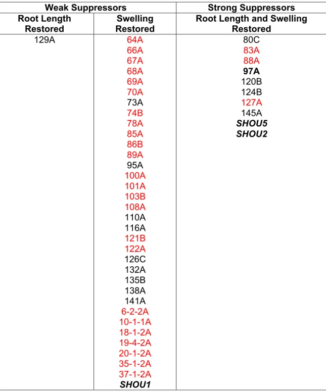
Eight independent suppressor lines that retested as robust fei2 fei2 suppressors
were identified from screening approximately 200,000 M2 seedlings representing
30,000 M1 seeds. We designated these suppressors shou mutations (the Chinese
word for thin). These suppressors represented seven distinct loci, two of which were
allelic and were designated shou2-1 and shou2-2. The fei1 fei2 shou2-1 and fei1 fei2
shou2-2 lines both had significantly fewer and shorter root hairs. The F1 of a
backcross to the parental fei1 fei2 line displayed a non-suppressed phenotype, and
the suppressor phenotype segregated in a ratio of 3 non-suppressed: 1 suppressed
in the F2 progeny of this backcross, consistent with shou2 acting as a single locus,
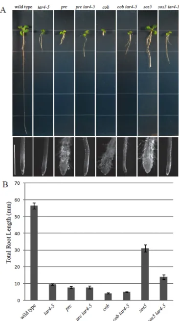
recessive mutation. In addition to the suppression of root length (Fig. 1B), the shou2
mutations also suppress the radial swelling (Fig. 1A) and the radial expansion of
cells in the elongation zone (Fig. 1C) in fei1 fei2 roots. We isolated the shou2-1
mutation by backcrossing to the wild type. This shou2-1 single mutant line displayed
fewer and shorter root hairs, similar to the fei1 fei2 shou2-1. Intriguingly, under the
sucrose), both the fei1 fei2 and shou2-1 parental seedlings displayed roots that were
significantly shorter that their wild-type counterparts, despite the fact that the fei1
fei2 shou2-1 triple mutants displayed nearly wild-type root elongation in the growth
condition that was used for the suppressor screen.
SHOU2 is allelic to IAR4
We used a map-based positional cloning approach to isolate the SHOU2
gene. The fei1 and fei2 mutations (isolated in the Columbia (Col) ecotype) were
introgressed six times into the Landsberg erecta (Ler) ecotype to generate a fei1 fei2
plant that was largely Ler except for small regions of DNA near the fei1 and fei2
mutations (see Methods). This line was crossed to fei1 fei2 shou2 to generate a
mapping population for shou2-1. Mapping with Col/Ler SSLPs indicated that SHOU2
was linked to the top of chromosome 1. Analysis of 350 fei1 fei2 F2 progeny with
additional molecular markers further delimited SHOU2 to a 47 kb interval between
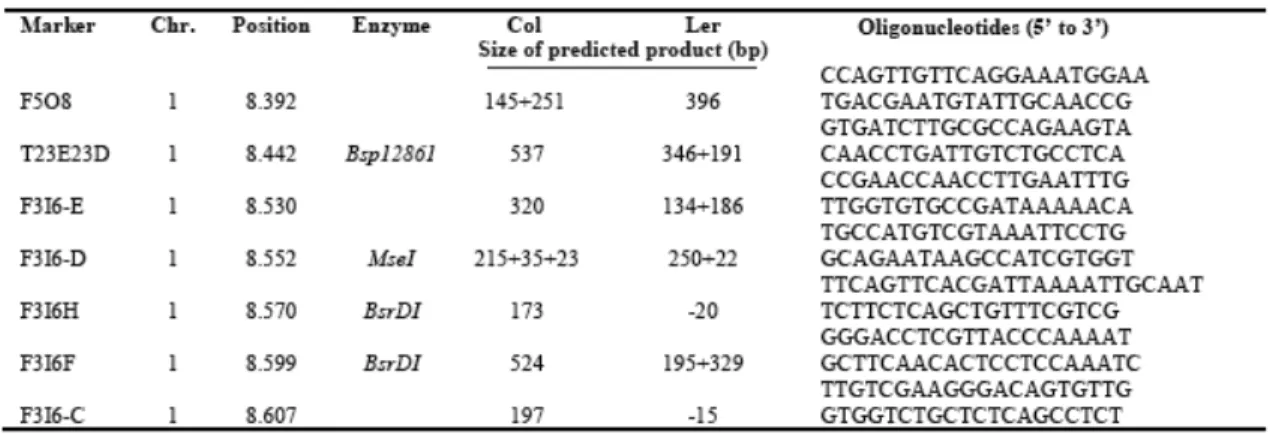
the F3I6.D and F3I6.F markers (Fig. 2A; Table 2.1). Sequencing of candidate genes
within this region identified missense mutations in the first and seventh exon of IAR4
(AT1G24180) of fei1 fei2 shou2-1 and fei1 fei2 shou2-2 respectively. The shou2-1
allele contains a C→T transition in the fifth exon of the coding region of IAR4, which
converts an arginine residue to a stop codon. The shou2-2 mutation is the result of a
G→A transition that is predicted to change a glutamate at position 366 to a stop
codon (Fig. 2B). To confirm that shou2 mutations correspond to AT1G24180, we
examined the ability of an independent DNA insertional allele that contains a
T-DNA insertion in the first exon of IAR4 (SALK_091909) to suppress fei1 fei2. This
phenotype of the roots was examined in non-permissive conditions (Fig. 2B). Similar
to the other alleles, shou2-3 suppressed the root swelling phenotype of fei1 fei2,
which confirms that mutations in IAR4 correspond to shou2. To avoid confusion and
be consistent with the prior studies, we re-named shou2-1, shou2-2, and shou2-3, to
iar4-5, iar4-6, and iar4-7 respectively.
A role for auxin in regulating cell wall function
As IAR4 is involved in the maintenance of auxin homeostasis and mutations
in IAR4 restore anisotropic growth in fei1 fei2, we hypothesized that a reduction in
the level of endogenous IAA would also suppress the loss of growth anisotropy in
fei1 fei2. To test this hypothesis, we examined the effect of mutations in the auxin
biosynthetic genes WEI8 and TAR2 on the fei1 fei2 root swelling phenotype. WEI8
and TAR2 are partially redundant genes that encode two of the five tryptophan
aminotransferases (TAA1) essential for the major auxin biosynthesis pathway in
plants. The level of IAA in the roots of double wei8 tar2 mutants is reduced by 50%
relative to the wild type (Stepanova et al. 2008), suggesting WEI8 and TAR2 are
required for auxin biosynthesis in roots. We generated a wei8 tar2 fei1 fei2
quadruple mutant to examine whether significant reductions in endogenous auxin
levels in the root suppressed growth isotropy in fei1 fei2. When grown under
restrictive conditions, the swelling of the root tip was suppressed in the quadruple
wei8 tar2 fei1 fei2 mutant (Fig. 3). The suppression of the fei1 fei2 phenotype by
wei8 and tar2 is similar to the suppression of fei1 fei2 by iar4 and suggests that
auxin is required for the radial cell expansion that occurs in response to decreases in
To further explore this hypothesis, we investigated the sensitivity of the iar4-5
mutant to the cellulose synthesis inhibitor isoxaben. Previous work has shown that
loss of growth anisotropy is exacerbated in cell wall mutants treated with isoxaben
(Desprez et al. 2002, Scheible et al. 2001). Consistent with these results, fei1 fei2 is
hypersensitive to isoxaben (Xu et al. 2008). However, in contrast to fei1 fei2, both
the triple fei1 fei2 iar4-5 and single iar4-5 are partially resistant to the effects of
isoxaben on root swelling (Fig. 4). The suppression of aberrant cell expansion by
iar4-5 suggests that the effect of the loss of cell wall integrity on root morphogenesis
can be attenuated by a reduction in auxin function.
The effect of iar4 on other fei1 fei2 phenotypes
We have previously shown that the FEI RLKs are required for proper
hypocotyl cell expansion in etiolated seedlings and in anchoring pectin in seed coat
mucilage to the seed surface (Harpaz-Saad et al. 2011). The hypocotyls of
dark-grown fei1 fei2 seedlings are significantly wider than those of the wild type (Xu et al.
2008). In addition, mutations in FEI2 lead to disruption of seed coat mucilage
structure (Harpaz-Saad et al. 2011). We examined whether mutations in IAR4 could
suppress these additional fei1 fei2 phenotypes. In contrast to its role in the root, iar4
did not suppress the increased hypocotyl width phenotype of fei1 fei2 (Fig. 5A and
5B). In fact, the iar4-5 mutant also had slightly wider hypocotyls and this effect was
additive with that of fei1 fei2. The additive nature of iar4-5 and fei1 fei2 on hypocotyl
width suggests that these genes may act in parallel to regulate cell wall function.
Unlike in the hypocotyl, mutations in iar4-5 did not affect the seed coat mucilage of
of fei1 fei2 indicating that mutations in IAR4 do not suppress this phenotype (Fig. 6A
and 6B).
An additional role for the FEI RLKs is to act additively with COBRA (COB) in
stamen filament elongation in the flower. COBRA encodes a GPI anchored protein
that associates with the cell wall and is required for the oriented deposition of
cellulose in rapidly expanding cells (Roudier et al. 2005). Like fei1 fei2, cob-1
mutants are deficient in cellulose and as a result display a short, swollen root
phenotype that is enhanced by elevated sucrose. Although neither the fei1 fei2 nor
1 mutants themselves display an obvious floral phenotype, a triple fei1 fei2
cob-1 mutant has short stamen filaments and as a result is partially infertile (Xu et al.
2008). Similar to root cells in the elongation zone, cells of the stamen filament also
undergo primarily longitudinal expansion. Therefore, we assessed the ability of
iar4-5to suppress the short stamen phenotype of fei1 fei2 cob-1 mutant. Analysis of a
quadruple fei1 fei2 cob-1 iar4-5mutant indicated that the iar4-5allele restores
stamen filament length and fertility in fei1 fei2 cob-1 (Fig. 5C). Thus, iar4-5
suppresses some, but not all of the fei1 fei2 phenotypes.
iar4 is a general suppressor of defects in cell wall synthesis
To ascertain whether loss-of-function mutations in IAR4 suppress defects in
cell expansion exhibited by other cell wall mutants or whether they are specific to the
FEI pathway, we crossed iar4-5 to sos5, procuste (prc; a hypomorphic allele of
cesa6), and a weak allele of cobra, cob-1. When grown in the presence of 4.5%
accompanied by radial expansion of cells in the root tip as a result of reduced
cellulose biosynthesis. As expected, iar4 suppressed the defects in cell expansion
that occur in the sos5 mutation, which acts in the FEI pathway. However, in contrast
to the fei1 fei2 iar4 triple mutant that displayed a substantial suppression of the root
elongation defect observed in both parental lines, the sos5 iar4 double mutant
retains a reduced root elongation phenotype. Thus, the root of the iar4 sos5 double
mutant is short, but not swollen and thus resembles the iar4 parental root
phenotype. iar4-5 also suppresses the swollen root phenotypes of both the cob-1
and prc mutants, both of which affect cellulose synthesis independent of the FEI
pathway (Fig. 7).
We next tested whether mutations in IAR4 could suppress the accumulation
of ectopic lignin in these mutants. Lignin is deposited ectopically into the cell wall in
response to decreased cellulose synthesis that occurs in cellulose deficient mutants.
Previous studies have shown that the roots of fei1 fei2, cob-1, and prc all
accumulate ectopic lignin (Caño-Delgado et al. 2003, Desprez, et al. 2002, Fagard
et al. 2000, Xu et al. 2008). Interestingly, when we assessed the roots of these cell
wall mutants in an iar4-5 background using a colorimetric stain, no ectopic lignin
deposition was observed (Fig. 8). This result is consistent with the suppression of
the root swelling defect in these mutants by iar4 and suggests that IAR4 is required
for the ectopic deposition of lignin that occurs in response to decreased cellulose
biosynthesis. Taken together, these observations suggest that iar4 is not specific to
the FEI pathway, but rather acts as a more general suppressor of defects in
DISCUSSION
We demonstrate that reducing auxin function, either through loss-of-function
mutations in IAR4 or in the auxin biosynthetic genes WEI8 and TAR2, suppresses
the root swelling that occurs in the fei1 fei2 mutant. Several lines of evidence
suggest that iar4, and by inference, auxin, acts not in the FEI pathway directly, but
rather independently to regulate cell wall function. First, iar4 acts additively with fei1
fei2 to increase hypocotyl width. Second, iar4 reverts the swollen root phenotype
and suppresses the accumulation of lignin ectopically in other cellulose synthesis
mutants such as cob-1 and prc, which act in parallel with the FEIs. Finally, the iar4
mutation confers resistance to the cellulose synthesis inhibitor, isoxaben. The data
support a model in which reduced auxin function acts to modulate cell wall function
in the root in some way to counteract the effects of reduced cellulose synthesis.
Previous studies have linked auxin to the regulation of cell wall function. The
acid-growth hypothesis attributes auxin-induced cell expansion to the acidification of
the cell wall and resulting increase in activity of the wall loosening enzymes
expansins (Hager 2003). Expansins disrupt the non-covalent bonds that form
between cellulose and hemicelluloses in the wall and thus promote cell expansion in
hypocotyls and modulate the growth of leaves, petioles, and roots (Cosgrove et al.
2000, Cosgrove et al. 2002, Hager 2003) Auxin Binding Protein (ABP1) may play an
important role in this response; ABP1 activates H+ ATPases and K+ channels at the
plasma membrane upon the perception of auxin and is required for cell elongation
enzymes such as xyloglucan hydrolases (XGH) and endotransglycosylases (XET),
which cleave and re-graft a major form of hemicellulose, xyloglucan, are also
activated upon acidification of the cell wall and in response to auxin (Lorences and
Zarra 1987). Consistent with these findings, the mechanical extensibility of
epidermal cells isolated from azuki bean epicotyls increases dramatically following
incubation with XGH (Kaku et al. 2002). These are among many studies that support
a role for auxin in increasing the extensibility of the cell wall in the shoot.
The role of auxin in roots is less well studied. The notably shorter root of the
auxin-insensitive, gain-of-function Aux/IAA mutant, axr3-1, coupled with the
repression of numerous genes involved in cell wall synthesis and remodeling
suggests a similar role for auxin in regulating cell wall function in the root to that of
the shoot. Among the genes that are de-regulated in axr3-1 seedlings treated with
IAA are those that encode arabinogalactan proteins (AGPs), expansins (EXP),
extensins, proline rich proteins (PRP), xyloglucan endotransglucosylase-hydrolases
(XTHs), and pectin methyl-esterases (PMEs) (Overvoorde et al. 2005). Although
extensins rigidify the cell wall, a disproportionate number of genes repressed in
axr3-1 encoded proteins that loosen the cell wall matrix and thus promote cell
elongation (Cosgrove 2005). Similarly, mutations in the auxin influx carrier, lax3,
prevent the induction of expansin expression in the root of Arabidopsis seedlings in
developing lateral roots. LAX3 is required for lateral root initiation and its expression
precedes the necessary changes in cell wall architecture that are predicted to play a
critical role in the emergence of lateral root primordium (Swarup et al. 2008). The
cell wall remodeling proteins in the axr3-1 and lax3 mutants provide further evidence
that auxin promotes wall loosening.
The growth of seedlings in the presence of auxin has been shown to lead to
root swelling in a manner independent of ethylene biosynthesis (Alarcon et al. 2012,
Eliasson et al. 1989). This suggests that exogenous auxin decreases the integrity of
the cell wall, leading to a loss of growth anisotropy. This is consistent with the
suppression of swelling in mutants defective in cellulose biosynthesis by reduction of
endogenous auxin that is described here. An important question is by what the
mechanism does exogenous auxin increases root swelling in wild-type roots, and
conversely, how does reduced endogenous auxin suppress swelling in
cellulose-deficient roots. One possibility is that auxin negatively regulates cellulose synthesis.
The suppression of the procuste mutant by iar4 makes this somewhat unlikely as
procuste is a null allele of CESA6. However, it is possible that reduced auxin levels
may elevate cellulose synthesis via alternative CESA complexes as CESA6 acts
redundantly with CESA2, CESA5, and CESA9 in some Arabidopsis tissues. This
scenario is unlikely, at least with respect to CESA5 because mutations in IAR4 do
not suppress the defects in seed coat mucilage production in fei1 fei2 where CESA5
is required for cellulose biosynthesis. A somewhat more plausible model is that
auxin modulates the rigidity of the wall not by regulating cellulose synthesis, but by
altering other properties of the wall, such as the crosslinking of cellulose microfibrils
or the activity of extensins or other cell wall modifying enzymes as described above.
Intriguingly, while both iar4 and fei1 fei2 mutant roots are short in the presence of
well as root elongation comparable to the wild type. In contrast, although mutations
in iar4 suppress the swollen root phenotypes of the other cell wall mutants, they do
not restore root elongation in prc, cob, or sos5. This suggests that iar4 does not
simply restore cellulose biosynthesis in these mutants, as this would rescue both the
swollen root and root length phenotypes. Additionally, it is conceivable the short root
phenotype of iar4 may be partially attributed to decreased cell division in the root
apical meristem as auxin has been shown to regulate both the activity and the size
of the meristem in the Arabidopsis root. However, although we isolated iar4 as a
suppressor of fei1 fei2, the short root phenotype of both fei1 fei2 and the iar4 mutant
but not the fei1 fei2 iar4 triple mutant raises an interesting question. How does fei1
fei2 suppress iar4? It is unlikely that it does so by suppressing any potential effects
of iar4 on cell division rates, and thus likely does so through modulation of cell wall
properties.
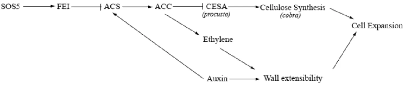
We propose a model that combines the previously characterized role of the
FEI receptor-like kinases in regulating cellulose synthesis with a role for auxin in
regulating cell wall rigidification in the root (Fig 9). We have previously shown that
ACC may act as a signal in the FEI pathway to regulate cellulose biosynthesis. The
fei1 fei2 mutations lead to radial cell expansion in the root as a result of decreased
cellulose synthesis, which alters wall function such that there is not sufficient force to
constrict radial expansion. One model consistent with the data is that decreased
auxin results in an increase in the rigidity of the cell wall, which can restrict radial cell
expansion in cellulose-deficient mutants. In most cases this increase in rigidity would
the length of the root. In the case of the fei1 fei2 iar4 line, this decreased cellulose
coupled with the increased rigidity caused by decreased auxin is precisely balanced,
leading to both a lack of swelling and near wild-type elongation. Auxin also increases
the expression of multiple ACS genes (Abel et al. 1995), which may also have an
effect on the FEI signaling pathway.
Alternatively, auxin could be involved in the signaling cascade linking
perception of perturbation of the cell wall to changes in cell wall synthesis.
Interestingly, a recent study has demonstrated that the inhibition of root cell
elongation that occurs in response to isoxaben is attenuated by mutations in the
tir1-1 auxin receptor and growth in the presence of the synthetic antagonist of TIRtir1-1,
PEO-IAA. Furthermore, results from this study indicated that inhibitors of the
precursor to ethylene, ACC, fully restore growth anisotropy in the presence of
isoxaben and this effect was shown to act independent of ethylene (Tsang et al.
2011). Consistent with this data, inhibitors of ACC, but not ethylene suppress the
swollen root phenotype of fei1 fei2 (Xu et al. 2008).
The characterization of iar4 in this study as a suppressor of defects in cell
wall synthesis provides evidence that auxin plays a key role in the regulation of
primary cell wall function and suggests that wall extensibility may be a major
determinant of cell expansion in the root. Whether auxin acts as a general regulator
of cell wall function throughout development or participates in the active signaling
processes that occur in response to perturbations in the cell wall remains an