OUTCOMES WITH A FOUR-IMPLANT SUPPORTED MONOLITHIC ZIRCONIA DENTAL
PROSTHESIS IN THE MAXILLA
Julie Ann Elpers
A thesis submitted to the faculty at the University of North Carolina at Chapel Hill in partial
fulfillment of the requirements for the degree of Master of Science in the School of Dentistry
(Prosthodontics)
Chapel Hill
2014
ii
iii
ABSTRACT
Julie Ann Elpers: Outcomes with a Four-Implant Supported Monolithic Zirconia Dental Prosthesis in the Maxilla (Under the direction of Lyndon F. Cooper)
A one-arm prospective clinical study undertaken in the spring of 2012 in the Department of Graduate
Prosthodontics aimed to document the treatment efficacy, fabrication and maintenance requirements, and change in patient quality of life with a 4-implant supported fixed prostheses for the edentulous maxilla. Four endosseous implants 6-8mm in length allowed patients previously confined to complete dentures or overdentures to enjoy the comfort and confidence of a fixed, esthetic dental prosthesis. The study sought to espouse a consistently
iv
TABLE OF CONTENTS
LIST OF TABLES ... V
LIST OF FIGURES ... VI
PART I: OUTCOMES WITH A FOUR-IMPLANT SUPPORTED MONOLITHIC ZIRCONIA
DENTAL PROSTHESIS IN THE MAXILLA: IMPLANT AND PROSTHETIC COMPLICATIONS ... 1
INTRODUCTION
... 1
MATERIALS AND METHODS
... 3
RESULTS
... 7
DISCUSSION
... 12
REFERENCES
... 17
PART II: OUTCOMES WITH A FOUR-IMPLANT SUPPORTED MONOLITHIC ZIRCONIA
DENTAL PROSTHESIS IN THE MAXILLA: PATIENT-CENTERED OUTCOMES ... 20
INTRODUCTION
... 20
MATERIALS AND METHODS
... 22
RESULTS
... 24
DISCUSSION
... 31
v
LIST OF TABLES
PART I: OUTCOMES WITH A FOUR-IMPLANT SUPPORTED MONOLITHIC ZIRCONIA
DENTAL PROSTHESIS IN THE MAXILLA: IMPLANT AND PROSTHETIC COMPLICATIONS… ... 1
TABLE 1.1 DEMOGRAPHIC DATA FOR THE STUDY POPULATION. ... 8
T
ABLE1.2
L
OCATION AND DIMENSION OF IMPLANTS PLACED... 9
T
ABLE1.3
D
ESCRIPTION OF IMPLANT FAILURES AND COMPLICATIONS... 10
T
ABLE1.4
D
ESCRIPTION OF NUMBER AND TYPE OF ABUTMENT USED,
BY LOCATION... 10
T
ABLE1.5.
C
OMPLICATIONS WITH DENTURE CONVERSION PROSTHESES,
MILLED INTERIM PROSTHESES,
AND TRY-
IN RESINS... 11
PART II: OUTCOMES WITH A FOUR-IMPLANT SUPPORTED MONOLITHIC ZIRCONIA
DENTAL PROSTHESIS IN THE MAXILLA: PATIENT-CENTERED OUTCOMES ... 20
T
ABLE2.1
D
EMOGRAPHIC DATA FOR THE STUDY POPULATION. ... 25
T
ABLE2.2
B
ASELINEOHIP-49
SEVERITY AND EXTENT SCORES,
BY DESCRIPTIVE GROUP. ... 27
T
ABLE2.3
P
OST-
TREATMENTOHIP-49
SEVERITY AND EXTENT SCORES,
BY DESCRIPTIVE GROUP. ... 27
T
ABLE2.4
B
ASELINEOHIP-49
SCORES BY DOMAIN... 29
T
ABLE2.5
P
OST-
TREATMENTOHIP-49
SCORES BY DOMAIN... 30
vi
LIST OF FIGURES
PART I: OUTCOMES WITH A FOUR-IMPLANT SUPPORTED MONOLITHIC ZIRCONIA
DENTAL PROSTHESIS IN THE MAXILLA: IMPLANT AND PROSTHETIC COMPLICATIONS ... 1
F
IGURE1.1
A AND B.
D
UPLICATED COMPLETE DENTURE PLACED FOR RADIOGRAPHIC/
SURGICAL GUIDE... 4
F
IGURE1.2.
E
XAMPLE OF PLANNED IMPLANT SITES IN NATIVE BONE. ... 4
F
IGURE1.3
A-
C.
S
URGICAL APPROACH FOR IMPLANT PLACEMENT... 5
F
IGURE1.4
A-
C.
F
INAL IMPRESSION TECHNIQUE. ... 6
F
IGURE1.5
A-
D.
P
ROSTHESIS FABRICATION WORKFLOW... 6
F
IGURE1.6
A AND B.
P
OSTOPERATIVE PANOREX FOR SINGLE ARCH AND DUAL ARCH CASES... 7
F
IGURE1.7
A AND B.
C
HIPPING OF THE VENEERING PORCELAIN... 11
PART II: OUTCOMES WITH A FOUR-IMPLANT SUPPORTED MONOLITHIC ZIRCONIA
DENTAL PROSTHESIS IN THE MAXILLA: PATIENT-CENTERED OUTCOMES ... 20
F
IGURE2.1
A-
C.
E
XAMPLES OF BASELINE DENTITION FOR INCLUDED PATIENTS... 23
1
CHAPTER I
OUTCOMES WITH A FOUR-IMPLANT SUPPORTED MONOLITHIC ZIRCONIA DENTAL PROSTHESIS IN THE MAXILLA: IMPLANT AND PROSTHETIC COMPLICATIONS
INTRODUCTION
Edentulism is a chronic disability that interferes with everyday tasks such as eating, speaking, and socializing (Roumanas 2009). Well-constructed, tissue-borne complete dentures remain an economical and relevant treatment option for the edentulous patient (Cooper 2009), yet complete dentures are not without disadvantages and there exist a significant proportion of patients who desire completely fixed prosthetics.
The long-term success (Adell et al 1990, Romeo et al 2004, Jemt et al 2006) and widespread acceptance of dental implants in the edentulous maxilla has made the full-arch fixed prosthesis a choice alternative to
conventional tissue-supported complete dentures and implant-retained removable appliances. Surgical protocols for the placement, loading, and restoration of osseointegrated implants in the edentulous maxilla have been described and evaluated in the literature (Lambert et al. 2009).
A multitude of approaches have been successfully used to rehabilitate the edentulous, atrophic maxilla (Toljanic et al 2009, Bidra et al 2012). While six or more implants were commonly advocated (Norton and Ferber 1999, Kinsel and Lamb 2003), four implants have been reported to be sufficient to support full-arch loading in the maxilla provided the anterior-posterior spread allows for cantilevers of less than 10mm anteriorly and posteriorly (Maló et al 2005, Jensen et al 2010). However, continued bone resorption and pneumatization of the sinuses often necessitates a modified approach to implant therapy to ensure sufficient distribution of the four fixtures. Broadly, these approaches may be arranged into three categories, and may be used in combination: 1)
augmentation of the edentulous ridge and/or sinus, 2) placement of dental implants into the zygoma or pterygomaxillary region, and 3) use of iplants with limited length/height.
2
or vertical augmentation by particulate or block grafting) continues to be an area of research requiring well-designed clinical trials with long-term follow-up (Atieh et al 2012). Preliminary results (Felice et al 2011) from a short-term study comparing 5-8.5mm implants vs. autogenous grafting procedures followed by 11.5mm implant placement suggest a lower rate of postoperative complications with short implants and similar implant success rates. Roughened-surface short implants do not show higher failure rates than implants >10mm, with one meta-analysis reporting implant survival rate in the range of 89.6%-100%, and no correlation between implant length and odds of failure (Sun et al. 2011). Additionally, shorter implants (8-9mm) have been shown to be successful in the long term (10 year) time horizon, and surface, location, prosthetic modality and bone quality do not seem to impact failure rates (Romeo et al 2006, Atieh et al 2012, Metens et al 2012).
Despite implant survival rates of 95-99%, long term data indicate survival rates for screw-retained fixed prostheses in the maxilla to be 82-84%, with complication-free survival as low as 9% (Mertens and Steveling 2011a and 2011b; Fischer and Stenberg 2011). A systematic review of 281 one-piece implant-supported complete dental prostheses (mean exposure time of 9.5 years) determined a rate of prosthetic complication to be 24.6% per 100 restoration-years. The cumulative rates of "prosthesis free of complications" after 5 and 10 years were 29.3% and 8.6%, respectively. The most common technical complication reported in the same systematic review was chipping of the veneering material, occurring 33.3% at 5 years and 66.6% at 10 years (Papaspyridakos et al 2012). Recent advances in materials and prosthesis design employing monolithic zirconia as well as lithium disilicate (Pozzi et al 2013, Limmer et al 2014) have reported far fewer incidences of chipping of the veneering material over the 1-3 year timeframe.
3
significant improvement in patient satisfaction and factors related to eating comfort, function and esthetics (Dierens et al 2009, Erkapers et al 2011, Limmer et al 2014).
The primary objectives of this one-arm prospective pilot study are to evaluate the survival of dental implants of limited width/length placed in native bone in the atrophic maxilla, and the long-term prosthetic outcome of fixed implant- supported maxillary complete prosthesis made of monolithic zirconia. The null hypothesis, therefore, can be stated as: a four-implant supported, screw-retained maxillary dental prosthesis made of monolithic zirconia will have implant and prosthetic complication rates comparable to those previously reported for implant-supported reconstructions in the maxilla.
MATERIALS AND METHODS
The details of the proposed study were submitted to the University of North Carolina at Chapel Hill (UNC-CH) Office of Human Research Ethics for IRB review, and the study was approved in March, 2012 (IRB# 12-0447). A total of 65 patients were screened from the patient pool at the UNC-CH School of Dentistry, as well as the community at large who responded to an online posting on the School’s website. Twenty patients who met the following inclusion/exclusion criteria were consecutively enrolled between April and October, 2012.
Inclusion Criteria
between 18-80 at time of enrollment
good physical health (ASA Class I or II)
edentulous in the maxilla or have a terminal dentition of eight or fewer teeth
maxillary vertical bone height of at least 6 mm and width of 4mm in the selected implant sites
no history of radiotherapy in head and neck region
willing to give informed consent
Exclusion Criteria
poor physical health or uncontrolled medical condition (ASA Class III or IV)
heavy smokers (in excess of 10 cigarettes/day)
vertical bone height less than 6mm and less than 4mm in width in any of the implant sites
severe Angle’s class II/III jaw relationships
inadequate vertical space for the definitive prosthesis
history of radiotherapy in head and neck region
issues in accepting a removable prosthesis (unwilling to wear dentures, severe gag reflex, etc.)
known alcohol and/or drug abuse
patient with unrealistic esthetic expectations
4
All patients with a terminal dentition were edentulated and allowed to heal for 3 months under an immediate denture. The remaining edentulous patients underwent fabrication of a new maxillary removable complete denture to optimize esthetics and function and aid in treatment planning for the future fixed implant-supported prosthesis. The maxillary denture was duplicated in radioopaque acrylic (BiocrylX, Great Lakes Orthodontics, Tonawanda, NY) to fabricate a radiolographic/surgical guide (Figure 1.1a and b). An 8cmx8cm Cone Beam
Computed Tomography (CBCT) volume (Sirona Orthophos XG 3D Dental CT Scanner, New York, NY), was obtained. Implant planning software (SimplantPro version 15, Materialise Dental, Waltham, MA) was used to map desired sites having both adequate bone for implant placement (6mm height x 4mm width) and congruence with tooth set-up (Figure 1.2). The surgical guide was marked and drilled by hand.
Figure 1.1 a and b. Duplicated maxillary complete denture placed for radiographic/surgical guide in (a) single-arch case opposing natural dentition and (b) dual single-arch case.
5
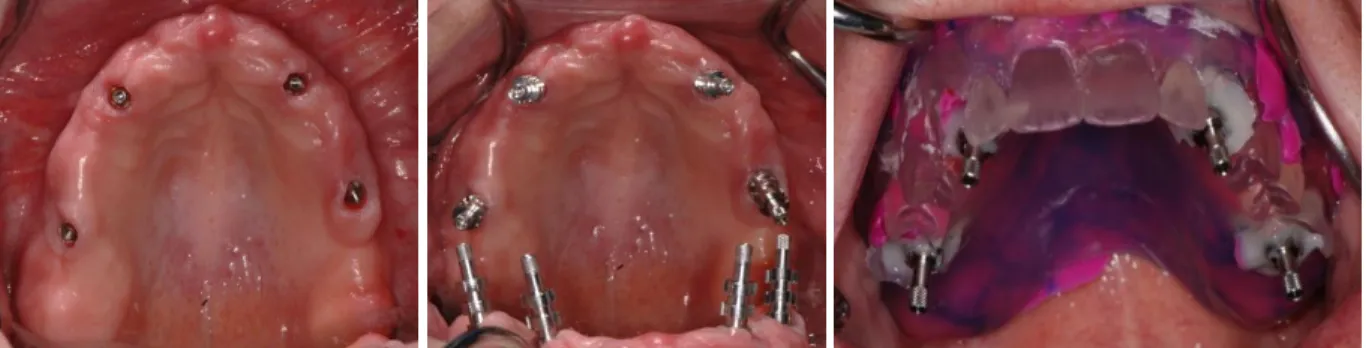
Under local anesthesia, sites were marked with the surgical guide and a crestal incision made to visualize the maxillary ridge (Figure 1-3a); all necessary alveolectomy was performed using a rongeur and/or e-cutter bur in a straight handpiece under copious irrigation. Four AstraTech Osseospeed™TX implants (Dentsply, Waltham, MA) were placed by standard protocol in planned locations of the maxilla and cover screws placed finger-tight (Figure 1.3b). Should a patient present with inadequate bone volume for implant placement in a particular site at the time of surgery, a plan was made for either a) placement into an alternate site, or b) the implant was placed in the planned site and any resulting dehiscence or fenestration was grafted by means of autogenous bone and bone substitute (Bio-Oss, Geistlich, Princeton, NJ) placed and secured with a collagen membrane (Bio-Gide, Geistlich, Princeton, NJ). Incisions were closed with 4-0 chromic gut suture (Figure 1.3c), the denture relined with soft tissue conditioner (Coe-comfort, GC America, Alsip, IL), and the patient followed up after two weeks to assess healing.
Figure 1.3a-c. Surgical approach for implant placement: (a) crestal incision made following marking of sites with surgical guide, (b) placement of cover screws, (c) incisions closed completely for at least 12 weeks of healing under complete denture.
6
Figure 1.4a-c. Final impression technique. UniAbutments torqued into implants following second-stage surgery (a), abutment-level impression pick-ups placed (b), and pick-ups secured into surgical guide washed with PVS (c).
As a first step in fabrication of the final prosthesis, a conversion prosthesis (Figure 1.5a) was made by picking up UniAbutment temporary cylinders (AstraTech, Dentsply, Waltham, MA) in the maxillary denture with quick-set autopolymerizing acrylic (Pro-Tech PLUS, Boynton Beach, FL). In cases where the denture was thin or the patient requested to remain in a removable prosthesis, this step was omitted. A mock-up of the final tooth set-up on the master cast was sent to the dental laboratory for digital design of the definitive prosthesis (Figure 1.5b). A milled resin prosthesis was fabricated, tried in, and adjusted for optimal phonetics, esthetics, and function (Figure 1.5c). This milled resin prosthesis was returned to the dental laboratory for refinement of the digital design and final fabrication of the monolithic zirconia prosthesis (Microdental, Morrisville, NC or ZirkonZahn GMBH, Gais, Italy). (Figure 1.5d).
7
Figure 1.5a-d. Prosthesis fabrication. The conversion prosthesis (a) is fabricated as both an interim restoration and to guide laboratory design of the final prosthesis (b) for this dual-arch case. Resin mock-ups are milled and tried in (c), returned to the laboratory, and the definitive monolithic zirconia prosthesis is milled, stained, sintered, and gingival porcelain applied (d).
At the delivery appointment, passive fit of the definitive prosthesis was evaluated using the one-screw test. All bridge screws were torqued to 15Ncm and access holes covered with cotton pellets and light-cure composite (Filtek, 3M ESPE). Any necessary intraoral occlusal adjustments were carried out sequentially with blue, pink, and grey polishing wheels (Dialite, Brasseler, Savannah, GA). A post-op Panorex was exposed at this point and evaluated (Figure 1.6a and b).
Figure 1.6a and b. Postoperative panorex for single arch (a) and dual arch (b) cases.
A hard acrylic occlusal guard was fabricated (Great Lakes Orthodontics, Tonawanda, NY) and fitted to the patient at a follow-up appointment 2-4 weeks after delivery. A 12-, 24-, and 60-month examination and
radiographic follow-up after prosthesis delivery will be completed to evaluate implant and prosthesis survival, complications and patient quality of life.
RESULTS
Sixty-five patients who were edentulous or had terminal dentition in the maxilla were screened. Twenty
patients were enrolled: fifteen (75%) were edentulous at baseline, the remaining five had a terminal dentition.
8
edentulated had severe (>5mm) periodontal attachment loss around most remaining teeth. Eleven patients (55%)
had been edentulated more than 2 years prior to enrollment.
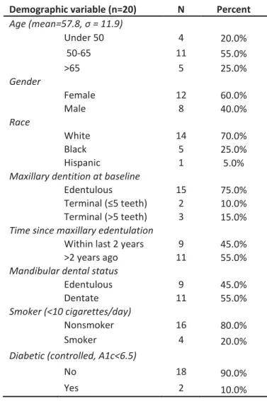
The study population (Table 1.1) can be described as mostly white (70%), smoking (80%), and
non-diabetic (85%). Twelve patients were female. In addition, nine patients had an existing or planned four-implant
supported mandibular prosthesis made of monolithic zirconia. The remaining cohort had either entirely natural
dentition (n=4), or some combination of implant restorations and natural dentition (n=7).
Table 1.1 Demographic data for the study population.
Demographic variable (n=20) N Percent
Age (mean=57.8, σ = 11.9)
Under 50 4 20.0%
50-65 11 55.0%
>65 5 25.0%
Gender
Female 12 60.0%
Male 8 40.0%
Race
White 14 70.0%
Black 5 25.0%
Hispanic 1 5.0%
Maxillary dentition at baseline
Edentulous 15 75.0%
Terminal (≤5 teeth) 2 10.0%
Terminal (>5 teeth) 3 15.0%
Time since maxillary edentulation
Within last 2 years 9 45.0%
>2 years ago 11 55.0%
Mandibular dental status
Edentulous 9 45.0%
Dentate 11 55.0%
Smoker (<10 cigarettes/day)
Nonsmoker 16 80.0%
Smoker 4 20.0%
Diabetic (controlled, A1c<6.5)
No 18 90.0%
9
Twenty patients underwent implant therapy in healed ridges using a two-stage approach (total number of
AstraTech Osseospeed TX implants placed: 85). Nearly half (48.7%) of implants were 4.0x6mm in dimension; the
remaining proportion were predominantly 4.0x8mm. There was no dimensional predilection for anterior/posterior
sites (Table 1.2). Almost all (94.1%) of implants were placed in native bone; five implants were placed in
previously-augmented sinus grafts with at least 6 months of healing prior to placement. Two patients received a
fifth implant in a central incisor site at the time of initial implant placement (to minimize anterior cantilever).
Panoramic radiographs were exposed following implant surgery and following final prosthesis placement.
The radiographs were evaluated for change in crestal bone height around implants. Those implants that
demonstrated a change in bone height on Panorex of at least 1mm were labeled “at risk” for future complications.
These findings are also summarized in Table 1.2.
Table 1.2. Location and dimension of implants placed, as well as location and dimension of implants determined to be “at risk” following loading (defined as bone height change ≥1mm following placement), as assessed by postoperative Panorex.
Implant Dimensions
4x6mm 4x8mm Total
Anterior sites (#6-11) 19 22 41
Implants at risk 3 2 5
Posterior sites(#3-5, #12-14) 21 20 41
Implants at risk 1 1 2
Total Implants loaded 40 42 82
Total implants at risk 4 3 7
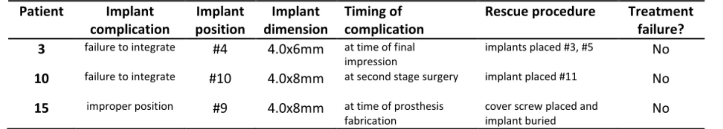
There were two early implant failures (2.4%), discovered at the time of second-stage surgery or shortly
thereafter (at the time of final impression). In another patient, implant malposition due to limited bone volume
prevented its incorporation into the prosthetic design, and the implant was buried (Table 1.3). Therefore, 82
implants were loaded with fixed, full-arch prostheses. Most implants (87.8%) were loaded prior to final fabrication
of monolithic zirconia restoration with an acrylic conversion or interim prosthesis. The average time since implant
10
Table 1.3. Description of implant failures and complications.
Patient Implant complication Implant position Implant dimension Timing of complication
Rescue procedure Treatment failure?
3 failure to integrate #4 4.0x6mm at time of final
impression
implants placed #3, #5 No
10 failure to integrate #10 4.0x8mm at second stage surgery implant placed #11 No
15 improper position #9 4.0x8mm at time of prosthesis
fabrication
cover screw placed and
implant buried No
For each osseointegrated implant, a 20- or 45-degree Uniabutment, or AstraTech Angled abutment, was
used. Twenty-degree UniAbutments of varying heights were placed in 70.7% of implants. Angle corrections were
largely required in the anterior sites, while posterior sites more often required use of 45-degree abutments to aid
in prosthesis draw (Table 1.4).
Table 1.4. Description of number and type of abutment used, by location.
20-degree UniAbutment 45-degree UniAbutment Angled Abutment
Anterior Implant sites 30 0 11
n=41
Posterior implant sites 28 10 3
n=41
Total 58 (70.7%) 10 (12.2%) 14 (17.1%)
Twelve treatment visits were planned per study protocol, and included both intake and all surgical and
prosthetic follow-ups. Table 1.5 enumerates the number of additional visits required between final impression
and prosthesis delivery, and complications with conversion prostheses, milled resin interim prostheses, and try-in
11
Table 1.5. Complications with denture conversion prostheses, milled interim prostheses, and try-in resins, assessed by number of additional patient appointments outside 12-visit treatment protocol.
Number of patients
Median number of additional appointments
per patient
Total number of additional appointments Additional try-in of new denture set-up or
lab-fabricated resin mock-up
12 1 (range= 1-3) 21
Abutment switch or new final impression 5 1 (range=0-1) 5
Interim prosthesis fracture or complication 6 1 (range=1-5) 14
Post-delivery adjustment 5 1 (range=1-2) 7
Total additional appointments 1.5 (range=0-8) 47
To date, the average time since definitive prosthesis delivery is 3.5 months (range 0.9-7.8 months). There
has been one prosthetic complication observed in one patient, which was discovered at the post-insertion visit.
The clinician noticed chipping of the porcelain veneering, which had not been noticed by the patient (Figure 1.7a).
This patient, a bruxer, was one of three patients who received a monolithic zirconia prosthesis framework with
facial porcelain veneering on the anterior 6 teeth.
12
DISCUSSION
The present study found a high level of osseointegration and demonstrated no treatment failures with up
to 1.3 years (mean: 9.1 months) of prosthetic loading and up to eight months after definitive monolithic zirconia
prosthesis delivery. Surgical revision for patients who experienced dental implant failures was managed by placing
an implant into an adjacent site the very day or day following discovery of failed osseointegration. It should further
be noted that both of the patients with primary failure of osseointegration of maxillary fixtures had a history of
severe periodontitis and implant failure in the mandible.
At the time of definitive prosthesis placement, all 82 implants were soundly integrated and without
evidence of peri-implant infection or chronic inflammation around abutments. An initial assessment of bone level
changes on Panorex was performed by visually comparing bone level around implants immediately following
placement and after final prosthetic loading. Generally, 8-12 months elapsed between these time points and the
implants had been loaded for an average of 5.7 months (range: 0-11.8 months) with an interim or conversion
prosthesis before delivery of the definitive prosthesis. Implants with bone level height changes of 1mm or more
were noted to be “at risk.” This “at risk” description was applied to 7 implants in five patients (8.5% at implant
level, 25% at the patient level), and no implant demonstrated more than 2mm of bone adaptation.
Though this data is short term, it is certainly indicative of the possibility that a “minimally invasive”
approach to implant therapy for a fixed, full-arch prosthesis may be of value. There are many approaches to
placement of dental implants in the atrophic maxilla, irrespective of final prosthetic design. Maló et al (2011)
discussed 5-year outcomes of a study cohort who underwent immediate loading in the maxilla on four implants: 25
out of 221 patients lost 41 implants, giving a patient-level survival rate of 88.7% and an implant-specific survival
rate of 95.8%. These outcomes are remarkably consistent with implant outcomes in this investigation to date. If
we are to operate on the premise that at least four dental implants are necessary for optimal reclamation of a
functional dental arch (Maló et al 2005; Jensen and Adams 2009), three approaches to the atrophic maxilla can be
broadly defined: bone grafting, employment of the zygoma or trans-sinus implants (Maló et al 2013), and use of
13
There have been two randomized controlled trials designed to address the use of short implants as
opposed to sinus augmentation for longer implants in the edentulous maxilla. Preliminary results from a
short-term study comparing the use of 5-8.5mm implants vs. autogenous iliac crest grafting and 11.5mm implants in the
edentulous maxilla suggested a lower rate of postoperative complications with short implants and similar implant
success rates. (Felice et al 2011). A second RCT used a split-mouth design to compare maxillary sinuses augmented
with particulate porcine bone via a lateral window (implants placed simultaneously) with 4x6mm implants in
native bone. Of the 20 patients treated, 15 patients preferred short implants, whereas 5 patients treated with
maxillary implants described both procedures as equally acceptable (Esposito et al 2012). This was largely
attributed to expedited treatment time and minimized surgical intervention.
The rate of early implant failures (2.4%) observed in this study was consistent with previously-reported
rates of failure for all types of short dental implants (Atieh et al 2012, Al-Hashedi et al 2014). Both patients who
experienced these “early failures” were edentulous in the maxilla for at least two years and exhibited severe
periodontal attachment loss around mandibular teeth. Parel and Phillips (2011) enumerated risk factors for dental
implant failure for a maxillary immediate-load protocol: opposing natural dentition, male gender, lack of bone density, the distal implant site, and parafunction. While this investigation is insufficiently powered to draw conclusions on risk factors for early implant failures, continued follow-up may reveal predictive factors for patients in whom this implant approach may be more risky.
Zitzmann and Marinello (2000) enumerated three broad prosthetic approaches to implant therapy for the
edentulous maxilla: implant-supported fixed partial dentures, the “hybrid” fixed [full-arch] implant restoration,
and the removable overdenture. The segmental fixed partial denture concept, applicable in cases of limited bone
resorption, is often championed for retrievability and cleansibility despite higher treatment costs. The full-arch
implant supported fixed denture, broadly defined, is capable of restoring soft and hard tissue deficits in the
edentulous maxilla. However, there are two major challenges inherent in this prosthetic approach. The first is the
patient’s lip position on animation, lip support, and tonus of the orofacial muscles. The prosthesis must be
carefully designed to address these issues and provide optimum dental and facial esthetics. The second major
challenge involves the architecture of the prosthesis with respect to the palatal anatomy and implant position.
14
implants; however, the contour of the palatal prosthesis as it abuts the tissue is often incongruent with optimal
phonetics and alveolar resorption may necessitate palatal positioning of the dental implants. The removable
overdenture, therefore, is often cited as a simple means by which to address these two contouring challenges and
removes the additional oral hygiene difficulties inherent in fully implant-supported fixed restorations (Fortin et al
2002). An acrylic denture base more readily accommodates soft and hard tissue deficiencies and is generally
considered less costly to replace.
This study attempted to address the above challenges in fixed maxillary prostheses and employed a
CAD-CAM fabrication process with potential for decreased manufacture costs. In terms of prosthesis design and
fabrication, the complexity and variability in conversion, try-in, and number of adjustments for this particular
approach cannot be underemphasized. The twelve treatment visits planned per study protocol allowed for a
four-appointment complete denture fabrication process and one resin-try-in for the lab-fabricated prosthesis.
However, the length of time during which patients were wearing an interim implant supported restoration (often
>6 months) necessitated multiple repairs, and in most cases multiple esthetic/phonetic try-ins were necessary. In
five cases the abutment selection required revision following CAD and a new final impression was needed.
Generally, the reason for delay in prosthesis fabrication was due to the intricacies of converting an “analog”
denture design into an esthetic, phonetic, and functional milled resin, and converting the approved resin mock-up
into a final esthetic prosthesis acceptable to the patient. A previous study (Limmer et al 2014), assessed the value
of a mandibular implant-supported fixed denture made of monolithic zirconia in the mandible. Issues with
phonetics, tissue seam morphology, and patient esthetic expectations inherent in maxillary fixed prostheses were
not addressed. While this CAD/CAM approach to dental prosthesis fabrication is certainly promising, there exists a
certain need for clinical attention to detail in maxillary full-arch prostheses, and additional treatment visits at the
try-in phase certainly underscores this issue.
Regarding the choice of dental material selected for this investigation, monolithic zirconia suprastructures
were selected based on the potential for good esthetic outcome without veneering, and high strength. Zirconia
has a monoclinic crystal structure from room temperature to 1170°C, tetragonal from 1170°C to 2370°C, and cubic
at temperatures above 2370°C. Upon cooling, a volume expansion of 3% to 5% is associated with the transition of
15
stabilize the tetragonal phase during cooling: in particular, yttria-stabilized zirconia has been developed specifically
for dental purposes. Transformation toughening refers to the transition from stabilized tertragonal to monoclinic
phase as a result of stress, surface treatment, or temperature changes (Al-Amleh, 2010). The volume expansion
resulting from the transformation can suppress cracks, increase toughness, or induce compressive surface stresses.
While transformation toughening would appear to address concerns with crack propagation and fatigue,
an accelerated ageing phenomenon is known as low-temperature degradation (LTD) causes a decrease in physical
properties by spontaneous phase transformation of the zirconia crystals from the tetragonal phase to the weaker
monoclinic phase in the presence of water (Flinn et al 2012). Conceivably, should a critical proportion of the bulk
material transition to the monoclinic phase, this would result in spontaneous catastrophic failure of the material.
Much has been made of the possibility of LTD: however, no data exist to demonstrate the frequency or time
course over which this may occur, or clinical relevance.
In this study, there were no observed fractures or failure of the monolithic zirconia component of the
implant-supported fixed dentures. While seventeen patients received purely monolithic prostheses without
veneer, three patients received prostheses with porcelain veneering along the facial of the incisors as means of
optimizing esthetics. One of these three patients, a bruxer, exhibited chipping of the veneer after only four weeks
in function. This underscores the major issues with veneer porcelain fracture reported in the literature (Guess et al
2012).
Because of the aforementioned limitations with regard to removal and hygiene with implant supported
fixed “hybrid” prostheses, it is worthwhile to note that zirconia has been found to be more biocompatible than
other ceramics, heat-cured resin, titanium, and metal alloys (Nakamura et al 2010). In vitro studies have
demonstrated significantly lower bacterial adhesion to polished zirconia surfaces when compared to titanium
(Rimodini et al 2004 and Scarano et al 2010). This is particularly important when the prosthesis is in direct contact
with the gingival tissue, as with an implant-supported full-arch maxillary prosthesis. This question has previously
been addressed for edentulous resin, metal, and acrylic prostheses. Kanao et al (2013) found that milled titanium
was superior to composite resin in terms of plaque adhesion to the intaglio surface of the fixed dental prosthesis
and resulted in lower levels of mucosal blood flow (and thereby inflammation). Another recent study (Abi Nader
16
tissue-surface area covered with plaque after 6 months despite rigorous oral hygiene instruction. This is certainly
of interest in the context of this study, as by and large patients wore an acrylic or resin interim prosthesis for 6
months or more following second-stage surgery. Given the fact that 8.5% of implants experienced some bone loss
from placement to definitive prosthesis delivery (up to 12 months), it is not lost on the investigators that the
manner in which patients are provisionalized may certainly matter. Anecdotally, a marked decrease in mucosal
inflammation and edema was noted between the day of prosthesis placement and follow-up 2-4 weeks later. As
clinicians and laboratories seek to improve and optimize the workflow in fabricating CAD/CAM zirconia
restorations, this provisionalization phase may be reduced in importance. Yet, for this investigation, it remains to
be seen whether the unanticipated complications in prosthesis try-in, design, and long term use of resin and acrylic
provisionals will have a significant negative effect on long-term tissue and implant health.
Certainly, this study served to describe a novel treatment approach and the challenges therein. While the
approach to implant therapy was indeed conservative with regard to implant number, dimension and loading,
design and management of the monolithic zirconia prosthesis proved complicated. All enrolled patients currently
have implant-supported full-arch monolithic zirconia prostheses supported by four or five implants delivered and
functioning, demonstrating a high level of clinical success in the short term. It is the objective of the accompanying
17
REFERENCES
Abi Nader S, Eimar H, Momani M, Shang K, Daniel NG, Tamimi F. Plaque Accumulation Beneath Maxillary All-on-4™ Implant-Supported Prostheses. Clin Implant Dent Relat Res. 2014 Jan 27
Adell R, Eriksson B, Lekholm U, Brånemark PI, Jemt T. Long-term follow-up study of osseointegrated implants in the treatment of totally edentulous jaws. Int J Oral Maxillofac Implants. 1990 Winter;5(4):347-59.
Al-Amleh B, Lyons K, Swain M. Clinical trials in zirconia: a systematic review. J Oral Rehabil. 2010 Aug;37(8):641-52.
Al-Hashedi AA, Ali TB, Yunus N. Short dental implants: An emerging concept in implant treatment. Quintessence Int. 2014 Mar 7.
Atieh MA, Zadeh H, Stanford CM, Cooper LF. Survival of short dental implants for treatment of posterior partial edentulism: a systematic review. Int J Oral Maxillofac Implants. 2012 Nov-Dec;27(6):1323-31.
Bassi F, Carr AB, Chang TL, Estafanous EW, Garrett NR, Happonen RP, Koka S, Laine J, Osswald M, Reintsema H, Rieger J, Roumanas E, Salinas TJ, Stanford CM, Wolfaardt J.Economic outcomes in prosthodontics. Int J Prosthodont. 2013 Sep-Oct;26(5):465-9. doi: 10.11607/ijp.3405. Review.
Bidra AS, Agar JR, Parel SM. Management of patients with excessive gingival display for maxillary complete arch fixed implant-supported prostheses. J Prosthet Dent. 2012 Nov;108(5):324-31.
Cooper, L. F. (2009), The Current and Future Treatment of Edentulism. Journal of Prosthodontics, 18: 116–122.
Dierens M, Collaert B, Deschepper E, Browaeys H, Klinge B, De Bruyn H. Patient-centered outcome of immediately loaded implants in the rehabilitation of fully edentulous jaws. Clin Oral Implants Res. 2009 Oct;20(10):1070-7. Epub 2009 Aug 30.
Erkapers M, Ekstrand K, Baer RA, Toljanic JA, Thor A. Patient satisfaction following dental implant treatment with immediate loading in the edentulous atrophic maxilla. Int J Oral Maxillofac Implants. 2011 Mar-Apr;26(2):356-64.
Esposito M, Cannizzaro G, Soardi E, Pistilli R, Piattelli M, Corvino V, Felice P. Posterior atrophic jaws rehabilitated with prostheses supported by 6 mm-long, 4 mm-wide implants or by longer implants in augmented bone. Preliminary results from a pilot randomised controlled trial. Eur J Oral Implantol. 2012 Spring;5(1):19-33.
Felice P, Soardi E, Pellegrino G, Pistilli R, Marchetti C, Gessaroli M, Esposito M. Treatment of the atrophic
edentulous maxilla: short implants versus bone augmentation for placing longer implants. Five-month post-loading results of a pilot randomised controlled trial. Eur J Oral Implantol. 2011 Autumn;4(3):191-202.
Fischer K, Stenberg T. Prospective 10-Year Cohort Study Based on a Randomized Controlled Trial (RCT) on Implant-Supported Full-Arch Maxillary Prostheses. Part 1: Sandblasted and Acid-Etched Implants and Mucosal Tissue. Clin Implant Dent Relat Res. 2011 Oct 18.
18
Flinn BD, deGroot DA, Mancl LA, Raigrodski AJ. Accelerated aging characteristics of three yttria-stabilized
tetragonal zirconia polycrystalline dental materials. J Prosthet Dent. 2012 Oct;108(4):223-30. doi: 10.1016/S0022-3913(12)60166-8.
Fortin, Y., Sullivan, R.M., Rangert, B.R. The Marius implant bridge: surgical and prosthetic rehabilitation for the completely edentulous upper jaw with moderate to severe resorption: a 5-year retrospective clinical study. (2002) Clinical implant dentistry and related research, 4 (2), pp. 69-77.
Guess PC, Att W, Strub JR. Zirconia in fixed implant prosthodontics. Clin Implant Dent Relat Res. 2012 Oct;14(5):633-45.
Jemt T, Johansson J. Implant treatment in the edentulous maxillae: a 15-year follow-up study on 76 consecutive patients provided with fixed prostheses. Clin Implant Dent Relat Res. 2006;8(2):61-9.
Jensen OT, Adams MW, Cottam JR, Parel SM, Phillips WR 3rd. The All-on-4 shelf: maxilla. J Oral Maxillofac Surg. 2010 Oct;68(10):2520-7.
Jensen OT, Adams MW. The maxillary M-4: a technical and biomechanical note for all-on-4 management of severe maxillary atrophy--report of 3 cases. J Oral Maxillofac Surg. 2009 Aug;67(8):1739-44.
Kanao M, Nakamoto T, Kajiwara N, Kondo Y, Masaki C, Hosokawa R. Comparison of plaque accumulation and soft-tissue blood flow with the use of full-arch implant-supported fixed prostheses with mucosal surfaces of different materials: a randomized clinical study.
Kinsel RP, Lamb RE. Gingival esthetics for immediately restored dental implants: prosthodontic and surgical procedures. Int J Oral Maxillofac Implants. 2003 Sep-Oct;18(5):760-1.
Lambert FE, Weber HP, Susarla SM, Belser UC, Gallucci GO. Descriptive analysis of implant and prosthodontic survival rates with fixed implant-supported rehabilitations in the edentulous maxilla. J Periodontol. 2009
Aug;80(8):1220-30. Clin Oral Implants Res. 2013 Oct;24(10):1137-43. doi: 10.1111/j.1600-0501.2012.02523.x. Epub 2012 Jul 18.
Limmer B, Sanders AE, Reside G, Cooper LF. Complications and Patient-Centered Outcomes with an Implant-Supported Monolithic Zirconia Fixed Dental Prosthesis: 1 Year Results. J Prosthodont. 2014 Jan 6.
MacEntee MI, Walton JN: The economics of complete dentures and implant-related services: a framework for analysis and preliminary outcomes. J Prosthet Dent 1998; 79 :24-30
Maló P, Rangert B, Nobre M. All-on-4 immediate-function concept with Brånemark System implants for completely edentulous maxillae: a 1-year retrospective clinical study. Clin Implant Dent Relat Res. 2005;7 Suppl 1:S88-94.
Maló P, Nobre Md, Lopes A. The rehabilitation of completely edentulous maxillae with different degrees of resorption with four or more immediately loaded implants: a 5-year retrospective study and a new classification. Eur J Oral Implantol. 2011 Autumn;4(3):227-43.
Maló P, Nobre Md, Lopes A. Immediate loading of 'All-on-4' maxillary prostheses using trans-sinus tilted implants without sinus bone grafting: a retrospective study reporting the 3-year outcome. Eur J Oral Implantol. 2013 Autumn;6(3):273-83.
19
Mertens C, Meyer-Bäumer A, Kappel H, Hoffmann J, Steveling HG. Use of 8-mm and 9-mm implants in atrophic alveolar ridges: 10-year results. Int J Oral Maxillofac Implants. 2012 Nov-Dec;27(6):1501-8.
Nakamura K1, Kanno T, Milleding P, Ortengren U. Zirconia as a dental implant abutment material: a systematic review. Int J Prosthodont. 2010 Jul-Aug;23(4):299-309.
Norton MR, Ferber C. The nonresilient hybrid removable prosthesis: treatment of choice for the atrophic maxilla. Int J Periodontics Restorative Dent. 1999 Apr;19(2):189-97.
Papaspyridakos P, Chen CJ, Chuang SK, Weber HP, Gallucci GO. A systematic review of biologic and technical complications with fixed implant rehabilitations for edentulous patients. Int J Oral Maxillofac Implants. 2012 Jan-Feb;27(1):102-10.
Parel SM, Phillips WR. A risk assessment treatment planning protocol for the four implant immediately loaded maxilla: preliminary findings. J Prosthet Dent. 2011 Dec;106(6):359-66
Pozzi A, Tallarico M, Barlattani A. Monolithic lithium disilicate full-contour crowns bonded on CAD/CAM zirconia complete-arch implant bridges with 3 to 5 years of follow-up. J Oral Implantol. 2013 Nov 4.
Rimondini L, Cerroni L, Carrassi A, Torricelli P. Bacterial colonization of zirconia ceramic surfaces: an in vitro and in vivo study. Int J Oral Maxillofac Implants. 2002 Nov-Dec;17(6):793-8.
Romeo E, Ghisolfi M, Rozza R, Chiapasco M, Lops D. Short (8-mm) dental implants in the rehabilitation of partial and complete edentulism: a 3- to 14-year longitudinal study. Int J Prosthodont. 2006 Nov-Dec;19(6):586-92.
Romeo E, Lops D, Margutti E, Ghisolfi M, Chiapasco M, Vogel G. Long-term survival and success of oral implants in the treatment of full and partial arches: a 7-year prospective study with the ITI dental implant system. Int J Oral Maxillofac Implants. 2004 Mar-Apr;19(2):247-59
Roumanas ED. The Social Solution—Denture Esthetics, Phonetics, and Function. Journal of Prosthodontics, 2009 18: 112–115.
Scarano A, Piattelli M, Caputi S, Favero GA, Piattelli A. Bacterial adhesion on commercially pure titanium and zirconium oxide disks: an in vivo human study. J Periodontol. 2004 Feb;75(2):292-6.
Sun HL, Huang C, Wu YR, Shi B. Failure rates of short (≤ 10 mm) dental implants and factors influencing their failure: a systematic review. Int J Oral Maxillofac Implants. 2011 Jul-Aug;26(4):816-25.
Toljanic JA1, Baer RA, Ekstrand K, Thor A. Implant rehabilitation of the atrophic edentulous maxilla including immediate fixed provisional restoration without the use of bone grafting: a review of 1-year outcome data from a long-term prospective clinical trial. Int J Oral Maxillofac Implants. 2009 May-Jun;24(3):518-26.
20
CHAPTER II
COMPLICATIONS WITH A FOUR-IMPLANT SUPPORTED MONOLITHIC ZIRCONIA DENTAL PROSTHESIS IN THE MAXILLA: PATIENT-CENTERED OUTCOMES
INTRODUCTION
Due to increased public health initiatives related to oral healthcare, it has previously been observed that edentulism is declining at a rate of about 1% per year in industrialized countries. While this fact may suggest that there will be a decreasing need for treatment interventions for edentulous patients, the average life expectancy and total population are increasing, and therefore the number of edentulous patients is remaining stable or slightly increasing. By 2020, is predicted that there will be approximately 37.9 million edentulous elderly adults in the United States (Douglass 2002). Edentulism is a chronic disability that interferes with everyday tasks such as eating, speaking, and socializing (Roumanas 2009). Well-constructed tissue-borne complete dentures remain an economical and relevant treatment option for the edentulous patient (Cooper 2009), yet complete dentures are not without disadvantages and there exist a significant proportion of patients who desire completely fixed prosthetics.
The cost of an implant-retained or implant-supported prosthesis has been calculated to be between 5 and 12 times more expensive than a conventional denture (MacEntee and Walton 1998). To justify this expense, as well as subsequent maintenance expenses, data must exist to indicate that the implant-supported prosthesis provides benefit to the patient congruent with the added expense. Bassi et al (2013) recently highlighted the paucity of data to support clinical decision-making in terms of initial and maintenance costs for prosthodontic care and their bearing on patient perceptions of benefit and satisfaction. To date, immediate full-arch
21
Any healthcare intervention should be systematically evaluated for a measureable improvement to improve the health of an individual or population. To this end, an instrument must be able to describe the nature, direction, and magnitude of change, and experimental studies may be designed to identify predictor of change and control for confounders. On a population scale, benefits include identification of sub-groups within the population requiring care, monitoring of at risk groups, targeting of financial resources and monitoring outcomes of clinical interventions.
Locker (1988) employed an existing model of oral health to define seven function domains and relate them hierarchically: disease can lead to impairment, then functional limitation, and ultimately physical, psychologic, and social disability/discomfort. Based on this, The Oral Health Impact Profile (OHIP-49) was specifically designed to measure the impact of oral health on psychosocial well-being (Slade and Spencer 1994). This questionnaire consists of 49 items that cover seven specific domains: functional limitation, physical pain, psychological discomfort, physical disability, psychological disability, social disability, and handicap. At the present time, measures of oral health-related quality of life have been mainly used in descriptive population studies, predominantly in older adults. These studies have established that the measures are reliable and show good concurrent validity when judged against clinical, subjective and behavioral criteria. A few studies (Awad et al 2000, Allen et al 2006, Brennan et al 2010) have employed versions of OHIP (including the shortened form, OHIP-14) to compare OHR-QoL between treatment groups: for example, conventional dentures vs. two-implant overdentures, or overdentures vs. implant-supported fixed prostheses. A recent review (de Souza et al 2013) highlighted the need for increased attention to patient-centered outcomes in removable prosthodontics trials.
22
Therefore, careful assessment and comparison of OHR-QoL pre- and post-treatment is of importance and provides a measure of treatment effectiveness for a given approach. Additionally, an understanding of patient factors as they relate to OHR-QoL pre- and post-treatment may further aid in clinical decision making. This study aims to assess baseline, post-treatment and change in OHR-QoL as measured by OHIP-49 for a specific treatment modality: placement of four dental implants into native bone by two-stage approach, and restoration with a monolithic zirconia implant-supported fixed denture.
MATERIALS AND METHODS
The details of the proposed study were submitted to the University of North Carolina at Chapel Hill (UNC-CH) Office of Human Research Ethics for IRB review, and the study was approved in March, 2012 (IRB# 12-0447). A total of 65 patients were screened from the patient pool at the UNC-CH School of Dentistry, as well as the
community at large who responded to an online posting on the School’s website. Twenty patients who met the following inclusion/exclusion criteria were consecutively enrolled between April and October, 2012.
Inclusion Criteria
between 18-80 at time of enrollment,
good physical health (ASA Class I or II)
edentulous in the maxilla or have a terminal dentition of eight or fewer teeth
maxillary vertical bone height of at least 6 mm and width of 4mm in the selected implant sites
no history of radiotherapy in head and neck region
willing to give informed consent
Exclusion Criteria
poor physical health or uncontrolled medical condition (ASA Class III or IV)
heavy smokers (in excess of 10 cigarettes/day)
vertical bone height less than 6mm and less than 4mm in width in any of the implant sites
severe Angle’s class II/III jaw relationships
inadequate vertical space for the definitive prosthesis
history of radiotherapy in head and neck region
issues in accepting a removable prosthesis (unwilling to wear dentures, severe gag reflex, etc.)
pregnant women
known alcohol and/or drug abuse
patient with unrealistic esthetic expectations
any other conditions that contraindicate dental implant therapy.
23
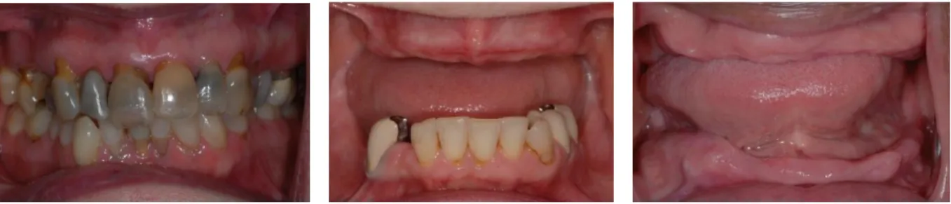
remaining edentulous patients (Figure 2.1b and c) underwent fabrication of a new maxillary removable complete denture to optimize esthetics and function and aid in treatment planning for the future fixed implant-supported prosthesis. A Cone Beam Computed Tomography (CBCT) volume (Sirona Orthophos XG 3D Dental CT Scanner, New York, NY) and implant planning software (SimplantPro version 15, Materialise Dental, Waltham, MA) was used to plan implant sites.
Figure 2.1a-c. Examples of baseline dentition for included patients: (a) natural teeth with terminal dentition in the maxilla, (b) existing maxillary edentulous condition opposing at least 5 natural teeth, (c) existing maxillary and mandibular edentulism.
Four AstraTech Osseospeed™TX implants (Dentsply, Waltham, MA) were placed by standard protocol in planned locations of the maxilla. Incisions were sutured and implants were completely covered for at least 12 weeks of submucosal healing. Postoperatively, the denture was relined with soft tissue conditioner (Coe-comfort, GC America, Alsip, IL), and the patient was seen for two-week follow-up to assess healing.
Following healing, second-stage surgery was conducted under local anesthesia, implants were evaluated for stability, and the appropriate AstraTech UniAbutment or Angled Abutment (Dentsply, Waltham, MA) was selected and torqued to 20 Ncm. An abutment-level impression was made for fabrication of the master cast, and a mock-up of the final tooth set-up on the master cast was sent to the dental laboratory for digital design of the final
prosthesis. A milled resin try-in prosthesis was fabricated, tried in, and adjusted for optimal phonetics, esthetics, and function. This milled resin was returned to the dental laboratory for final fabrication of the monolithic zirconia prosthesis (Microdental, Morrisville, NC or ZirkonZahn GMBH, Gais, Italy).
The definitive prosthesis (Figure 2.2a and b) was evaluated for fit and then delivered to the patient. A hard acrylic occlusal guard was fabricated (Great Lakes Orthodontics, Tonawanda, NY) and fitted to the patient at a follow-up appointment 2-4 weeks after delivery. At this time, the follow-up OHIP-49 questionnaire was
60-24
month examination and radiographic follow-up after prosthesis delivery will be completed to evaluate implant and prosthesis survival, complications and patient quality of life.
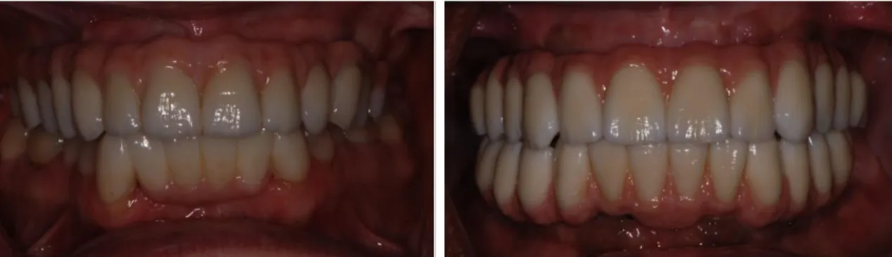
Figure 2.2a and b. Examples of maxillary single arch (a) and dual arch (b) monolithic zirconia implant-supported fixed dentures.
Two methods were used for OHIP-49 scoring: the severity score was calculated as the sum total of responses on an ordinal scale (0-4) for the 49 items. The response “very often” was scored as 4 and “never” was scored as 0, meaning that severity scores range from 0-196 with “0” considered ideal. The extent score was a count of the number of items to which the response “very often” or “fairly often” was given (range 0-49). In addition, items were scored according to functional domain to attempt to identify which parameters of OHR-QoL were most important at baseline and post-treatment. Statistical analysis of OHIP-49 data was carried out using IBM SPSS Statistics for Windows, Version 22.0 (IBM Corp, Armonk, NY). No correction for multiple tests was used as the baseline and post-treatment observations were generally one year apart. Because of the small sample size and highly variable responses, normality in OHIP-49 scores could not be assumed and nonparametric tests were applied when assessing for differences in median OHIP-49 severity and extent scores at the two time points and between demographic groups. Univariate ANOVA was applied, however, when examining variance in the change
in severity and extent scores at baseline and post-treatment. Significance level was set at 0.05.
RESULTS
Sixty-five patients who were edentulous or had terminal dentition in the maxilla were screened. Twenty
patients were enrolled: fifteen (75%) were edentulous in the maxilla at baseline, the remaining five had terminal
25
patients who were edentulated had severe (>5mm) periodontal attachment loss around most remaining teeth.
Eleven patients (55%) had been edentulated more than 2 years prior to enrollment.
The study sample (Table 2.1) can be further described as mostly white (70%), non-smoking (80%), and
non-diabetic (85%). Twelve patients were female. In addition, nine patients had an existing or planned
four-implant supported mandibular prosthesis made of monolithic zirconia. The remaining cohort had either entirely
natural dentition (n=4), or some combination of implant restorations and natural dentition (n=7).
Table 2.1 Demographic data for the study population.
Demographic variable (n=20) N Percent
Age (mean=57.8, σ = 11.9)
Under 50 4 20.0%
50-65 11 55.0%
>65 5 25.0%
Gender
Female 12 60.0%
Male 8 40.0%
Race
White 14 70.0%
Black 5 25.0%
Hispanic 1 5.0%
Maxillary dentition at baseline
Edentulous 15 75.0%
Terminal (≤5 teeth) 2 10.0%
Terminal (>5 teeth) 3 15.0%
Time since maxillary edentulation
Within last 2 years 9 45.0%
>2 years ago 11 55.0%
Mandibular dental status
Edentulous 9 45.0%
Dentate 11 55.0%
Smoker (<10 cigarettes/day)
Nonsmoker 16 80.0%
Smoker 4 20.0%
Diabetic (controlled, A1c<6.5)
No 18 90.0%
Yes 2 10.0%
Twenty patients underwent implant therapy in healed ridges using a two-stage approach (total AstraTech
26
placed in two patients with previously-augmented sinus grafts with at least 6 months of healing prior to
placement. Two early implant failures (2.4%) were discovered at the time of second-stage surgery or shortly
thereafter (at the time of final impression). In another patient, implant malposition due to limited bone volume
prevented its incorporation into the prosthetic design and the implant was buried. Therefore, 82 implants were
loaded with fixed, full-arch prostheses. Most implants (87.8%) were loaded prior to final fabrication of monolithic
zirconia restoration with an acrylic conversion or interim prosthesis. To date, average time since implant loading is
9.1 months (range 1.2-14 months); average time since definitive prosthesis delivery is 3.5 months (range 0.9-7.8
months).
At baseline, the median OHIP-49 severity score was 59 (range: 5-161), and median extent score was 9
(range: 0-38). The median post-treatment severity score was 8 (range: 0-29), median post-treatment extent score
was 0 (range: 0-4). Related-samples Wilcoxon signed rank test demonstrated significant differences between
baseline and post-treatment OHIP-49 severity and extent scores (p=0.001 for both cases, 2-sided test).
Five variables were assessed for differences in severity and extent scores: age group (under 50, 50-65,
over 65), gender, edentulous vs. terminal dentition at baseline, time since maxillary edentulation (within last 2
years), and mandibular dental status (Tables 2.2 and 2.3). No statistically significant differences in OHIP-49 severity
or extent scores at baseline or post-treatment between groups separated by age group (independent samples
Kruskal-Wallis test), gender, edentulous/terminal dentition, or time since edentulation (independent-samples
Mann-Whitney U test). At baseline, patients edentulous in the maxilla opposing natural dentition had significantly
higher OHIP-49 severity and extent scores when compared to those who had either existing or planned mandibular
full-arch implant-supported fixed dentures (independent-samples Mann-Whitney U test). However, there was no
significant difference post-treatment between the single-arch and dual-arch cases in terms of OHIP-49 severity or
27
Table 2.2. Baseline OHIP-49 severity and extent scores, by descriptive group.
N
Median OHIP-49
severity score p-valuea
Median
OHIP-49 extent score p-valuea
Age
under 50 4 83.5 (12-161) 0.747b 15.0 (3-38) 0.486b
50-65 11 60 (5-132) 10 (0-28)
>65 5 58 (11-86) 5 (0-13)
Gender
female 12 69.5 (5-161) 0.057c 11.5 (0-38) 0.082c
male 8 48.5 (11-86) 4 (0-13)
Maxillary dentition at baseline
Edentulous 15 58 (5-161) 0.866c 8 (0-38) 0.553c
Terminal dentition 5 60 (50-114) 10 (5-25)
Time since maxillary edentulation
Within last 2 years 9 53 (12-114) 0.331c 8 (0-25) 0.776c
>2 years ago 11 77 (5-161) 10 (0-38)
Mandibular dental status
Fixed prosthesis 9 53 (5-86) 0.046c
5 (0-13) 0.046c
Dentate 11 77 (11-161) 13 (0-38)
aExact significance (2-sided test) b
Independent-Samples Kruskal-Wallis Test
cIndependent-Samples Mann-Whitney U Test
Table 2.3. Post-treatmentOHIP-49 severity and extent scores, by descriptive group.
N
Median OHIP-49
severity score p-valuea
Median OHIP-49
extent score p-valuea
Age
under 50 4 15 (15-18) 0.347b 0 (0-4) 0.521b
50-65 11 6 (0-24) 0 (0-2)
>65 5 4 (0-29) 0 (0-1)
Gender
female 12 6 (0-29) 0.442c 0 (0-4) 0.657c
male 8 13.5 (0-27) 0 (0-2)
Maxillary dentition at baseline
Edentulous 15 8 (0-29) 0.530c 0 (0-2) 0.665c
Terminal dentition 5 10.5 (5-22) 0 (0-4)
Time since maxillary edentulation
Within last 2 years 9 16.5 (5-27) 0.020c 0 (0-4) 0.177c
>2 years ago 11 3 (0-29) 0 (0-0)
Mandibular dental status
Fixed prosthesis 9 7 (0-24) 0.778c 0 (0-2) 0.0840c
Dentate 11 9 (0-29) 0 (0-4)
a
Exact significance (2-sided test)
b
Independent-Samples Kruskal-Wallis Test
28
Participants who had been edentulated within the past 2 years had a significantly higher median OHIP-49
severity scores following treatment (Mann-Whitney U, p=0.02). There were no other significant differences
between OHIP-49 severity and extent scores following treatment between the descriptive groups.
Data were further analyzed for differences in median OHIP-49 across the seven functional domains (Table
2.4 and 2.5). Generally, there were no differences between the descriptive groups at baseline and post-treatment
for physical pain, physical disability, psychological disability, social disability, and handicap. However, women and those who had natural dentition in the lower arch had significantly higher scores (Mann-Whitney U, p=0.02 and
0.025, respectively) for the domain Psychological Discomfort. Also, those who had natural dentition in the lower
arch had significantly higher scores in the Functional Limitation domain when compared to those with lower
implant-supported prostheses (Mann-Whitney U, p=0.025). Following treatment, for patients who had been
edentulous in the maxilla for less than 2 years, post-treatment OHIP-49 was significantly higher across the
Functional Limitation Mann-Whitney U, p=0.002) and Physical Disability (Mann-Whitney U, p=0.02) domains.
Finally, median change in OHIP-49 severity and score was found to be different for males (33,
range=-6-86) and females (62, range=38-146; Mann-Whitney U, p=0.031), and median change in OHIP-49 extent score was
different among those edentulous in one arch only (13, rangle=0-38)compared to those with upper andlower ISFDs
(3, range -2-13; Mann-Whitney U, p=0.038). Univariate ANOVA (Table 2.6) found gender to be significant to
explain the variance in OHIP-49 change from baseline to post-treatment for both scoring methods (p=0.041,
R-squared=0.211, and p=0.048, R-squared=.200), respectively. Additionally, post-treatment mandibular dental status
29
Table 2.4. Baseline OHIP-49 scores for domains with significant differences between descriptive groups.
OHIP-49 Domain*
Functional Limitation (0-36)
Psychological discomfort (0-20)
N Median p-valuea Median p-valuea
Age
under 50 4 22.5 (4-33) 0.621b 14(0-20) 0.758b
50-65 11 16 (3-31) 9 (0-20)
>65 5 11 (6-23) 11 (0-14)
Gender
female 12 21.5 (3-33) 0.115c 14.5 (0-20) 0.020c
male 8 14.5 (4-23) 5.5 (0-14)
Maxillary dentition at baseline
Edentulous 15 16 (3-33) 0.800c 11 (0-20) 0.735c
Terminal dentition 5 16 (13-29) 9 (8-20)
Time since maxillary edentulation
Within last 2 years 9 16 (4-29) 0.456c
8 (0-20) 0.230c
>2 years ago 11 22 (3-33) 14 (0-20)
Mandibular dental status
Fixed prosthesis 9 16 (3-23) 0.025c 8 (0-14) 0.025c
Dentate 11 22 (6-33) 15 (0-20)
*For simplicity, only domains for which significant differences between groups were detected are shown.
a
Exact significance (2-sided test)
bIndependent-Samples Kruskal-Wallis Test c
30
Table 2.5. Post-treatment OHIP-49 scores for domains with significant differences between descriptive groups.
OHIP-49 Domain*
Functional Limitation (0-36)
Physical Disability (0-36)
N Median p-valuea Median p-valuea
Age
under 50 4 6 (5-6) 0.206b 3 (2-7) 0.357b
50-65 11 3 (0-8) 1 (0-7)
>65 5 2 (0-10) 2 (0-1)
Gender
female 12 3 (0-6) 0.272c 2 (0-10) 0.351c
male 8 5 (0-10) 5 (0-7)
Maxillary dentition at baseline
Edentulous 15 2 (0-10) 0.262c 2 (0-10) 0.665c
Terminal dentition 5 5 (3-6) 2.5 (0-7)
Time since maxillary edentulation
Within last 2 years 9 6 (3-10) 0.002c
5 (0-7) 0.020c
>2 years ago 11 2 (0-5) 0 (0-10)
Mandibular dental status
Fixed prostheis 9 3 (0-8) 0.778c 2 (0-7) 0.840c
Dentate 11 4 (0-10) 2 (0-10)
*For simplicity, only domains for which significant differences between groups were detected are shown.
a
Exact significance (2-sided test)
bIndependent-Samples Kruskal-Wallis Test c
31
Table 2.6. Univariate ANOVA for changes in OHIP-49 severity and extent scores among descriptive groups.
N
Mean change in OHIP-49
severity score p-valuea
R-squared Mean change OHIP-49 extent score p-valuea R-squared Age
under 50 4 73.0 0.575 0.063 16.7 0.313 0.128
50-65 11 60.8 10.8
>65 5 40.6 5.6
Gender
female 12 74.9 0.041 0.211 14.5 0.048 0.200
male 8 33.1 5.0
Maxillary dentition at baseline
Edentulous 15 58.2 1.000 0.000 10.7 0.981 0.000
Terminal dentition 5 58.2 10.8
Time since maxillary edentulation
Within last 2 years 9 42.2 0.163 0.105 8.1 0.340 0.051
>2 years ago 11 71.2 12.8
Mandibular dental status
Fixed prosthesis 9 37.8 0.069 0.172 5.0 0.026 0.246
Dentate 11 74.9 15.4
a
Exact significance (2-sided test)
DISCUSSION
In assessing the effectiveness of an oral health intervention, the OHIP-49 can be employed philosophically in a number of ways. It provides descriptive information on OHR-QoL among the study cohort in their current dental state. It can also be employed as means of measuring the impact of a treatment intervention by comparing pre-and post-treatment OHIP-49 scores (Awad et al 2000, Allen et al 2006, Erkapers et al 2011, Limmer et al 2014). Along with the primary objective of describing a novel treatment approach to the edentulous maxilla, this study sought to define, to some extent, a population for which this treatment approach may be most useful. This is based on the premise that a significant decrease in OHIP-49 severity and extent scores is indicative of improved oral-health related quality of life and, by extension, overall quality of life.
This study found that the median OHIP-49 severity score was 59 (range: 5-161) at baseline, and 8 (range:
0-29) post-treatment. The baseline and post-treatment medians were significantly different, and the median
change in OHIP-49 score from baseline to post-treatment was 54 points (range: -6-194). This is certainly indicative
32
heterogeneity in responses. While all but one patient showed a positive change in baseline and post-treatment
OHIP-49 severity scores, it is interesting to assess which aspects of OHR-QoL changed significantly over the course
of the study, and for which patients the change was most marked.
In the present study, the additive method was used to score the OHIP-49, wherein each item response was coded (0=never, through 4=very often) and the responses summed. This was chosen from among the three most common methods of scoring in the literature: additive, simple count, and weighted score. Simple count assigns a dichotomy to responses and sums the total number of items indicating disability/discomfort/limitation, etc. The weighted score is based on the Thurstone paired-comparison technique and assigns weights to items before summing them (five of the 49 items have a weight >2, with the balance weighted between 1.106-1.925). Allen and Locker (1997) found that additive and weighted scores are highly correlated and distinguish equally well between oral health status variables (edentulous/dentate, dry mouth, <20/≥20 teeth, self-rated oral health, perceived oral health need). The correlations of scores and ability of the simple count method to distinguish between groups were far lower. The authors further concluded that weighting did not substantially improve performance of the OHIP-49.
The extent score is another important aspect to OHR-QoL assessment. At baseline, the median extent score was 9 (range: 0-38), and median post-treatment extent score was 0 (range: 0-4). It is reassuring to the clinician that the study population experienced an overall, consistent decrease in number of OHIP-49 items to which “very often” and “fairly often” were considered responses and by the end of the study only four patients had extent scores greater than zero. This is indicative of the relatively low number of patients with significant OHR-QoL issues in the weeks post-treatment, and is probably to be somewhat expected in the 2 to 4 weeks following prosthesis delivery.