EFFECTS OF LOCAL MUSCLE VIBRATION ON CARTILAGE DEFORMATION IN ANTERIOR CRUCIATE LIGAMENT RECONSTRUCTED INDIVIDUALS ACUTELY AFTER WALKING
Allison S Barnes
A thesis submitted to the faculty at the University of North Carolina at Chapel Hill in partial fulfillment of the requirements for the degree of Master of Arts in the Department of Exercise
and Sport Science (Athletic Training).
Chapel Hill 2019
ABSTRACT
Allison S Barnes: Effects of local muscle vibration on cartilage deformation in anterior cruciate ligament reconstructed individuals acutely after walking
(Under the direction of Troy Blackburn)
Objectives: To examine the effects of local muscle vibration (LMV) on cartilage
deformation acutely after walking in individuals with anterior cruciate ligament reconstruction (ACLR). Design: Cross-over study. Participants: 12 ACLR individuals ages 18-35.
Interventions: Participants performed isometric squats while being exposed to LMV or no
vibration (control). Interventions were delivered in a counterbalanced design. Main outcome measures: Cartilage cross-sectional area (CSA) (mm2), quadriceps and hamstring
TABLE OF CONTENTS
LIST OF FIGURES ... v
LIST OF TABLES... vi
LIST OF ABBREVIATIONS ... vi
CHAPTER 1: INTRODUCTION ... 1
CHAPTER 2: LITERATURE REVIEW... 5
Introduction ... 5
Knee Osteoarthritis... 6
Definition, Prevalence & Consequences... 6
Gait Mechanics ... 6
Proprioception ... 8
Co-contraction & Absorption of Forces ... 9
Effects of Altered Gait Mechanics on Cartilage ... 12
Cartilage Deformation and Viscoelasticity ... 13
Cartilage Imaging ... 14
Local Muscle Vibration ... 15
Rationale ... 17
CHAPTER 3: METHODS... 24
Experimental Design ... 24
Participants ... 24
Data Collection Procedures ... 25
EMG Data Collection ... 25
Ultrasonographic Image Analysis... 26
Treadmill Walking Protocol... 26
Local Muscle Vibration ... 27
Data Reduction ... 27
Statistical Analysis ... 28
CHAPTER 4: RESULTS ... 29
CHAPTER 5: DISCUSSION ... 32
LIST OF FIGURES
Figure 1 – Preparatory Co-activation Graph………30
Figure 2 – Heelstrike Co-activation Graph………...30
Figure 3 – Weight Acceptance Graph………...…30
LIST OF TABLES
LIST OF ABBREVIATIONS
ACLD—anterior cruciate ligament deficient ACLR—anterior cruciate ligament reconstruction AMI—arthrogenic muscle inhibition
LMV—local muscle vibration
PTOA—post traumatic osteoarthritis RPP—reproduction of passive positioning
CHAPTER 1: INTRODUCTION
Anterior cruciate ligament tears are the most common and most serious ligamentous injury to the knee, and can have irreversible long-term consequences despite successful surgical reconstruction (ACLR) and rehabilitation.1 Post-traumatic osteoarthritis (PTOA), a form
of osteoarthritis that occurs following traumatic injury to a joint, is a debilitating disease affecting as many as 25% of patients just 5 years post-ACLR.2 PTOA presents as degeneration of
cartilage and bone in a joint, and leads to subsequent symptoms of pain, stiffness and functional disability, which can dramatically affect quality of life.3,4 Much research has evaluated risk
factors that contribute to the initiation and progression of primary OA, but less research has focused on prevention of PTOA following ACL injury.
Degradation of cartilage structure following a traumatic injury can potentially lead to PTOA. Cartilage structure can be assessed by measuring its cross-sectional area and the change in cross-sectional area (i.e. strain), and is representative of the overall health of the joint.5 In healthy individuals, acute loads placed upon a joint result in cartilage deformation;
however, aberrant loads may have detrimental effects.6 Cartilage imaging can be performed
using diagnostic ultrasound techniques, and has proven to be a reliable and sensitive tool for evaluating cartilage thickness changes acutely.7 This cartilage degradation is influenced by a
variety of factors, some of which may be modifiable. With the end goal being a decrease in cartilage degradation and hopeful lessening of PTOA consequences, it is necessary to establish and evaluate the contributing variables that play a role in deterioration of joint health.
with altered biomechanics.8 Similar to individuals with primary OA, individuals with ACLR tend to
display lesser knee flexion angles, greater knee adduction angles, and slower walking speeds during walking compared to healthy matched controls.9–14 These alterations, in combination, can lead to greater loading magnitudes and rates through the knee.11 These greater loading rates
and magnitudes are associated with greater cartilage breakdown, suggesting that greater loading rates may be linked to the development of PTOA.15–17
While the factors that contribute to aberrant gait biomechanics are poorly understood, altered somatosensory function and muscle activation patterns have been implicated.
Proprioception is a measure of somatosensory function that assesses the central nervous system’s ability to sense how a joint is positioned in space, and is reliant on joint receptors and other mechanoreceptors around the joint (e.g. tenomuscular and cutaneous receptors) to provide necessary information.18 In the case of injury, decreases in proprioception are common,
partially due to the loss of receptors and subsequent decline in neuromuscular control. The ACL is a key structure in providing not only mechanical stability to the knee joint, but also significant proprioceptive input. When the ACL is ruptured, neural pathways that would normally send vital information to the central nervous system are no longer able to do so.18,19 When sensory
feedback is not functioning at its optimal level, neuromuscular control is suboptimal and may contribute to aberrant gait biomechanics, such as heightened co-contraction and decreased knee flexion during gait.18,20–22 ACLR individuals have been seen to have heightened co-activation in their ACLR limb compared to contralateral limb, as well as when compared to healthy controls. (Blackburn manuscript in review) ACLR individuals have a perceived sense of instability, which in turn, may cause this heightened co-activation of the quadriceps and
When the neuromuscular system is not functioning properly, muscle activation is directly affected. In ACLR individuals, as well as those with primary OA, a deficit of voluntary activation (VA) of the quadriceps can be seen.24,25 This quadriceps dysfunction directly impacts gait
biomechanics. When the quadriceps is not functioning at its optimal level, it becomes unable to effectively eccentrically load upon weight acceptance and single-leg support during walking. In combination with sensory instability, dysfunction of the quadriceps can further increase co-activation and subsequent aberrant gait biomechanics. Without the ability to quickly produce force through the quadriceps, the individual lands more stiffly, with less knee flexion to absorb and attenuate forces, which likely places loads of greater magnitude on the articular
cartilage.23,24,26–28
Restoring full quadriceps function is rarely adequately achieved, despite being a primary goal of rehabilitation following ACLR. Other strategies to improve overall muscle function are necessary. Vibratory stimulation has become a popular supplement to exercise, as it is reported to improve activation, strength, power, EMG activity, and overall function.29–33 Vibratory stimuli have been shown to improve neuromuscular deficits that are often seen in ACLR individuals; quadriceps activation and strength, as well as proprioception, as vibration directly stimulates sensory receptors and muscle fibers.20,34 Studies have looked at vibration and its impact on
proprioception and neuromuscular function, however no studies have looked at direct correlations between vibration and factors related to PTOA, such as cartilage imaging. By targeting these variables and restoring neuromuscular function, improvements in gait
devices can be more portable, cost effective, clinically applicable, while being just as effective as WBV interventions.
The purpose of this study was to evaluate the effects of local muscle vibration (LMV) on cartilage deformation in the knee acutely after walking. In theory, vibratory stimuli will enhance proprioception and gait mechanics, leading to better absorption of forces, and increased acute cartilage deformation.
The specific aims of this study included the following:
1. To determine the acute effect of LMV on co-activation during walking in individuals with ACLR. We hypothesize that co-activation of the quadriceps and hamstrings will decrease following LMV compared to a control (no intervention) condition.
2. To determine the acute effect of LMV on cartilage deformation after walking in individuals with ACLR. We hypothesize that cartilage deformation will increase following LMV compared to a control (no intervention) condition.
CHAPTER 2: LITERATURE REVIEW
Introduction
Anterior cruciate ligament tears are the most common and most serious ligamentous injury to the knee, particularly in the active population. Many individuals undergo reconstructive surgery in order to regain stability of the knee, allowing them to return to prior functional levels. Especially in the athletic community, the main goal tends to be return to participation as early as possible. Equally as important, however, are the long-term joint health considerations,
particularly post-traumatic osteoarthritis (PTOA). As many as 25% of individuals with an ACL injury have osteoarthritic changes in their knee within 5 years of injury, with many displaying changes within 1-2 years.39,40 Regardless of radiographic changes, 48% of patients self-report
an abnormal knee 6 months surgery, while 59% report symptomatic issues 14 years post-ACLR.15 While considerable research has evaluated risk factors associated with PTOA,
evidence regarding preventative strategies is limited.41–45
Osteoarthritis is a disease that affects the articular cartilage in joints, thus evaluating factors that influence cartilage degradation following ACLR is warranted.42 It is theorized that
Knee Osteoarthritis
Definition, Prevalence & Consequences
Osteoarthritis (OA) is defined as the “progressive disease of synovial joints that represents failed repair of joint damage that results from stresses that may be initiated by an abnormality of the synovial joint tissues.”42 This degeneration of cartilage and bone leads to
subsequent symptoms of pain, stiffness and functional disability, which dramatically affect the quality of life in many adults.46 Much research has evaluated risk factors that contribute to the
initiation and progression of OA, some of which include age, sex, BMI, joint injury, and joint alignment. Less research has been done with regards to prevention and reduction of the incidence of OA following ACL injury.41 Injury to a joint can significantly alter joint stability,
kinematics, and articular contact mechanics, among other variables. The long-term
consequence of these alterations is post-traumatic osteoarthritis (PTOA), a subcategory of OA that is initiated by damage to either bone, ligament, or cartilage within a joint. Determining the difference between primary OA and PTOA on a radiograph is impossible, as they are the same disease and present in the same ways. PTOA accounts for approximately 10% of all cases of knee OA, which is the highest of any joint.3,4 While surgical reconstruction of the ACL
re-establishes stability of the knee, evidence suggests that ACL reconstruction in itself does not prevent or reduce the incidence of OA. Louboutin et al.44 and Bates et al.47 found that both
reconstructed and untreated ruptures have an increased risk of OA compared to the uninjured individual. In addition to knee PTOA being a physical detriment, it also poses a large financial burden of up to $17 billion annually.48
Gait Mechanics
mechanical instability is likely a main initiator of PTOA in a ligament injured patient. There is a connection between an unstable joint and the amount of sliding that occurs between joint surfaces. With increased sliding, the efficiency of surrounding musculature is reduced, leading to altered joint mechanics.43,44 Even if an individual undergoes reconstruction to restore
mechanical stability, he/she still may exhibit functional instability, along with altered
biomechanics hypothesized to increase risk of PTOA. A study comparing walking kinematics of ACLD and ACLR individuals to healthy controls showed significant deficiencies in knee
extension as well as greater knee varus and internal tibial rotation moments in those without an intact ACL.8 In a systematic review exploring knee kinematics following ACLR, it was found that
individuals less than 6 months post-surgery demonstrated greater knee flexion angles during walking than their healthy controls. However, as time increased following reconstruction (6 months to 3 years), knee flexion angles decreased, resulting in a significant difference between groups.9 Another systematic review found similar results, with decreased knee flexion angle,
along with increased knee adduction moment being common characteristics in ACLR
individuals. These gait alterations have been identified up to five years after reconstruction, and it is unknown whether they fully return to normal.10 Noehren et al11 investigated gait deviations in
females with ACLR and found that along with smaller knee flexion angles, ACLR subjects also exhibited greater initial vertical impact forces and loading rates. Similar to individuals with ACLR, those with primary OA demonstrate the same alterations of decreased knee flexion moment and increased adduction moment compared to healthy controls.12,13 In addition to joint
mechanics, it has also been observed that a slower walking speed may be linked to OA. In a longitudinal study, individuals with slower preferred walking speeds were associated with a greater rate of radiographic and symptomatic knee OA.14 The present research seems to be in
whole can result in greater loading rates and magnitudes. These greater loading rates and magnitudes are associated with greater cartilage degradation, suggesting the idea that greater loading rates may be linked to the development of PTOA.15–17 While these contributing factors seem to be numerous, there are two primary variables that can potentially be targeted via intervention to influence gait alterations; proprioception, and consequently muscle co-activation.9–14,16,17
Proprioception
Injuries often lead to decreases in proprioceptive acuity, partially due to loss of receptors in the joint. Joint receptors and mechanoreceptors throughout muscles and skin provide
information to the central nervous system about joint position sense. This sensory input is integrated with vision and other senses to provide feedback about surroundings to the nervous system. With injury, some receptors are lost, and some are altered, which distorts the
information sent to the nervous system. Without adequate sensory input from all receptors, the brain does not have optimum joint position sense, making it difficult for the neuromuscular system to function properly.20,21 Specifically, there are active proprioceptive receptors within the
intact ACL, which are damaged and no longer functional following rupture. Upon measuring direct electrical activity occurring upon stimulation of the ACL during arthroscopic procedures, it was found that stimulation of the middle substance of the ACL provided the greatest amount of electrical activity in the cerebral cortex.18 In a study of rat ACLs, it was found that the nerve
endings of the ACL project onto the cerebrum, therefore playing a role in the afferent pathways of those receptors.19 Without one piece of the sensory puzzle, the brain is unable to fully sense
the position of the joint. While intramuscular receptors are still intact around the joint, both muscular and tendinous receptors are necessary to maintain full proprioceptive abilities.49
positioning (RPP). Wojtys et al50 found that those with knee injuries showed demonstrable, yet
treatable neuromuscular impairments after injury, including decreased quadriceps strength.50
A study examining time from ACL injury and its effect on proprioception and postural stability yielded important results. Using measurements of stability along with RPP, it was concluded that proprioception was significantly poorer in those greater than 3 months after injur y.51 Similar
results were found in individuals with primary knee OA compared to healthy controls.52,53
Somatosensory deficits may also exist more distally in the limb. One study found that ACLR individuals had cutaneous deficits at the first metatarsal and medial malleolus in comparison to healthy controls.54 Without addressing neuromuscular and proprioceptive impairments, the joint
is left without its protective mechanism, which leaves it vulnerable to increased shear forces, and may eventually lead to cartilage degradation in the form of OA.45 It has also been found that
individuals who have significant sensory loss present with higher co-activation ratios during walking.22 This suggests that proprioceptive deficits are a contributing factor to the previously
discussed compensations seen in ACLR individuals, the most significant of which being decreased knee flexion moment upon weight acceptance. This somatosensory deficit can ultimately play a role in the stiffened knee landing and increased joint forces often seen in ACLR individuals.11
Co-contraction & Absorption of Forces
Of the factors contributing to altered gait mechanics, co-contraction, a simultaneous contraction of the quadriceps and hamstrings, seems to be the last part of the chain that ultimately drives up the forces acting on the knee joint. As previously discussed, the loss of proprioception within the joint is in part to blame for this heightened co-activation. ACLR
joint, which in turn recruits the quadriceps and hamstrings as a compensatory mechanism. By increasing contraction, compression forces through the joint increase. This adds unnecessary increases in stability, which can be detrimental to cartilage health.23,55
Another significant factor contributing to co-contraction is the dysfunction of the
quadriceps that is commonly seen following ACLR. Despite the quadriceps being a target during rehabilitation protocols, deficits often persist well beyond return to participation. In order to create appropriate rehabilitation methods to target this problem of dysfunction, it is important to understand the underlying mechanisms by which the quadriceps remain weakened even after compliance with rehabilitation programs. Evidence supports the notion that arthrogenic muscle inhibition (AMI), which is an impairment caused by ongoing reflex inhibition of the musculature surrounding the joint, is partially responsible for the deficits in quadriceps strength. This is also referred to as voluntary activation failure, and often presents bilaterally even with unilateral ACL injury. In addition to AMI, the loss of receptors throughout the ACL itself is also thought to disrupt the normal interaction between the ACL and quadriceps. An individual who is unable to actively recruit all necessary motor neurons in the quadriceps will undoubtedly present with strength deficits. In addition to AMI, general muscle atrophy is seen, but not fully understood, following reconstruction.56 When looking at how this dysfunction has functional impacts,
alterations can be seen in landing tasks as well as in gait. In movements such as jump landings, a healthy individual will rely mostly on eccentric quadriceps activity to decelerate during landing, while using relatively little hamstring activation. In ACL injured individuals, however, increased co-contraction is seen, as there is decreased quadriceps activation, combined with an increase in hamstring activation.57,58 Rudolph et al.26 demonstrated gait adaptations in ACL-deficient
individuals. Both copers (stable during daily activities) and non-copers (instability with daily activities) showed reduced knee flexion during walking due to co-contraction, attributed to a greater relative contribution from the hamstrings. Further research suggests that ACLR
as well as those with primary OA. All three groups tend to have quadriceps weakness and higher muscle co-contraction, which leads to less knee flexion motion during weight acceptance and single-limb support than a control group. Quad strength as well as knee instability displayed high predictive values of these compensations.23,24,26,27,59,60 Upon examining quadriceps function
following ACL-R, Blackburn et al28 found that poor function, specifically the rate of torque
development, is related to gait alterations, such as increased ground reaction forces and loading rates, that are theorized to lead to cartilage degradation. Other researchers have looked at similar variables found corroborating results, suggesting that quadriceps weakness and central activation deficits following knee injury can contribute to PTOA.24,25 Similar to the adaptations in
ACLD individuals, two different case control studies,61,62 found that OA afflicted individuals also
had significant increases in muscle co-activation during walking when compared to a healthy control group. ACLR individuals have also been seen to have heightened co-activation in their ACLR limb compared to contralateral limb, as well as when compared to healthy controls. (Blackburn manuscript in review) This co-activation was the result of increased hamstring muscle activation. It is theorized that this abnormal muscle activation can impact load
distribution in the knee, contributing to disease progression. In a study looking at the effect of temporary quadriceps paralysis via femoral nerve block, those without functioning quadriceps sustained weight acceptance forces more than twice as great as those with normal function.63
This further confirms the idea that deficits in quad function can dramatically impact the forces sustained through the knee joint.
adequate strength along with perceived instability lead to increased joint contact forces and a stiffened knee response. This is associated with dysfunctional biomechanics that have been linked to a decline in joint health following ACLR, suggesting that increases in co-activation may play a role in the development of PTOA.23
Effects of Altered Gait Mechanics on Cartilage
One of the defining factors of OA is the degeneration of articular cartilage, which eventually leads to a decrease in cartilage thickness.42 Being one of the structures associated
with this disease, research should be done to evaluate cartilage in the knee, and how it is impacted by neuromuscular deficits and poor gait mechanics. In a healthy individual, loads placed upon a joint cause cartilage deformation within a healthy range. In fact, a primary function of the articular cartilage is to deform in response to loading such that the load transmitted to the underlying bone is minimized. Abnormally excessive loads hypothetically have a detrimental influence on cartilage health. When there are deficits in biomechanics that lead to alterations in joint articulation and loading, the efficiency of force attenuation is
decreased. Increased forces are placed on areas of cartilage that were previously optimally loaded, while areas of cartilage that would typically experience higher loads are absorbing less. Consequently, degenerative effects take place if the tissue is not able to adapt to the new loading pattern.6 In addition to altered location of cartilage loading, individuals with ACLR also
demonstrate an increased rate of loading. Studies done using animal models showed that high rates of loading led to increased damage to cartilage than low rates of loading.16 Excessive
loads that are not able to be attenuated through the eccentric contraction of the quadriceps upon weight acceptance are taken up by the articular cartilage.24 As seen in the animal models,
produced lower loading rates on impact. The impulsive loaders had greater ground reaction forces on contact, specifically in the ACLR limb. Along with increased forces, these individuals had increased loading rates, which, as stated from the previous study, has been shown to correlate with increased cartilage damage.17
Cartilage Deformation and Viscoelasticity
Joint space narrowing is one of the main signs of cartilage damage and OA progression. This implies that those who are impulsive loaders would display thinning of the cartilage
following a short bout of an everyday task such as walking. However, due to the biomechanics of cartilage, this is likely not the case. Cartilage is made up of 60-80% water and 20-40% solid material (collagen, interfibrillar proteoglycan gel, chondrocytes). Due to this makeup, cartilage has viscoelastic properties that allow it to function as a force attenuator and joint lubricator. Under normal loading conditions, cartilage will deform and “bounce back”. The weight acceptance and loading causes synovial fluid to flow out of the cartilage matrix, while subsequent unloading allows the cartilage to resaturate.64,65 Because of its viscoelastic
properties, cartilage thinning and degeneration is a gradual process. “Cartilage damage” that occurs with every instance of loading does not directly cause thinning of the cartilage. Instead, in response to excessive loading rates and magnitudes, cartilage becomes stiff. In a healthy individual with appropriate loading, the synovial fluid held within the articular cartilage is given time to be squeezed out into the synovial space. When load is removed, the fluid is sucked back into the cartilage like water would be taken up by a sponge. Individuals with altered gait
on smaller areas of cartilage result in a gradual thinning of the cartilage.66 Because the cartilage
is incapable of performing its role as a shock absorber, it and the subchondral bone ultimately suffer. Over time, with thousands of these microtraumatic events occurring each day, the layer of articular cartilage degenerates, leading to the disease of OA.
Cartilage Imaging
Throughout the literature, radiographs are seen as the gold standard technique used to visualize joint space narrowing and diagnose OA. While radiographs show space between bony articulations, they cannot give a visualization of the articulating cartilage. With the use of MRI, it has been shown that there are associations between loss of cartilage thickness and
radiographic progression of OA in the knee. With an MRI, cartilage thickness can be measured, which makes it a more reliable source of information regarding OA than a radiographic image. However, MRI imaging is often a cost-prohibitive and time-intensive method of viewing articular cartilage.67,68
Ultrasonography has become a common technique used to obtain images of articular cartilage within a joint, such as the hip or knee. Compared to MRI or x-ray, ultrasound is relatively low cost, as well as a quick and easy option to view and measure cartilage thickness. It does have its limitations; for example, it has a small visual window, which makes it impossible to look at the joint as a whole in one image. Ultrasonography is also unable to locate intrinsic bone abnormalities, including marrow lesions or sclerosis, which are associated with
osteoarthritis.69–71 Despite its limitations, ultrasonography has proven to be both a reliable and sensitive modality.
decreases. The medial tibiofemoral cartilage, specifically, and a reduction in its thickness, has been significantly associated with radiographic progression of OA.68 By studying the
mechanisms of cartilage deformation, methods to alter these mechanisms can be devised. Successful solutions to remediate abnormal cartilage deformation could be the missing piece that is needed to reduce the development of PTOA.72
Harkey et al.7 evaluated medial femoral cartilage deformation using ultrasonographic
imaging. Decreases in cartilage thickness were observable acutely following 30-minute sessions of running or walking.7 It was found that there was greater cartilage deformation after walking
and running compared to a control (no loading) condition. There was no difference, however, seen between the walking and running conditions. This study provided evidence to support the notion that ultrasonography can be a useful modality for detecting acute changes in cartilage thickness.
Local Muscle Vibration
It has been proposed that vibratory stimuli can positively impact individuals who undergo ACLR by improving proprioception and muscle activation, which in turn may reestablish normal gait biomechanics. With restoration of gait biomechanics, articular cartilage loading may return to normal levels, prolonging or even preventing the onset of PTOA.
Vibratory stimulation has become a popular supplement to exercise, as it is reported to improve activation, strength, power, EMG activity, and overall function.30–33,73 Vibratory stimuli have been shown to improve neuromuscular deficits that are often seen in ACLR individuals; quadriceps activation and strength, as well as proprioception, as vibration directly stimulates sensory receptors and muscle fibers.20,34 Studies have looked at vibration and its impact on
The majority of vibration research has been conducted using whole body vibration techniques. Interventions of LMV may be equally effective in restoring quadriceps function in ACLR individuals. Pamukoff et al35 demonstrated that both LMV and WBV interventions can be
equally useful modalities for restoring quadriceps function in ACLR individuals. In terms of clinical feasibility, LMV may be a more practical approach. LMV devices can be more portable, cost effective, clinically applicable, while being just as effective as WBV interventions.
Local muscle vibration (LMV) provides low-to-moderate oscillating vibrations directly to the quadriceps while the individual performs an unloaded isometric half-squat. It works by initiating the stretch reflex, activating spindles within the muscle to cause a reflexive contraction, as it has been shown that vibration to the quadriceps facilitates excitability of spinal reflexes. With the device vibrating anywhere from 20-60 Hz (oscillations per second), the muscle is forced to then reflexively contract and relax the same number of times per second. This external stimulation has been shown to not only increase motor unit recruitment, but also activate
sensory and joint receptors as well.74
It has been shown that LMV can increase quadriceps activation, especially in those with prior dysfunction. In a study conducted by Pamukoff et al.35 looking at the effects of WBV and
LMV on quadriceps function, both interventions showed positive results acutely. Vibratory stimuli, either local or whole body, have been shown to enhance quadriceps activation in healthy individuals,32,36 as well as those with ACL-R,35,37 and even those with artificially induced
quadriceps arthrogenic inhibition.38
In addition to the direct effect on motor control, LMV has also been shown to have a positive impact on somatosensory function. A randomized controlled trial investigating the impacts of a vibration intervention on individuals with primary OA indicated that whole body vibration (WBV) on a stable platform increased muscle strength, while WBV on a balance board improved TDPM values.34 Individuals who have undergone ACL reconstruction show
nervous system is receiving, the muscular component will also be positively impacted. When the neuromuscular system is able to work optimally, the gait alterations typically following ACLR will hypothetically be corrected. Properly functioning quadriceps will work to absorb and attenuate forces as they allow the knee to flex during weight acceptance. Dynamically absorbed forces will take the stresses off static structures, reducing the deformation of articular cartilage.32,34–38,75
Given the effects of vibration on proprioception, muscle activation, and co-activation, this intervention may be helpful in reducing the risk of PTOA following ACL injury.29–33 By targeting the improvement of neuromuscular function of the quadriceps through the use of LMV,
improvements in gait biomechanics along with proper loading and deformation of cartilage may take place. These present findings show that vibratory stimuli may be useful in rehab as a way to facilitate neuromuscular control and activation of the quadriceps. It is unclear how muscle vibration will directly impact cartilage deformation, which is the purpose for the current study.
Rationale
Professionals in the sports medicine field, particularly athletic trainers, are generally focused on returning an athlete to pre-injury participation as promptly as possible. The
predominant thought is not always about the long-term outcomes for the athlete. However, it is important to consider the consequences that injuries sustained at a young age can have later in life. Ruptures of the ACL are not only devastating at the time of the injury but can also impact the individual in the form of osteoarthritis. This is why it is important to gain a better
cartilage lead to degeneration and osteoarthritis. Local muscle vibration can increase both proprioception and quadriceps function. Therefore, the purpose of this study is to look at the effects of local muscle vibration on the articular cartilage of the knee to determine if the
CHAPTER 3: METHODS
Experimental Design
In this cross-over study, femoral articular cartilage CSA was measured prior to and after walking on a treadmill under two conditions (LMV and control). A repeated-measures design was used, in which each participant completed each condition during different data collection sessions separated by at least 1 day. The order of the two conditions was counterbalanced to ensure no order effect. Ultrasound images were obtained in the ACLR limb. Co-activation of the quadriceps and hamstrings was assessed during walking with electromyography sensors. Statistical analyses were conducted to determine the effect of vibration on co-activation and cartilage deformation, as well as the association between co-activation and cartilage
deformation. Participants
Data were obtained from 12 subjects with a history of unilateral ACLR recruited from a sample of convenience within the local University community. Subjects participated in the study only if they were between the ages of 18-35 years, had undergone unilateral ACLR at least 6 months but no more than 5 years prior to participation, with no revision or second surgery, and were currently physically active at least 20 minutes 3 times per week. Participants were
Data Collection Procedures
Participants arrived at the Gait Biomechanics Laboratory at the University of North Carolina at Chapel Hill for each data collection session. Upon arrival, participants rested in a long-sit position (knees in full extension) for 45 minutes to unload the knee cartilage. EMG electrodes were placed over vastus medialis and lateralis, as well as the medial and lateral hamstrings. Ultrasound images of the femoral cartilage in the ACLR limb were obtained prior to and following each intervention, as well as following treadmill walking, during which EMG data were sampled.
EMG Data Collection
Surface EMG electrodes (Trigno, DelSys Inc., Natic, MA) were placed parallel to the muscle fibers of the vastus medialis, vastus lateralis, as well as medial and lateral hamstrings of the ACLR limb to evaluate the electrical activity of these muscles during treadmill walking. The investigator identified the area of greatest muscle bulk for each muscle which was then shaved, lightly abraded, and cleaned with isopropyl alcohol. Electrodes were secured to the skin with adhesive tape parallel to the direction of action potential propagation. Electrode placements were verified through manual muscle testing and observing the signal on an oscilloscope.
Ultrasonographic Imaging
After sitting for 45 minutes and being fitted with EMG electrodes, ultrasound images were obtained. While in a seated position with the back against the wall, the knee to be imaged was positioned in 140° of flexion, measured with a manual goniometer. A tape measure was secured to the table to measure placement of heel with the knee in 140° of flexion and to ensure accurate repositioning for the post-test assessment.7 Four images were acquired prior to and
reflection of the ultrasound beam off the articular cartilage surface. A transparency grid was placed over the screen to aid in reproducibility of the probe placement. With the intercondylar notch centered on the grid, the positioning of the lateral and medial condyles on the grid was recorded and replicated in subsequent assessments. Ultrasound images of the femoral cartilage were obtainedimmediately following the walking protocol. Using the tape measure secured to the treatment table, the participant was placed in the same position as during the
pre-intervention US assessment, the US probe was repositioned using the transparency grid, and 3 images of the femoral cartilage were obtained via the same procedures. All post-loading US images were captured within 5 minutes following the walking protocol to minimize fluid rebound of the cartilage.
Ultrasonographic Image Analysis
Ultrasound images were processed using ImageJ software (National Institutes of Health, Bethesda, MD). To determine cartilage CSA, a perpendicular line in ImageJ was drawn on the surface of the cartilage to the bony boundary of the cartilage at the intercondylar notch, the lateral femoral condyle, and the medial femoral condyle. Cartilage area was also calculated in ImageJ by outlining the cartilage via segmentation. Values were obtained for two of the four images for each time point (pre and post-intervention, post walking) and averaged for statistical analysis. The individual conducting image analysis was blinded to condition and time point. Additionally, the individual who collected images was not the individual who analyzed images.
Treadmill Walking Protocol
was set to match this overground walking speed. Participants wore pedometer to determine correct number of steps taken.
Instrumented split-belt treadmill was utilized for walking protocol. Force data were collected via two force plates integrated into the treadmill (Bertec Corp., Columbus, OH).
Heelstrike and toe off were determined by vGRF curve measured from forceplates embedded in treadmill. The vGRF was used to determine stance phase of gait. This data was used as timing references for EMG data.
Local Muscle Vibration
With the participant in a double-leg half squat with knees flexed approximately 40°, a custom-made LMV device was placed over the quadriceps tendon. Vibration was applied at 30 Hz and 2g acceleration32 for 1 minute intervals with 2 minutes of rest. This procedure was
repeated 6 times. The control condition followed the same procedures without the LMV device attached.38
Data Reduction
EMG sensor data were sampled at 2000 Hz over three separate 60 second intervals: 1) after 5 minutes of walking, 2) after the midpoint of each participant’s walking protocol, and 3) during the last minute of walking. EMG data were corrected for DC bias, bandpass (20-350 Hz) and notch (59.5-60.5 Hz) filtered (4th order Butterworth), full wave rectified, and lowpass filtered
at 10 Hz (8th order Butterworth) to create a linear envelope.55 Quadriceps/hamstrings
co-contraction indices were calculated as described by Schmitt and Rudolph.23 The EMG linear
envelope was normalized to the peak amplitude during each stance phase averaged across all stance phases within the three given sampling intervals, and the co-contraction index was evaluated via the following equation:
𝐶𝑜 − 𝑐𝑜𝑛𝑡𝑟𝑎𝑐𝑡𝑖𝑜𝑛 𝑖𝑛𝑑𝑒𝑥 =
∑
𝐸𝑀𝐺𝐿
𝑖𝐸𝑀𝐺𝐻
𝑖(𝐸𝑀𝐺𝐿
𝑖+ 𝐸𝑀𝐺𝐿𝐻
𝑖)
𝑛𝑖=0
EMGL and EMGH are the EMG activity of the least active and more active muscle between the two antagonists, respectively. The co-activation index was calculated over three distinct
intervals: 1) preparatory – the 100ms prior to heel strike, 2) the 200ms interval centered on heel strike, and 3) weight acceptance phase – the 1st 50% of the stance phase between both the
lateral musculature (vastus lateralis and long head of the biceps femoris) and medial musculature (vastus medialis and medial hamstring muscles).
Statistical Analysis
Statistical analyses were performed using SPSS. A dependent samples t-test was used to compare co-activation of the ACLR limb between the control condition and the LMV condition. A dependent samples t-test was also used to compare the change in cartilage CSA between the two conditions (%∆ = cartilage deformation= ((meanpost-meanpre)/meanpre))*100). A partial
CHAPTER 4: RESULTS
Participants included 9 females and 3 males (age 20.5 ± 2.9 years; height 1.61 ± 0.12m; mass 65.9 ± 12.66 kg; time from surgery 34.17 ± 13.82 months), with two having hamstring tendon grafts and 10 having patellar tendon grafts.
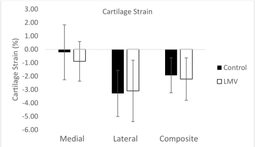
No statistical differences in cartilage deformation were noted between groups; lateral compartment (LMV = -3.1% vs. CON = -3.3%; P = 0.950, observed power = 0.05), medial compartment (LMV = -0.9% vs. CON = -0.2%; P = 0.785, observed power = 0.06), total area (LMV = -2.2% vs. CON = -1.9%; P = 0.882, observed power = 0.05). In addition, there were no differences in pre to post-measures of cartilage strain in the control condition, either laterally (CONpre = 0.5% vs CONpost = 0.4%; P = 0.073, observed power = 0.44), medially (CONpre =
0.4% vs CONpost = 0.4%; P = 0.742, observed power = 0.06), or in total area (CONpre = 0.8% vs
CONpost = 0.8%; P = 0.109, observed power = 0.36); or in the LMV condition, laterally (LMVpre =
0.4% vs LMVpost = 0.4%; P = 0.165, observed power = 0.27), medially (LMVpre = 0.4% vs LMVpost
= 0.4%; P = 0.516, observed power = 0.09), or in total area (LMVpre = 0.8% vs LMVpost = 0.8%; P
= 0.170, observed power = 0.27).
0.00 0.10 0.20 0.30 0.40 0.50 0.60 Co -A ct iva tion Index Heelstrike Co-Activation Control LMV 0.00 0.10 0.20 0.30 0.40 0.50 Co -A ct iva tion Index Preparatory Co-Activation Control LMV 0.00 0.20 0.40 0.60 0.80 1.00 1.20 Co -A ct iva tion Index
Weight Acceptance Co-Activation
Control LMV
Similarly, no significant correlations were noted between co-activation and cartilage deformation (Table 1).
Table 1: Correlation between co-activation and cartilage deformation
Cartilage
Region
Composite co-activation Medial co-activation Lateral co-activation
Prep HS WA Prep HS WA Prep HS WA
Total R = 0.400
P = 0.223
R = 0.215 P = 0.526
R = 0.245 P = 0.468
R = 0.071 P = 0.835
R = 0.174 P = 0.609
R = 0.192 P = 0.571
R = 0.446 P = 0.169
R = -0.157 P = 0.644
R = -0.241 P = 0.475
Medial R = 0.217
P = 0.521
R = 0.125 P = 0.715
R = 0.095 P = 0.781
R = 0.032 P = 0.925
R = 0.058 P = 0.866
R = -0.004 P = 0.99
R = 0.292 P = 0.384
R = 0.009 P = 0.979
R = -0.138 P = 0.686
Lateral R = 0.352
P = 0.288
R = 0.211 P = 0.534
R = 0.26 P = 0.44
R = 0.09 P = 0.793
R = 0.224 P = 0.507
R = 0.288 P = 0.39
R = 0.328 P = 0.324
R = -0.2 P = 0.555
R = -0.176 P = 0.604 Partial Pearson correlation, controlling for gait speed, between co-activation and cartilage deformation of the
control condition. Co-activation broken down into medial, lateral and composite; cartilage deformation also broken
down into medial, lateral and total area.
-6.00 -5.00 -4.00 -3.00 -2.00 -1.00 0.00 1.00 2.00 3.00
Medial
Lateral
Composite
Car til ag e St rai n (% ) Cartilage Strain Control LMV
CHAPTER 5: DISCUSSION
The current study was designed to simultaneously examine co-activation values and cartilage deformation in response to a vibratory intervention. Overall, this pilot study found no statistically significant results. This lack of significant data is not meaningless, however. While no differences were noted in cartilage deformation, trends were noted suggesting that LMV reduces co-activation. Vibration has been shown previously to have an impact on activation, strength, power, EMG activity, proprioception and overall function.20,30–34,73 These trends found in our data are consistent with previous research and provide justification for further studies with a larger sample size.
A single session of LMV produced no statistically significant improvements in activation. While not adequately powered, a trend in the data suggested a decrease in co-activation in the LMV condition. At heelstrike, which showed the greatest downward trend, effect sizes for medial, lateral, and composite co-activations were 0.498, 0.432, and 0.450,
respectively. Figures 1-3 in chapter 4 illustrate this downward trend.
This decrease in co-activation could be due to a variety of factors. The EMG source for changes in co-activation is unclear, as an increase in quadriceps activity or a decrease in hamstrings activity (or vice versa) could produce similar changes in the co-activation ratio. LMV may improve proprioception of the affected limb.20,34 Previous studies26,76,77 have attributed the
plays a role in the heightened co-activation following ACLR. Typically following ACLR, there are deficits in quadriceps function compared with control subjects.78–81 Quadriceps activation is decreased in these individuals, as seen during gait.82,83 Greater hamstring co-activation has also
been seen in these ACLR individuals who have quadriceps deficits, suggesting that these two factors go hand-in-hand. With that in mind, the ALCR individual with quadriceps activation deficits would have an increase in overall co-activation27 because the calculation, referenced in
chapter 3, takes the ratio of hamstrings: quadriceps activation. The smaller the number in the denominator (quadriceps activation), the larger the overall number.23 Several studies30–33,73 have
demonstrated improvements in quadriceps function following LMV. As such, improvements in quadriceps function following LMV may explain the trends for decreasing co-activation.
Additionally, Pamukoff et al.27 suggested that deficits in quadriceps function could be due in part
to reciprocal inhibition from heightened hamstrings co-activation in individuals with ACLR. Applying that idea to the current study, it could be suggested that with a decrease in hamstrings co-activation, quadriceps activation may reciprocally increase, resulting in an overall decrease in co-activation. These findings imply potential benefits to using LMV as an adjunct to traditional rehabilitation programs post ACLR. However, future work is necessary to identify the
mechanism(s) by which co-activation is altered. Further research is needed to target the root cause in order to better implement this intervention successfully in rehabilitation programs.
There were no statistically significant differences in cartilage strain between the LMV or control conditions. In addition to there being no difference between conditions, there was also no difference from pre to post-measures of cartilage cross-sectional area in either condition, meaning that no statistically significant deformation of cartilage was observed. The lack of statistically significant results could be due to a variety of factors. This pilot study evaluated the pathological limb only, and it is possible that the ultrasound assessment may not be sensitive enough to detect changes occurring in pathological cartilage. While Harkey et al.7 found acute
population with no history of osteoarthritis or knee injury. In the pathological population, cartilage may not respond in the same way that healthy cartilage would84. Under pathologic
conditions, chondrocytes exhibit an imbalance of synthesis and degradation. Chondrocyte pericellular matrix is enlarged and less stiff in pathological cartilage as compared to healthy cartilage, which disrupts the overall micromechanical environment of the cartilage.85 In addition,
Harkey et al. obtained ultrasound images at the same time of day (±2 h) to control for diurnal variation in femoral cartilage thickness. Previous research86,87 has shown significant
compressive strain during the day, likely due to a loss of interstitial water due to joint loading. Our study did not take that confounding variable into consideration. While some sessions occurred early in the morning, others occurred later at night. While our participants sat for 45 minutes to unload cartilage prior to imaging, it is unclear if this approach accounted for diurnal influences. Another explanation for the lack of change could be that participants did not take enough steps to produce a change in cartilage structure. While we standardized the number of steps taken by participants, Harkey et al.7 used a 30 min walking protocol, and did not specify
the number of steps that were taken. In our study, overall walking times varied, as participants walked at a self-selected speed. Additionally, this study only evaluated the acute effects of vibration. LMV may not have enough of an acute effect to impact cartilage deformation, but changes may occur when using vibration more often such as embedded into ACLR
rehabilitation, or after tracking cartilage structure longitudinally. Based on these findings, it is unclear whether LMV influences cartilage deformation or overall cartilage health.
No statistically significant correlations were observed between co-activation and
cartilage deformation. This finding is likely attributable in part to the limited variability in cartilage deformation, and suggests that other variables have a greater impact on cartilage deformation. Stiffer loading responses with higher loading rates more directly contribute to cartilage
degradation over time.16,17,24 Vibration potentially minimizes these factors by promoting greater
our study due to the lack of associated gait biomechanics data. It is possible that co-activation values may change, but due to the fact that we are unsure to what extent the hamstrings and quadriceps activation individually change in this calculation, we cannot predict how
biomechanics would be affected. Future research could be conducted to evaluate the impact of vibration directly on walking biomechanics to determine if there is a correlation between co-activation and knee flexion angles, as decreased knee flexion angles are common in the ACLR population and are linked with greater loading rates.9–13 These increased loading rates have been associated with greater cartilage degradation in animal models,16 which suggests that
greater loading rates in ACLR individuals may play a role in PTOA development.17 In addition,
ACLR individuals display lesser external knee flexion moments.82 These decreased knee flexion
angles have also been associated with PTOA development.88 While vibration may not directly
impact cartilage strain, it may have a more direct impact on altered biomechanics.
Future studies in this area should evaluate changes in cartilage over a longer period of walking. While 3,000 steps did not produce significant deformation, differences may be
REFERENCES
1. Lyman S, Koulouvaris P, Sherman S, Do H, Mandl LA, Marx RG. Epidemiology of Anterior Cruciate Ligament Reconstruction. J Bone Jt Surgery-American Vol. 2009;91(10):2321-2328. doi:10.2106/JBJS.H.00539.
2. Eckstein F, Wirth W, Lohmander LS, Hudelmaier MI, Frobell RB. Five-year followup of knee joint cartilage thickness changes after acute rupture of the anterior cruciate ligament. Arthritis Rheumatol. 2015;67(1):152-161. doi:10.1002/art.38881. 3. Svoboda SJ. ACL Injury and Posttraumatic Osteoarthritis. Clin Sports Med.
2014;33(4):633-640. doi:10.1016/j.csm.2014.06.008.
4. Thomas AC, Hubbard-Turner T, Wikstrom EA, Palmieri-Smith RM. Epidemiology of Posttraumatic Osteoarthritis. J Athl Train. 2017;52(6):491-496. doi:10.4085/1062-6050-51.5.08.
5. Eckstein F, Collins JE, Nevitt MC, et al. Cartilage thickness change as an imaging biomarker of knee osteoarthritis progression - data from the fnih OA biomarkers consortium. Arthritis Rheumatol (Hoboken, NJ). 2015;67(12):3184-3189.
doi:10.1002/art.39324.
6. Chaudhari AMW, Briant PL, Bevill SL, Koo S, Andriacchi TP. Knee kinematics, cartilage morphology, and osteoarthritis after ACL injury. Med Sci Sports Exerc. 2008;40(2):215-222. doi:10.1249/mss.0b013e31815cbb0e.
7. Harkey MS, Blackburn JT, Davis H, Sierra-Arévalo L, Nissman D, Pietrosimone B. Ultrasonographic assessment of medial femoral cartilage deformation acutely following walking and running. Osteoarthr Cartil. 2017;25(6):907-913.
doi:10.1016/j.joca.2016.12.026.
8. Gao B, Zheng N (Nigel). Alterations in three-dimensional joint kinematics of anterior cruciate ligament-deficient and -reconstructed knees during walking. Clin Biomech. 2010;25(3):222-229. doi:10.1016/j.clinbiomech.2009.11.006.
9. Hart HF, Culvenor AG, Collins NJ, et al. Knee kinematics and joint moments during gait following anterior cruciate ligament reconstruction: a systematic review and meta-analysis. Br J Sports Med. 2016;50(10):597-612. doi:10.1136/bjsports-2015-094797. 10. Gokeler A, Benjaminse A, van Eck CF, Webster KE, Schot L, Otten E. Return of normal
gait as an outcome measurement in acl reconstructed patients. A systematic review. Int J Sports Phys Ther. 2013;8(4):441-451.
http://www.pubmedcentral.nih.gov/articlerender.fcgi?artid=3812834&tool=pmcentrez&ren dertype=abstract.
11. Noehren B, Wilson H, Miller C, Lattermann C. Long-term gait deviations in anterior cruciate ligament-reconstructed females. Med Sci Sports Exerc. 2013;45(7):1340-1347. doi:10.1249/MSS.0b013e318285c6b6.
12. Landry SC, McKean KA, Hubley-Kozey CL, Stanish WD, Deluzio KJ. Knee biomechanics of moderate OA patients measured during gait at a self-selected and fast walking speed.
J Biomech. 2007;40(8):1754-1761. doi:10.1016/j.jbiomech.2006.08.010.
13. Zeni JA, Higginson JS. Differences in gait parameters between healthy subjects and persons with moderate and severe knee osteoarthritis: A result of altered walking speed?
Clin Biomech. 2009;24(4):372-378. doi:10.1016/j.clinbiomech.2009.02.001.
slower walking speed with incident knee osteoarthritis- related outcomes. Arthritis Care Res. 2012;64(7):1028-1035. doi:10.1002/acr.21655.
15. Barenius B, Ponzer S, Shalabi A, Bujak R, Norlén L, Eriksson K. Increased Risk of Osteoarthritis After Anterior Cruciate Ligament Reconstruction. Am J Sports Med. 2014;42(5):1049-1057. doi:10.1177/0363546514526139.
16. Ewers BJ, Jayaraman VM, Banglmaier RF, Haut RC. Rate of blunt impact loading affects changes in retropatellar cartilage and underlying bone in the rabbit patella. J Biomech. 2002;35(6):747-755. doi:10.1016/S0021-9290(02)00019-2.
17. Blackburn JT, Pietrosimone B, Harkey MS, Luc BA, Pamukoff DN. Inter-limb differences in impulsive loading following anterior cruciate ligament reconstruction in females. J Biomech. 2016;49(13):3017-3021. doi:10.1016/j.jbiomech.2016.07.030.
18. Pitman MI, Nainzadeh N, Menche D, Gasalberti R, Song EK. The intraoperative
evaluation of the neurosensory function of the anterior cruciate ligament in humans using somatosensory evoked potentials. Arthroscopy. 1992;8(4):442-447.
http://www.ncbi.nlm.nih.gov/pubmed/1466702. Accessed November 30, 2017. 19. Park H Bin, Koh M, Cho SH, Hutchinson B, Lee B. Mapping the rat somatosensory
pathway from the anterior cruciate ligament nerve endings to the cerebrum. J Orthop Res. 2005;23(6):1419-1424. doi:10.1016/j.orthres.2005.03.017.
20. Brunetti O, Filippi GM, Lorenzini M, et al. Improvement of posture stability by vibratory stimulation following anterior cruciate ligament reconstruction. Knee Surgery, Sport Traumatol Arthrosc. 2006;14(11):1180-1187. doi:10.1007/s00167-006-0101-2.
21. Lustosa LP, Ocarino JM, Andrade MAP de, Pertence AE de M, Bittencourt NFN, Fonseca ST. Muscle co-contraction after anterior cruciate ligament reconstruction: Influence of functional level. J Electromyogr Kinesiol. 2011;21(6):1050-1055.
doi:10.1016/j.jelekin.2011.09.001.
22. Thoumie P, Mevellec E. Relation between walking speed and muscle strength is affected by somatosensory loss in multiple sclerosis. J Neurol Neurosurg Psychiatry.
2002;73(3):313-315. doi:10.1136/jnnp.73.3.313.
23. Schmitt LC, Rudolph KS. Influences on knee movement strategies during walking in persons with medial knee osteoarthritis. Arthritis Care Res. 2007;57(6):1018-1026. doi:10.1002/art.22889.
24. Palmieri-Smith RM, Thomas AC. A Neuromuscular Mechanism of Posttraumatic Osteoarthritis Associated with ACL Injury. Exerc Sport Sci Rev. 2009;37(3):147-153. doi:10.1097/JES.0b013e3181aa6669.
25. Urbach D, Nebelung W, Becker R, Awiszus F. Effects of reconstruction of the anterior cruciate ligament on voluntary activation of quadriceps femoris a prospective twitch interpolation study. J Bone Jt Surg. 2001;83(8):1104-1110. doi:10.1302/0301-620X.83B8.11618.
26. Rudolph KS, Axe MJ, Buchanan TS, Scholz JP, Snyder-Mackler L. Dynamic stability in the anterior cruciate ligament deficient knee. Knee Surgery, Sport Traumatol Arthrosc. 2001. doi:10.1007/s001670000166.
28. Troy Blackburn J, Pietrosimone B, Harkey MS, Luc BA, Pamukoff DN. Quadriceps Function and Gait Kinetics after Anterior Cruciate Ligament Reconstruction. Med Sci Sports Exerc. 2016;48(9):1664-1670. doi:10.1249/MSS.0000000000000963.
29. Roelants M, Verschueren SMP, Delecluse C, Levin O, Stijnen V. Whole-Body-Vibration– Induced Increase in Leg Muscle Activity During Different Squat Exercises. J Strength Cond Res. 2006;20(1):124. doi:10.1519/R-16674.1.
30. Bosco C, Cardinale M, Tsarpela O. Influence of vibration on mechanical power and electromyogram activity in human arm flexor muscles. Eur J Appl Physiol.
1999;79(4):306-311. doi:10.1007/s004210050512.
31. Cardinale M, Lim J. Electromyography Activity of Vastus Lateralis Muscle During Whole-Body Vibrations of Different Frequencies. J Strength Cond Res Natl Strength Cond Assoc J Strength Cond Res. 2003;17(173). https://insights.ovid.com/crossref?an=00124278-200308000-00032. Accessed April 24, 2018.
32. Pamukoff DN, Ryan ED, Troy Blackburn J. The acute effects of local muscle vibration frequency on peak torque, rate of torque development, and EMG activity. J Electromyogr Kinesiol. 2014;24(6):888-894. doi:10.1016/j.jelekin.2014.07.014.
33. Bazett-Jones DM, Finch HW, Dugan EL. Comparing the effects of various whole-body vibration accelerations on counter-movement jump performance. 2008;7(1):144-150.
https://www.scopus.com/record/display.uri?eid=2-s2.0-40749153588&origin=inward&txGid=ce77da04bd23ea2dadfa131e4d4786c9. Accessed April 24, 2018.
34. Trans T, Aaboe J, Henriksen M, Christensen R, Bliddal H, Lund H. Effect of whole body vibration exercise on muscle strength and proprioception in females with knee
osteoarthritis. Knee. 2009;16(4):256-261. doi:10.1016/j.knee.2008.11.014.
35. Pamukoff DN, Pietrosimone B, Lewek MD, et al. Whole-Body and Local Muscle Vibration Immediately Improve Quadriceps Function in Individuals with Anterior Cruciate Ligament Reconstruction. Arch Phys Med Rehabil. 2016;97(7):1121-1129.
doi:10.1016/j.apmr.2016.01.021.
36. Pamukoff DN, Pietrosimone B, Lewek MD, et al. Immediate effect of vibratory stimuli on quadriceps function in healthy adults. Muscle and Nerve. 2016;54(3):469-478.
doi:10.1002/mus.25081.
37. Costantino C, Bertuletti S, Romiti D. Efficacy of Whole-Body Vibration Board Training on Strength in Athletes After Anterior Cruciate Ligament Reconstruction. Clin J Sport Med. June 2017:1. doi:10.1097/JSM.0000000000000466.
38. Blackburn JT, Pamukoff DN, Sakr M, Vaughan AJ, Berkoff DJ. Whole body and local muscle vibration reduce artificially induced quadriceps arthrogenic inhibition. Arch Phys Med Rehabil. 2014;95(11):2021-2028. doi:10.1016/j.apmr.2014.07.393.
39. Eckstein F, Wirth W, Lohmander LS, Hudelmaier MI, Frobell RB. Five-year followup of knee joint cartilage thickness changes after acute rupture of the anterior cruciate
ligament. Arthritis Rheumatol (Hoboken, NJ). 2015;67(1):152-161. doi:10.1002/art.38881. 40. Culvenor AG, Collins NJ, Guermazi A, et al. Early knee osteoarthritis is evident one year
following anterior cruciate ligament reconstruction: a magnetic resonance imaging evaluation. Arthritis Rheumatol (Hoboken, NJ). 2015;67(4):946-955.
doi:10.1002/art.39005.
2015;27(3):276-283. doi:10.1097/BOR.0000000000000161.
42. Lane NE, Brandt K, Hawker G, et al. OARSI-FDA initiative: defining the disease state of osteoarthritis. Osteoarthr Cartil. 2011;19(5):478-482. doi:10.1016/j.joca.2010.09.013. 43. Fleming BC, Hulstyn MJ, Oksendahl HL, Fadale PD. Ligament Injury, Reconstruction and
Osteoarthritis. Curr Opin Orthop. 2005;16(5):354-362.
http://www.ncbi.nlm.nih.gov/pubmed/17710194. Accessed October 12, 2017.
44. Louboutin H, Debarge R, Richou J, et al. Osteoarthritis in patients with anterior cruciate ligament rupture: A review of risk factors. Knee. 2009;16(4):239-244.
doi:10.1016/j.knee.2008.11.004.
45. Ratzlaff CR, Liang MH. New developments in osteoarthritis. Prevention of injury-related knee osteoarthritis: opportunities for the primary and secondary prevention of knee osteoarthritis. Arthritis Res Ther. 2010;12(4):215. doi:10.1186/ar3113.
46. Le TK, Montejano LB, Cao Z, Zhao Y, Ang D. Healthcare Costs Associated with Osteoarthritis in US Patients. Pain Pract. 2012;12(8):633-640. doi:10.1111/j.1533-2500.2012.00535.x.
47. Bates NA, Myer GD, Shearn JT, Hewett TE. Anterior cruciate ligament biomechanics during robotic and mechanical simulations of physiologic and clinical motion tasks: a systematic review and meta-analysis. Clin Biomech (Bristol, Avon). 2015;30(1):1-13. doi:10.1016/j.clinbiomech.2014.12.006.
48. Mather RC, Koenig L, Kocher MS, et al. Societal and economic impact of anterior cruciate ligament tears. J Bone Joint Surg Am. 2013;95(19):1751-1759.
doi:10.2106/JBJS.L.01705.
49. Cossich V, Mallrich F, Titonelli V, de Sousa EB, Velasques B, Salles JI. Proprioceptive deficit in individuals with unilateral tearing of the anterior cruciate ligament after active evaluation of the sense of joint position. Rev Bras Ortop (English Ed. 2014;49(6):607-612. doi:10.1016/j.rboe.2013.07.003.
50. Wojtys EM, Huston LJ. Longitudinal Effects of Anterior Cruciate Ligament Injury and Patellar Tendon Autograft Reconstruction on Neuromuscular Performance. Am J Sports Med. 2000;28(3):336-344. doi:10.1177/03635465000280030901.
51. Lee D-H, Lee J-H, Ahn S-E, Park M-J. Effect of Time after Anterior Cruciate Ligament Tears on Proprioception and Postural Stability. Eynon N, ed. PLoS One.
2015;10(9):e0139038. doi:10.1371/journal.pone.0139038.
52. Shanahan CJ, Wrigley T V, Farrell MJ, Bennell KL, Hodges PW. Proprioceptive impairments associated with knee osteoarthritis are not generalized to the ankle and elbow joints. Hum Mov Sci. 2015;41:103-113. doi:10.1016/j.humov.2015.02.008.
53. Knoop J, Steultjens M, Van Der Leeden M, et al. Proprioception in knee osteoarthritis: a narrative review. Osteoarthr Cartil. 2011;19:381-388. doi:10.1016/j.joca.2011.01.003. 54. Hoch JM, Perkins WO, Hartman JR, Hoch MC. Somatosensory deficits in post-ACL
reconstruction patients: A case–control study. Muscle and Nerve. 2017;55(1):5-8. doi:10.1002/mus.25167.
55. Collins AT, Blackburn JT, Olcott CW, Dirschl DR, Weinhold PS. The effects of stochastic resonance electrical stimulation and neoprene sleeve on knee proprioception. J Orthop Surg Res. 2009;4(1):3. doi:10.1186/1749-799X-4-3.
Reconstruction. Clin Sports Med. 2008;27(3):405-424. doi:10.1016/j.csm.2008.02.001. 57. Kellis E, Arabatzi F PC. Muscle co-activation around the knee in drop jumping using the
co-contraction index. J Electromyogr Kinesiol. 2003.
58. Elias ARC, Hammill CD, Mizner RL. Changes in Quadriceps and Hamstring
Cocontraction Following Landing Instruction in Patients With Anterior Cruciate Ligament Reconstruction. J Orthop Sport Phys Ther. 2015;45(4):273-280.
doi:10.2519/jospt.2015.5335.
59. Slemenda C, Brandt KD, Heilman DK, et al. Quadriceps weakness and osteoarthritis of the knee. Ann Intern Med. 1997;127(2):97-104.
http://www.ncbi.nlm.nih.gov/pubmed/9230035. Accessed November 9, 2017.
60. Baker KR, Xu L, Zhang Y, et al. Quadriceps weakness and its relationship to tibiofemoral and patellofemoral knee osteoarthritis in Chinese: The Beijing osteoarthritis study.
Arthritis Rheum. 2004;50(6):1815-1821. doi:10.1002/art.20261.
61. Hortobágyi T, Westerkamp L, Beam S, et al. Altered hamstring-quadriceps muscle balance in patients with knee osteoarthritis. Clin Biomech. 2005;20(1):97-104. doi:10.1016/j.clinbiomech.2004.08.004.
62. Childs JD, Sparto PJ, Fitzgerald GK, Bizzini M, Irrgang JJ. Alterations in lower extremity movement and muscle activation patterns in individuals with knee osteoarthritis. Clin Biomech. 2004;19(1):44-49. doi:10.1016/j.clinbiomech.2003.08.007.
63. Radin EL, Yang KH, Riegger C, Kish VL, O’Connor JJ. Relationship between lower limb dynamics and knee joint pain. J Orthop Res. 1991;9(3):398-405.
doi:10.1002/jor.1100090312.
64. Cohen NP, Foster R), Mow VC, Foster RJ. Composition and Dynamics of Articular Cartilage: Structure, Function, and Maintaining ~ealthy State.
https://www.jospt.org/doi/pdf/10.2519/jospt.1998.28.4.203. Accessed January 28, 2018. 65. LU XL, MOW VC. Biomechanics of Articular Cartilage and Determination of Material
Properties. Med Sci Sport Exerc. 2008;40(2):193-199. doi:10.1249/mss.0b013e31815cb1fc.
66. Kd B, Dieppe P, El R, Brandt KD, Dieppe P. Regular Order ID : Group ID :
Etiopathogenesis of Osteoarthritis. Rheum Dis Clin North Am. 2011;34(3):531-559. doi:10.1016/j.rdc.2008.05.011.
67. Eckstein F, Wirth W, Nevitt MC. Recent advances in osteoarthritis imaging—the Osteoarthritis Initiative. Nat Rev Rheumatol. 2012;8(10):622-630.
doi:10.1038/nrrheum.2012.113.
68. Eckstein F, Collins JE, Nevitt MC, et al. Cartilage thickness change as an imaging biomarker of knee osteoarthritis progression - data from the fnih OA biomarkers consortium. Arthritis Rheumatol (Hoboken, NJ). 2015;67(12):3184-3189.
doi:10.1002/art.39324.
69. Keen HI, Conaghan PG. Usefulness of Ultrasound in Osteoarthritis. Rheum Dis Clin North Am. 2009;35(3):503-519. doi:10.1016/j.rdc.2009.09.002.
70. Naredo E, Acebes C, Moller I, et al. Ultrasound validity in the measurement of knee cartilage thickness. Ann Rheum Dis. 2009;68(8):1322-1327.
doi:10.1136/ard.2008.090738.