Investigation of Probe Substrates to Assess Hepatic Transport Function
by Brandon Swift
A dissertation submitted to the faculty of the University of North Carolina at Chapel Hill in partial fulfillment of the requirements for the degree of Doctor of
Philosophy in the Eshelman School of Pharmacy
Chapel Hill 2009
ABSTRACT Brandon Swift
Investigation of Probe Substrates to Assess Hepatic Transport Function (Under the direction of Kim L.R. Brouwer, Pharm.D., Ph.D.)
99mTc-MIBI as probe substrates to predict the hepatic clearance of the anticancer
agent, sorafenib studies were conducted to confirm similar mechanisms of hepatic uptake. Lastly, the pharmacokinetics and hepatic exposure of 99mTc-MIBI and 99mTc-MEB were compared in a patient with hepatocellular carcinoma and Child’s Pugh B cirrhosis vs. healthy human volunteers. Pharmacokinetic models were constructed to describe the distribution and elimination of 99mTc-MEB and 99mTc-MIBI, to compare alterations in key rate constants representing hepatic
ACKNOWLEDGEMENTS
I want to thank Dr. Kim L. R. Brouwer for the opportunity to work on this exciting project and for guiding my personal and scientific development. Dr. Brouwer is the reason I considered graduate school and was a motivating factor in my progression as a student and Ph.D. candidate. I also want to thank all the members of my doctoral dissertation advisory committee who have been
instrumental in the progression of this project: Dr. Gary M. Pollack who has been a wonderful chair of this committee, very supportive, always giving invaluable advice, and a great advocate; Dr. Marijana Invanovic who was always available, for her help in improving the hepatic scintigraphy methods and a few long days sitting in front of the computer staring at the Siemens software; Dr. Dhiren
Thakker for his scientific advice and lectures in drug metabolism; Dr. Roy Hawke for always being available as a source of advice; and Dr. Bert O’Neil for allowing me to work with him on the clinical study even though we completed one subject successfully during my time here.
Poole, as well as fellow graduate students Rong Zhao, Jeannie Padowski, Will Proctor, and Shawn Watson. I am indebted to my friend Xianbin Tian with whom I had the pleasure of collaborating on our fexofenadine studies and our
unforgettable trip to Fox Chase Cancer Center in Philadelphia, Pennsylvania. I would like to express my gratitude and appreciation to Drs. Gary Kruh and Martin Belinsky for their collaboration, hospitality at Fox Chase Cancer Center, and generosity in sharing their gene knockout mice. I also want to thank Elaine
Kimple for her administrative support and making life at school and in the lab flow smoothly.
Most of all I am very grateful for the support of my wife Julie who
TABLE OF CONTENTS
LIST OF TABLES………vii LIST OF FIGURES………..……..viii CHAPTER
1. Introduction………1
2. Integration of Preclinical and Clinical Data with Pharmacokinetic Modeling and Simulation to Evaluate Fexofenadine as a Probe for Hepatobiliary Transport Function ………76
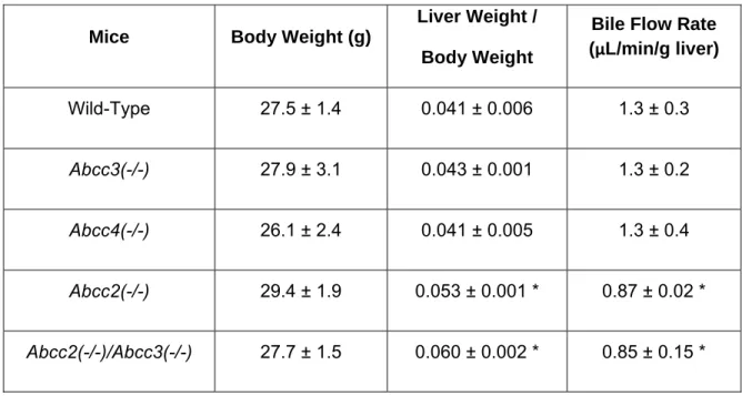
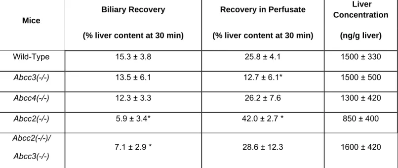
3. Impact of Basolateral Mrp3 (Abcc3) and Mrp4 (Abcc4) on the Hepatobiliary Disposition of Fexofenadine in Perfused Mouse Livers……….………….…...113
4. Evaluation of 99mTechnetium-Mebrofenin and 99m Technetium-Sestamibi as Specific Probes for Hepatic Transport Protein
Function in Rats and Human ………134
5. Hepatobiliary Disposition of Sorafenib in Human Hepatocytes...178
6. Pharmacokinetics and Hepatic Exposure of 99m Technetium-Sestamibi and 99mTechnetium-Mebrofenin in a Patient with Hepatocellular Carcinoma and Child's Pugh B Cirrhosis versus Healthy Human Volunteers: An Exploratory Analysis ….…..…...211
7. Conclusions and Future Work………255 APPENDIX
LIST OF TABLES
Table 1.1: Clinical Transport Protein Probe Substrates……….74 Table 2.1: Pharmacokinetic parameters governing fexofenadine disposition in healthy humans ……….……….106 Table 2.2: Accumulation, BEI and in vitro Clbiliary of 3H-taurocholate, 3H-digoxin and fexofenadine in human sandwich-cultured hepatocytes ………...107 Table 2.3: Fexofenadine disposition in single-pass perfused TR- rat liver……..108 Table 3.1: Body weight,liver weight normalized for body weight, and bile flow rate in wild-type and transporter gene knockout mice………...130 Table 3.2: Recovery of fexofenadine in perfusate and bile during the washout phase of mouse liver perfusions, andliver concentrations after the washout phase ……….131 Table 5.1:Mass balance of sorafenib in incubation medium and hepatocyte cell lysates in human sandwich-cultured hepatocytes ………..………..….204 Table 6.1:Summary of individual and mean pharmacokinetic parameter
estimates and associated variability governing 99mtechnetium-sestamibi
LIST OF FIGURES
Figure 2.1: Model scheme depicting fexofenadine disposition in healthy humans ……….109 Figure 2.2: Mean fexofenadine disposition in healthy humans. The curves
represent the best fit of the pharmacokinetic model based on the scheme
depicted in Fig. 2.1 to the data obtained from Shimizu et al. 2006 ………110 Figure 2.3: Effects of modifications in hepatic uptake and efflux processes on the plasma concentration-time profile and hepatic mass-time profile of fexofenadine ……….111 Figure 2.4: Bile flow rates, fexofenadine concentrations in outflow perfusate, and biliary excretion rates of fexofenadine in single pass perfused livers from Mrp2-deficient TR- rats ………. ………..……….112 Figure 3.1. Fexofenadine concentrations in outflow perfusate of single pass perfused livers from wild-type C57BL/6 and gene-disrupted mouse livers ……132 Figure 3.2. Biliary excretion rate and cumulative biliary excretion of fexofenadine in perfused livers from wild-type C57BL/6 and gene-disrupted mouse livers …133 Figure 4.1 Initial uptake rate of 99mtechnetium-sestamibi, 99m
technetium-mebrofenin, 3H- estradiol-17-β-D-glucuronide and 14C- tetraethylammonium in suspended wild-type rat hepatocytes ………..171 Figure 4.2 Initial uptake rate of 99mtechnetium-mebrofenin, 3H- estradiol-17-β -D-glucuronide and 14C- tetraethylammonium in suspended human hepatocytes..173 Figure 4.3 99mTechnetium-mebrofenin basolateral and canalicular efflux and hepatocellular accumulation in rat [wild-type and TR-(Mrp2-deficient)] and human sandwich-cultured hepatocytes ……… 175 Figure 4.4 99mTechnetium-sestamibi basolateral and canalicular efflux and
Figure 5.2 Initial uptake of 14C-sorafenib in suspended human hepatocytes ....206 Figure 5.3 Uptake of 14C-sorafenib and 14C- tetraethylammonium in CHO-hOCT1 cells incubated in the absence and presence of 10 µM decynium 22 …………208 Figure 5.4 Accumulation, biliary excretion and in vitro Clbiliary of sorafenib,
sorafenib N-oxide and sorafenib glucuronide in human sandwich-cultured
hepatocytes ……… 209 Figure 6.1 Model scheme depicting 99mTechnetium-sestamibi disposition in a patient with HCC and Child’s Pugh B cirrhosis and healthy humans ..………. 249 Figure 6.2 Semi-physiological model scheme depicting 99m
Technetium-mebrofenin disposition in a patient with HCC and Child’s Pugh B cirrhosis and healthy humans……….. 250 Figure 6.3 Representative 99mtechnetium-sestamibi disposition in a patient with HCC and Child’s Pugh B cirrhosis and healthy human volunteers. The curves represent the best fit of the pharmacokinetic model based on the scheme depicted in Fig. 6.1 to the subject with HCC and Child’s Pugh B cirrhosis and healthy volunteers (n=7) data obtained from Ghibellini et al. 2007 ……….……251 Figure 6.4 Dose normalized hepatic mass-time profile of 99m
technetium-sestamibi in a patient with HCC and Child’s Pugh B cirrhosis and simulated hepatic mass-time profile using final parameter estimates listed in table 6.1in healthy human volunteers ………..252 Figure 6.5 Representative 99mtechnetium-mebrofenin disposition in a patient with HCC and Child’s Pugh B cirrhosis and healthy human volunteers. The curves represent the best fit of the semi-physiological based pharmacokinetic model in the scheme depicted in Fig. 6.2 to the subject with HCC and Child’s Pugh B cirrhosis and healthy volunteers (n=3)
data………..………. 253 Figure 6.6 Dose normalized hepatic mass-time profile of 99m
technetium-mebrofenin in a patient with HCC and Child’s Pugh B cirrhosis and healthy
human volunteers ……….. 254 Figure A.1.1. Effect of seeding density and extracellular matrix on cell
Figure A.1.2. Effect of seeding density and extracellular matrix on cell
morphology and bile canalicular network formation in day 4 mouse sandwich-cultured hepatocytes ... 328 Figure A.1.3. Influence of seeding density, extracellular matrix and day in culture on transport protein levels in mouse sandwich-cultured hepatocytes.
Representative immunoblots of Bcrp, Ntcp, Mrp4, Mrp3, Mrp2, Bsep, and Mdr1a/1b in mouse hepatocytes cultured in BiocoatTM/MatrigelTM (BC/MG) or BiocoatTM/gelled-collagen (BC/GC) sandwich configuration in six-well plates and maintained with DMEM for 3-4
days………....330 Figure A.1.4. Relative expression of Mrp4 protein compared with β-actin in
CHAPTER 1
Transport proteins are responsible for drug absorption, distribution and excretion. Transporters govern movement of endogenous compounds and xenobiotics across biological membranes to maintain cellular and physiologic solute concentrations and fluid balance, or to protect the body from dietary and environmental toxins. Most transport proteins are expressed in tissues with barrier functions such as the liver, kidney, intestine, placenta and brain, where they often play a key role in determining bioavailability, therapeutic efficacy and pharmacokinetics of drugs. Transport proteins are classified into two superfamilies, the adenosine triphosphate (ATP)-binding cassette (ABC) protein family and the solute carrier (SLC) protein family. ABC transport proteins function unidirectionally and actively efflux substrates directly utilizing the energy of ATP hydrolysis. The SLC family transporters are facilitated transporters, secondary or
tertiaty active transporters, which function bidirectionally allowing drugs/molecules to move across cell membranes utilizing the electrochemical potential gradient or ion gradient, and thus do not directly consume any chemical energy. Proteins in the SLC and ABC familes often work in concert as a vectorial process to remove compounds from the blood and excrete them into bile or urine.
impact on clinical pharmacokinetics, efficacy and safety of drugs. Subsequently, probes that have been used to assess specific transport protein function will be discussed.
ABC Transport Proteins
Multidrug Resistance (MDR1) P-glycoprotein (P-gp)
P-gp was one of the first ABC proteins to be studied because of its role in the development of multidrug-resistance to chemotherapeutic agents. Like many other ABC proteins, P-gp comprises two membrane-bound domains, each made up of six transmembrane helices (7, 8). P-gp is encoded by the ABCB1 gene, and is expressed at low levels in most tissues, but is found in much higher amounts at the apical surface of epithelial cells lining the colon, small intestine, pancreatic ductules, bile ductules (hepatocytes), kidney proximal tubules and the adrenal gland (9). Tissue localization suggests that P-gp plays a physiological role in the protection of susceptible organs such as the brain and testis from toxic xenobiotics, the secretion of metabolites and xenobiotics into bile, urine and the lumen of the gastrointestinal tract, and possibly the transport of hormones from the adrenal gland. P-gp represents the most widely studied ABC transport protein, and is responsible for the transport of predominantly bulky hydrophobic and cationic substrates including many chemotherapeutic agents, cardiac glycosides, cyclosporine A, and HIV-1 protease inhibitors.
BCRP is encoded by the ABCG2 gene. ABCG2 is considered a half-transporter that forms a homodimer in order to function, as opposed to other ABC half-transporters that engage in heterodimeric association to form a functional transporter such as ABCG5/ABCG8 (10). Little is known about the molecular mechanism(s) controlling expression; recent studies suggest that expression may be regulated by sex hormones or hypoxia (11-13). Localization of BCRP is very similar to P-gp; the highest expression of ABCG2 mRNA is in placental tissue, with lower levels in brain, prostate, small intestine, testis, ovary, colon and liver (14, 15). BCRP was discovered based on its affinity to mitoxantrone and shares some substrate overlap with P-gp. BCRP also transports a variety of anticancer drugs such as SN-38, topotecan and doxorubicin (16-18). Other BCRP substrates include sulfate conjugates (19) the tyrosine kinase inhibitors (CI1033, gefitinib and imatinib (20-22)) and fluoroquinolone antibiotics
(grepafloxacin, ulifloxacin, ciprofloxacin and ofloxacin (23)).
Multidrug Resistance Proteins (MRP)
substances across the plasma membrane (1). MRP2 was first localized to the canalicular membrane of rat and human hepatocytes (25, 26). Since then, MRP2 also has been identified on the apical membrane of kidney proximal tubules, small intestine, colon, gallbladder, bronchi and placenta (27-29). The exclusive apical localization in these polarized cells underscores MRP2’s role in the excretion and detoxification of endogenous and xenobiotic organic anions. MRP2 substrates include many endogenous compounds such as leukotriene C4, as well as other glutathione, glucuronide, and sulfate conjugates of substrates such as S-glutathionyl 2,4-dinitrobenzene, mono- and bisglucuronosyl bilirubin, 17β-glucuronosyl estradiol and estrone 3-sulfate (30-33). The function and substrate specificity of MRP2 has been studied extensively by taking advantage of the naturally occurring Mrp2 deficient rats: Eisai hyperbilirubinemic (EHBR) rats and the CY/TR- mutant rats (25, 26, 34). Furthermore, these hereditary hyperbilirubinemic mutant rats helped to elucidate the upregulation of basolateral Mrp3, a compensatory mechanism that enables the excretion of Mrp2 substrates into the systemic circulation when Mrp2 function is compromised, thus avoiding excessive accumulation of organic anions in the hepatocytes (35, 36). A similar hereditary disorder has been discovered in humans; the absence of a functional
ABCC2 protein on the canalicular membrane leads to conjugated
constitutive in other organs (42). Several hereditary and acquired liver disorders lead to increased ABCC3 protein levels including Dubin-Johnson syndrome (35), progressive familial intrahepatic cholestasis type 3 (42), icteric primary biliary cirrhosis (43), and obstructive cholestasis (44).
SLC Transport Proteins
Organic Anion-Transporting Polypeptides (OATPs)
The OATPs are encoded by the SLCO genes that contain 12
transmembrane helices and are expressed in various epithelial cells. There are 11 human isoforms of which OATP1A2, OATP2A1, OATP2B1, OATP3A1 and OATP4A1 are expressed widely among human tissues while others appear to have tissue specific expression, such as OATP1B1 and OATP1B3 in liver, OATP4C1 in kidney and OATP6A1 in testes (45, 46). OATPs generally are considered to function in a bidirectional manner, as dictated by the solute
angiotensin II receptor antagonists, angiotensin converting enzyme inhibitors and cardiac glycosides. Although a large number of OATPs have been identified, clinical relevance and drug-drug interactions related to OATPs have been noted primarily for OATP1A2, mediating intestinal absorption, and the liver-specific OATP1B1 and OATP1B3 isoforms, mediating hepatic uptake of drugs (50, 51). Furthermore, OATP1B1 and OATP1B3 have important physiological roles in the hepatic uptake of unconjugated and conjugated bilirubin (52, 53) and bile acid homeostasis based on direct transcriptional regulation by FXR (54) and down-regulation in expression levels during cholestatic disease (55). Both OATP1B1 and OATP1B3 have been shown to be affected by drugs such as rifampin (56, 57).
Organic Cation Transporters (OCTs)
prostate, adrenal gland, salivary gland and many other tissues (65). OCTs mediate the uptake of low molecular weight relatively hydrophilic organic cations (type I) such as the prototypical cations tetraethylammonium (TEA), the
neurotoxin N-metyl-4-phenylpyridine (MPP+) and the endogenous compound N-methylnicotinamide (NMN) (58, 66, 67). Drugs that are substrates of the OCTs include metformin, famotidine, and ranitidine (67-69). Most of the drug-drug interactions involving OCT proteins have been shown to occur for drugs predominantly excreted in urine by active renal secretion (70-72).
Organic Anion Transporters (OATs)
OATs are in the same SLC22A family as the organic cation transporters. Five human isoforms have been identified (OAT1, OAT2, OAT3, OAT4 and URAT1), which are located primarily in the kidneys (73). The uptake of organic anions is driven by the concentration gradient of intracellular dicarboxylates, such as α-ketoglutarate. The high concentration gradient of dicarboxylates is
maintained by the sodium-dicarboxylate cotransporter, which in turn is driven by the inwardly directed sodium gradient across the basolateral membrane
diuretics, β-lactam antibiotics, methotrexate and nonsteroidal anti-inflammatory drugs (75).
Importance of Transport Protein Probe Substrates
Systemic concentrations typically correlate with drug efficacy and toxicity, and therapeutic concentration ranges have been established for a large number of drugs. However, significant interindividual variability in the concentration-response and/or concentration-toxicity relationships may exist for some drugs. Genetic polymorphisms and deficiencies in transport proteins are a well
recognized source of variability resulting in altered pharmacokinetics and subsequent pharmacologic and toxicological effects of certain drugs (3). Recently, a high impact paper was published documenting that a common
variant in SLCO1B1, the gene encoding OATP1B1, was strongly associated with increased risk of statin-induced myopathy (76). In addition to genetic
polymorphisms, various disease states such as cholestasis and cirrhosis may modify transport protein expression and/or localization, thereby affecting the pharmacokinetics and/or pharmacodynamics of some drugs (77, 78). Therefore, utilizing a probe substrate to phenotype the functional changes in specific
Probe substrates are also crucial for elucidating drug-drug interactions at the transport level, and would be useful for preventing potential adverse events in patient populations that require the use of many concomitantly administered drugs. A clinically important factor that may alter the efficacy and/or toxicity of a drug or endogenous compound is transporter-based drug interactions, which may result in inhibition, induction, or a mixed effect on the uptake and/or efflux of transport proteins (79). To date, many reviews have documented the
considerable number of transporter-based clinically important drug interactions (6, 79-81). In fact, the US Food and Drug Administration (FDA) has issued a guidance on performing drug-drug interaction studies to determine P-gp inhibition
in vivo (82). Probe substrates are an important tool that can be used to clarify the impact that a specific transport protein may have on a drug’s disposition, interaction, efficacy and safety; information generated using probe substrates may lead to drug label changes that provide important mechanistic information useful for drug therapy. This same approach can be applied in the drug
development or in vitro setting to facilitate evaluation of transport/interaction mechanisms for new drug candidates in which transport processes are relatively unknown. Utilizing a specific probe substrate in a cell-based, intact organ or in vivo model in the presence and absence of an investigational drug could aid in elucidating whether the investigational drug is an inhibitor, which often suggests the investigational drug is a substrate for that protein as well.
The aim of this review is to present an overview of potential probe
tissue concentrations of probe substrates to elucidate transporter-based drug-drug interactions that may not be reflected by systemic concentrations will be highlighted. In addition, the challenges in identifying specific probe substrates and inhibitors will be discussed.
What Makes a Good Probe Substrate?
First and foremost, a probe substrate must be specific for a transport protein, and the pharmacokinetics of the probe substrate must be sensitive to functional changes in the transport protein. Ideally, a transporter probe substrate would display little or predictable metabolism. Furthermore, the probe would need to be safe for healthy human volunteer studies. Another important
elucidating the distribution of the radiolabeled probe and determining organ exposure.
Identification of specific probe substrates of transport proteins has proved to be difficult. There are many limitations including the broad and overlapping substrate specificity of the specific isoforms of each transport protein subfamily. Similarly, species differences in the transport proteins primarily involved in the disposition of some substrates require assessment of the function, or evaluation of drug interactions, in the appropriate species. This is the case for fexofenadine in which Mrp2 is primarily responsible for biliary excretion in mice whereas P-gp appears to play a more important role in rats (84, 85). Another difficulty is determining the exact mechanism of interaction in the vectorial transport of substrates in polarized cells when uptake and efflux processes are involved. For example, digoxin has been touted as a good P-gp probe substrate(82). However, an OATP-mediated interaction that inhibits the active uptake of digoxin into the hepatocytes or renal tubular cells before gaining access to P-gp could confound interpretation of data generated in a P-gp drug interaction study (86). There is also evidence for multiple binding sites for P-gp (87, 88) and MRP2/Mrp2 (89, 90); the binding of one compound may affect the binding of a second compound, regardless of whether the modulating compound is a substrate of that specific transport protein. A comparison can be made with cytochrome P450 (CYP) 3A4 and the need to use two compounds, midazolam and testosterone, as probe substrates for different binding sites to assess drug-drug interactions (51).
substrate in a particular tissue without affecting the blood or plasma
concentration of the substrate. For example, the P-gp inhibitor cyclosporine A significantly increased the distribution of verapamil, a P-gp probe substrate into the brain, without significantly affecting the plasma or blood concentration-time profile of verapamil (91). It is important to consider whether the clearance of a probe substrate reflects the function of a transport protein. For instance, a probe with a high hepatic extraction will have a hepatic clearance that is reflective of changes in blood flow; thus, it may not be an appropriate probe for a transport protein unless the probe is validated to confirm that it is sensitive to perturbation in function of the transport protein while accounting for differences in blood flow.
In vivo Probe Substrates
P-gp
For instance, when P-gp is inhibited in rat the metabolism of the dual CYP3A/P-gp substrates tacrolimus and digoxin were increased (96-98). Therefore, inhibition of P-gp leads to increased metabolism reducing any potential accumulation of the P-gp substrate.
Digoxin is the recommended probe in the US FDA draft guidance (http://www.fda.gov/cder/quidance/index/htm) for assessing P-gp inhibition. Digoxin is considered the gold standard P-gp probe because it is metabolically stable in humans (99, 100), and pharmacokinetic changes in absorption and clearance can be attributed primarily to modulation in P-gp function in the gut and kidney/liver, respectively(101, 102). In addition, many studies have assessed P-gp mediated drug-drug interactions using digoxin as the probe substrate(103). However, one limitation with digoxin is that close monitoring is required for untoward signs of digitalis-mediated toxicity due to the narrow therapeutic
window of this drug; furthermore, concomitant administration of numerous drugs (e.g. quinidine, amiodarone, and verapamil) may lead to toxicity (104-106), and represents an important exclusion criteria. Another difficulty with digoxin is in separating interactions involving inhibition of P-gp function in the gut (105, 107) from those in the kidney and liver(108, 109). However, digoxin can be
administered both intravenously and orally, allowing the impact of P-gp on digoxin bioavailability to be dissected from its impact on systemic or renal
and kidney, which has been shown with the specific P-gp/Bcrp inhibitor
GF120918 in vitro(86), in addition to interactions at the level of P-gp mediated efflux.
One limitation with most probe drugs is that concentrations can only be measured in the systemic circulation in vivo. However, potential drug-drug interactions may result in tissue accumulation with minimal impact on systemic exposure. This has been demonstrated with the use of pharmacokinetic
modeling of pravastatin(112) and fexofenadine(85) in humans. To overcome this problem, tissue concentrations should be determined in addition to blood
concentrations with the use of imaging technologies such as single-photon computed tomography (SPECT), PET and magnetic resonance imaging (MRI).
One such example is 99mTechnetium (99mTc)-labeled sestamibi, a
and kidney in the presence of the specific P-gp inhibitors PSC833 and
XR9576(121-123). In fact, 66 patients genotyped for the G2677T/A and C3435T single nucleotide polymorphisms (SNPs) in ABCB1 exons 21 and 26,
respectively, exhibited a significant reduction in the hepatic elimination rate constant of 99mTc-sestamibi(124). This suggests that 99mTc-sestamibi hepatic scintigraphy may provide a useful indicator of P-gp-mediated drug clearance. However, there is limited evidence in vitro and in vivo in rats that 99mTc-sestamibi is a substrate of Mrp1 and Mrp2(117, 125) but the overall significance in humans is unknown.
Another imaging agent utilized as a P-gp probe that has an advantage over 99mTc-sestamibi is the PET agent 11C-verapamil, which has been used for quantitative dynamic imaging studies. Abundant data from in vitro and preclinical species studies have indicated that verapamil is a P-gp substrate(126-130); verapamil is also lipophilic so that it diffuses readily across the blood-brain barrier. Therefore, any possible interaction is most likely due to P-gp inhibition, and not by other uptake mechanisms. Working with a short-lived isotope such as 11C has it advantages and disadvatages. One advantage is that only a tracer
generating the predominant metabolites norverapamil, D-620, and D-617 . However, norverapamil and D-620 would result in the loss of the 11C label while D-617 and several other minor metabolites would retain the label contributing to the imaging (133). The majority of these unconjugated metabolites of verapamil have been shown to be substrates of P-gp and should mimic the disposition of 11C-verapamil (91, 134). Sasongko et al., successfully demonstrated a modest
inhibition of P-gp in humans at the blood brain barrier utilizing 11C-verapamil PET imaging to quantify the AUCbrain to AUCblood ratio in the absence and presence of cyclosporine without affecting metabolism or protein binding (91).
Other studies in vitro and in preclinical species have investigated the clinical use of the following PET tracers as P-gp probes: 11C-colchicine (135), 11C-daunorubicin (126), 18F-fluorpaclitaxel (136), 11C-carazolol (137), 18
F-fluorocarazolol (137), 11C-carvedilol (138), 18F-MPFF (137), 11C-WAY100635 (137), 11C-GR218231 (139), 11C-TMSX (140), 11C-MPDX (140), 11C-flumazenil (140), 11C-donepezil (140), 11C-loperamide (141), and 11C-carfentanyl (141).
ideal P-gp probes. At one point, fexofenadine was considered an optimal replacement(142), but has since been shown to be a substrate of multiple transport proteins(84, 85, 143). Use of radiolabeled P-gp probes such as 99m Tc-sestamibi and 11C-verapamil allow for quantitation of tissue concentrations that are inaccessible with typical sampling, but are instrumental in demonstrating drug-drug interactions involving P-gp function at the canalicular membrane of hepatocytes in the liver, or the endothelial cells making up the blood brain barrier. More work is necessary to validate these and other radiolabeled probes.
BCRP
Bcrp expression and substrates are similar to P-gp, suggesting that this protein plays a similar role in drug absorption and disposition. However, this ABC half transporter is a relatively novel transporter, and its clinical relevance in drug absorption and elimination has not been characterized completely. The ideal BCRP-selective clinical probe substrate remains to be identified; many BCRP substrates lack a safety profile acceptable for use in healthy volunteer studies(6). There are several nonsynonymous SNPs in the human ABCG2 gene that are associated with decreased BCRP function including C421A, C376T, G34A, T1291C and T623C; these may contribute to the intersubject variability observed for drugs that are BCRP substrates(144-148). The most widely studied SNP is C421A, which has been used to screen potential BCRP probe substrates.
Nitrofurantoin is a suitable Bcrp/BCRP substrate based on in vitro
vivo pharmacokinetic studies have shown that Bcrp affects the absorption and biliary excretion of nitrofurantoin utilizing Bcrp-knockout mice, which exhibited a 4-fold higher plasma AUC after oral administration. After intravenous
administration, the nitrofurantoin AUC was 2-fold higher, and biliary excretion decreased 48-fold in Bcrp-knockout compared to wild-type mice (149).
BCRP/Bcrp is the primary transport protein responsible for the active secretion of nitrofurantoin into milk in mice and humans, and Bcrp limits the fetal distribution of substrates in pregnant mice (149, 151, 152). Nitrofurantoin is almost
completely absorbed in humans, suggesting that BCRP efflux does not
substantially influence intestinal absorption of this permeable compound(83). Nitrofurantoin elimination occurs primarily by renal (~30-60% unchanged) and biliary excretion, with some reductive metabolism by enzymes in human tissue and intestinal bacterial flora(83, 153). Nitrofurantoin exhibits a short half life in humans of ~46 – 58 min(83, 154). Recently, nitrofurantoin was assessed as a potential BCRP probe in healthy Chinese male subjects with the ABCG2
polymorphism C421A, which is associated with reduced transport activity(154). Surprisingly, no significant differences in nitrofurantoin pharmacokinetics among the genotypic cohorts were observed. One explanation for this finding may be that the C421A polymorphism affects the pharmacokinetics of specific
substrates, as is the case for topotecan and diflomotecan in which the plasma exposure increased only 1.4-fold for oral topotecan vs. 4-fold for intravenous diflomotecan(155, 156). Another explanation may be due to a smaller
compared to in vitro experiments in human BCRP-transfected cell lines and in vivo rodent studies. These data suggested that nitrofurantoin is not a suitable clinical probe substrate for assessing BCRP activity.
Sulfasalazine is another probe that has been used clinically to phenotype BCRP function. The bioavailability of sulfasalazine is relatively low, with only one-quarter to one-third of the drug absorbed; extensive metabolism by intestinal bacteria results in the release of sulfapyridine and 5-aminosalicylate(157, 158). Less than 10% of the absorbed sulfasalzine dose is excreted unchanged in urine, with the remainder excreted unchanged in bile(157, 158). Therefore,
sulfasalazine may be useful for assessing BCRP function in most tissues
including the intestine. Importantly, the plasma AUC of sulfasalazine increased 111-fold in Bcrp-knockout compared to wild-type mice and was not significantly different in Mdr1a knockout mice, suggesting that sulfasalazine was sensitive and selective for Bcrp (159). Urquhart et al., demonstrated that sulfasalazine was a substrate of human BCRP in the Caco-2 system, and evaluated
sulfasalazine as an in vivo probe in 17 healthy individuals with either the ABCG2
genotypes 34GG/421CC (wild-type) or 34GG/421CA (variant SNP)(160). Remarkably, subjects with the 34GG/421CA genotype exhibited a 2.4-fold
greater sulfasalazine AUC compared to subjects with the wild-type 34GG/421CC genotype after administration of a 1000 mg oral suspension(160).
substrate of multiple transport proteins including the OATPs, NTCP, P-gp and MRP2 (164, 165). Therefore, rosuvastatin relies on multiple mechanisms for clearance, suggesting that it would not be sensitive to modulation of a specific clearance pathway. Rosuvastatin, an HMG-CoA reductase inhibitor whose target site is the liver, is excreted extensively into bile with ~90% of the dose recovered in feces (76.8% unchanged); these characteristics suggest that rosuvastatin would be a good probe to assess hepatic transport protein function(162). After intravenous administration of rosuvastatin, renal and nonrenal (hepatic)
clearance accounted for 28 and 72% of total systemic clearance, respectively (166). Zhang et al., evaluated the pharmacokinetics of rosuvastatin in individuals with the ABCG2 C421A SNP, excluding those with the SLCO1B1 521T>C variant and CYP2C9*1/*3, which would be expected to cause decreased hepatic uptake and metabolism, respectively. Their findings demonstrated that rosuvastatin disposition was significantly altered by the ABCG2 C421A SNP (AUC was increased 80%; Cmax was increased 90%; oral clearance was decreased 43%) after exclusion of individuals with the SLCO1B1 T521C and CYP2C9*3 SNPs to get a clear understanding of the impact of the BCRP (421C>A) genetic
may be involved in an interaction, including inhibition of uptake, metabolism and/or efflux.
One of the most widely studied BCRP substrates is the anticancer drug topotecan; a 2.2-fold increase in plasma AUC was reported in cancer patients when coadministered with the dual BCRP/P-gp inhibitor GF120918(167, 168). However, this interaction was not due solely to BCRP because topotecan is also a P-gp substrate(169, 170). Furthermore, topotecan cannot be administered to healthy humans due to its cytotoxic effects.
To date there are few candidate BCRP probe substrates identified, but sulfasalazine appears to be the most promising. Other than the extensive metabolism in the gut, sulfasalazine is amenable to assessing BCRP function in the kidneys and liver. Furthermore, a clinical study in 17 subjects demonstrated increased systemic exposure in patients expressing the variant SNP 421CA (160). The difficulty in identifying specific probes for efflux proteins is the
substrate overlap with uptake transport proteins. This is one of the reasons that rosuvastatin is not an ideal BCRP probe. More work is necessary to identify and validate probes that are sensitive to changes in BCRP function.
MRP2
MRP2 mediates the efflux of many endogenous and xenobiotic organic anions including glutathione, glucuronide and sulfate conjugates. The
MRP2 function is that most substrates are likely substrates for the uptake proteins residing on the basolateral membrane (i.e. OATs and OATPs) of polarized cells in which MRP2 is localized on the apical membrane (i.e. renal tubular cells and hepatocytes). Furthermore, considerable substrate overlap between MRP2 and MRP3 exists. MRP3 is upregulated as a compensatory mechanism when MRP2 function is reduced in hepatocytes. Therefore, the combination of uptake and efflux in both directions makes it difficult to ascertain which protein the probe is assessing. For example, an increase in systemic exposure could be the result of either uptake inhibition, or MRP2 inhibition with increased MRP3 basolateral efflux. This complex scenario could be resolved with the use of a radiolabeled probe that enabled external quantitative imaging and direct measurement of increased tissue exposure due to decreased MRP2 efflux.
99mTc-Mebrofenin is a candidate for phentoyping MRP2 function in vivo. 99mTc-Mebrofenin is a substrate of OATP1B1, OATP1B3, MRP2 and MRP3 in
transfected Xenopus oocytes and HEK293 cells (171). In fact, case reports have documented a failure to visualize the hepatobiliary tree after 99mTc-disofenin (DISIDA), the diisopropyl analog of acetanilidoiminodiacetic acid, when
administered to patients with Dubin-Johnson syndrome (MRP2-deficient) (172, 173). 99mTc-HIDA, another analog of acetanilidoiminodiacetic acid, exhibited delayed biliary secretion in TR- rats (Mrp2-deficient) compared to wild-type rats, demonstrating the importance of Mrp2 in canalicular transport (125). A
greater than its analogs when serum bilirubin levels are as high as 20 mg/dL, suggesting that hepatic uptake of 99mTc-mebrofenin would not be inhibited by other OATP substrates (174). 99mTc-mebrofenin is a high hepatic extraction drug, with more than 98% of an intravenous dose taken up by the liver; the hepatic excretion half-life is rapid (16 min) and only ~1.5% of the dose is recovered in urine after 24 hours in humans (174). Assuming well stirred
conditions hepatic clearance would be limited by blood flow not uptake transport proteins (175) and the rapid excretion into bile (174) demonstrates the
importance of MRP2-mediated biliary excretion compared to MRP3-mediated basolateral efflux. Therefore, 99mTc-mebrofenin is a potential hepatic probe of MRP2 and perhaps MRP3 in specific hereditary and acquired liver disorders including progressive familial intrahepatic cholestasis, cirrhosis and cholestasis (42-44).
minor degree of metabolism with 89% of the unchanged form circulating in plasma and 85% excreted into feces as the unchanged form after oral
administration (181, 182). Similar to olmesartan, hepatic clearance of valsartan accounts for 70% of total clearance; valsartan is a substrate of OATP1B1,
OATP1B3 and MRP2, but not P-gp or BCRP in transfected HEK293 and MDCKII cells (178). CYP2C9 is the only isoform involved in the 4-hydroxylation of
valsartan; many prototypical inhibitors (i.e. furafylline, S(+)-mephenytoin,
quinidine and troleandomycin) did not affect CYP2C9-mediated 4-hyroxylation of valsartan, thus plasma concentrations and systemic clearance would not be effectively altered with concomitant administration of CYP2C9 inhibitors(183). Both drugs, however, have not been established as clinical probes, or
investigated in patients with genetic variants in the ABCC2 gene which encodes for MRP2.
Another approach to phenotyping MRP2 function has been to take advantage of the role of MRP2 in the biliary clearance of phase II conjugates. For example, mycophenolate mofetil, an ester prodrug, is rapidly and extensively (95%) hydrolyzed by esterases to the active metabolite mycophenolic acid (184). Mycophenolic acid is conjugated by glucuronidation primarily in the liver by
UGT1A9 and UGT2B7, but also by UGT1A8 in the gastrointestinal tract (185, 186). The glucuronide conjugates of mycophenolic acid are excreted into bile by MRP2 (187, 188), where they undergo bacterial deconjugation and extensive enterohepatic circulation, which accounts for 10 to 61% of the AUC of
in urine (190). Many studies have investigated the effect of ABCC2
polymorphisms (C24T and G1249A) on the pharmacokinetics and
pharmacodynamics of mycophenolic acid; however, the contribution of variants in the SLCO1B3 and UGT1A9 genes confound data interpretation (191-194). Overall, many mechanisms are involved in the clearance of mycophenolic acid, suggesting that the phenolic glucuronide may not be the best probe to assess MRP2 function.
Due to MRP2localization on the apical membrane (i.e. renal tubular cells and hepatocytes), and the substrate overlap with many uptake proteins (i.e. OATs and OATPs), identifying specific probe substrates in which systemic concentrations are altered will be difficult. Currently there are no identified probes for this protein, and some that have promise (i.e. valsartan and
olmesartan) always will be limited by the potential for multiple sites of interaction altering the probe substrate pharmacokinetics. For this reason 99mTc-mebrofenin is the most promising probe to assess hepatic MRP2 function due to its high hepatic extraction and the ability to directly assess systemic and tissue concentrations of this probe.
OATPs
Isoform-specific substrates would be useful tools to define the contribution of specific OATP isoforms to drug disposition in vitro and in vivo.
Mechanisms of hepatic transport have been studied extensively for the statins, a class of compounds where the target organ is the liver. Except for pravastatin, most statins undergo negligible urinary excretion(196), and with the exception of pravastatin(197) and rosuvastatin(163, 198), most statins are metabolized by CYPs. Thus, many statins are susceptible to metabolism-mediated drug-drug interactions(196). Administration of potent CYP3A4, CYP2C9 and CYP2C19 inhibitors had no significant effects on the
pharmacokinetics of pravastatin in healthy volunteers(199, 200). Transport pathways are particularly important for pravastatin clearance. However,
pravastatin is a substrate of multiple transport proteins including BCRP, MRP2, BSEP, OATP1A2, OATP1B1, OATP2B1, and OAT3 (201-205). Despite multiple mechanisms of transport, pravastatin uptake is the rate-limiting step in the overall hepatic clearance of this statin in the rat (206, 207). Furthermore, pravastatin disposition has been studied in various ethnic groups and in patients exhibiting multiple genotypes. Ho et al. investigated the effects of polymorphisms in the hepatic drug uptake transport protein OATP1B1 (SLCO1B1), and the efflux proteins MRP2 (ABCC2), BSEP (ABCB11) and BCRP (ABCG2), following a single 40 mg oral dose of pravastatin in healthy European-Americans and African-Americans (208). The AUC and Cmax were significantly higher in the subjects with the SLCO1B1 T521C polymorphism while neither ABCC2, ABCB11
pharmacokinetics. Other studies using pravastatin as an in vivo probe for
SLCO1B1 activity have demonstrated altered pharmacokinetics in Japanese and European Caucasian individuals who possess variant SLCO1B1 alleles *1b (A388G), *5 (T521C) and *15 (combination of *1b and *5) (209-211).
Rosuvastatin, another potential probe, is metabolically stable, but is a substrate for many hepatic uptake and efflux transport proteins including
OATP1B1, OATP1B3, OATP1A2, OATP2B1, NTCP, P-gp and MRP2 (164, 165). As discussed above, rosuvastatin undergoes minimal metabolism. However, rosuvastatin relies on multiple transport proteins for clearance, suggesting that it would not be a good phenotypic probe specific for a given transport protein. Pharmacokinetic differences in rosuvastatin systemic exposure have been investigated in subjects with polymorphisms in the SLCO1B1 gene. Following a 10 mg oral dose of rosuvastatin administered to 30 healthy Koreans, the AUC and Cmax were significantly higher in the variant group SLCO1B1*15 compared to the control SLCO1B1*1a group (212). Similarly, 32 young healthy white
volunteers of European descent genotyped for the SLCO1B1 521T>C exhibited an increase of 65 and 79% in AUC and Cmax, respectively, compared to those with the CC genotype (213) following a 10 mg oral dose of rosuvastatin. Therefore, rosuvastatin disposition is sensitive to changes in OATP1B1 and BCRP function as a result of genetic polymorphism, but this drug is not the best probe due to the involvement of multiple transport mechanisms.
excretion of pitavastatin(215). Unlike pravastatin and rosuvastatin, pitavastatin undergoes more extensive metabolism by CYP2C9 (216, 217), and
glucuronidation by UGTs, that results in spontaneous lactonization by the elimination of the glucuronide moiety (218). The lactone form can be converted back to the parent drug by a reversible reaction(218). Two studies demonstrated that the SLCO1B1*15 allele resulted in a significant increase in AUC and Cmax of pitavastatin in Korean and Japanese healthy volunteers (219, 220).
Furthermore, the nonsynonymous ABCG2 variant C421A was not associated with altered pharmacokinetics of pitavastatin (220). Therefore, OATP1B1 is a determinant of pravastatin, pitavastatin and rosuvastatin hepatic uptake affecting their pharmacokinetic profiles in humans.
Fexofenadine is also a substrate of the OATPs, specifically OATP1B1, OATP1B3 and OATP1A2 in humans (221-224). However, much like other OATP substrates, fexofenadine is also a substrate of many efflux proteins such as P-gp, MRP2, MRP3, BSEP, and MATE1 (143, 222, 223, 225). Fexofenadine
mechanism(s) resulting in modulation of fexofenadine pharmacokinetics may not be straightforward. In fact, when fexofenadine is administered with known P-gp modulators such as diltiazem (233) or verapamil (234), or in individuals with P-gp polymorphisms (C3435T and G2677T) (230, 235, 236), no significant change in fexofenadine pharmacokinetics has been observed. However, significant changes in fexofenadine AUC have been reported in drug-drug interaction
studies with known OATP modulators such as fruit juices (237), and in individuals with polymorphisms in the SLCO1B1 gene (238), suggesting that fexofenadine may be more suitable for assessing OATP function. None the less fexofenadine is not an optimal probe due to the involvement of multiple transport proteins in uptake and excretion.
Most of the aforementioned compounds are primarily substrates of OATP1B1. The other liver-specific OATP isoform, OATP1B3, is involved
predominantly in the hepatic uptake of telmisartan in humans. Ishiguro et al., did not observe significant uptake of telmisartan in OATP1B1 expressing HEK293 cells, but telmisartan displayed a high affinity for OATP1B3 with a Km of 0.81 µM
(239). Furthermore, 30 µM estrone-3-sulfate inhibited, the OATP substrate,
estradiol 17β-D-glucuronide uptake, but did not inhibit the uptake of telmisartan in cyropreserved human hepatocytes(239). Telmisartan is a fairly lipophilic
compound (log P 3.2) that is metabolized to an inactive acyl glucuronide by UGTs in the intestine and liver. The acyl glucuronide is excreted rapidly into bile and accounts for 10% of the circulating drug-related material 1 h after oral
C-labeled mass balance studies, 98% of the telmisartan was recovered in feces, and less than 1% was recovered in urine (240, 241). One challenge in
establishing an OATP1B3 probe is the lack of identified specific inhibitors in vitro,
or especially in vivo, and the rare prevalence of functional polymorphisms in the
SLCO1B3 gene.
The difficulties in identifying OATP probe substrates are the overlapping specificity of the different isoforms and the involvement of efflux proteins. As mentioned above, statins distribute into the liver and may be useful for assessing OATP1B1 and OATP1B3 function. Although it appears that pravastatin is uptake rate-limited, as with all of the probe drugs discussed, many efflux proteins also are involved in pravastatin hepatobiliary disposition. Thus, assuming that alterations in the pharmacokinetics of the probe drug are due to decreased function of one specific transport protein may not be correct. Therefore, better probes need to be identified although currently, fexofenadine has been used extensively and may be suitable for assessing transport protein-mediated interactions in the intestine. With the improvement and increased availability of
in vitro systems to study hepatic uptake, the differential involvement of OATP1B1 vs. OATP1B3 will continue to be elucidated and better probes will be identified. OATs
their involvement in the kidney because they are thought to play a minor role in the hepatic uptake of drugs. The potential for drug-drug interactions exists for drugs that are secreted actively by the OATs and whose renal clearance is a significant component of total clearance. For example, the decrease in renal excretion caused by probenecid co-administration with penicillins, cephalosporins and antivirals can be attributed, at least in part, to active tubular secretion via OATs (244-246). In general, the clinical significance of an OAT-mediated drug-drug interaction resulting in marked inhibition of active renal clearance has a very minimal impact on systemic exposure or AUC (247). An exception to this
depends on the therapeutic margin of the OAT substrate of which the only clinically significant example is methotrexate when coadministered with other OAT inhibitors, probenecid and nonsteroidal anti-inflammatory drugs (248, 249). This may be due to the fact that renal elimination (a combination of glomerular filtration, active secretion, and in some cases reabsorption) typically accounts for 50% or less of total clearance, and therefore, other routes of clearance can compensate for reduced OAT function due to a genetic polymorphism or competitive inhibition (5, 79).
on the observation that renal clearance exceeds that of creatinine clearance in patients (254). Tenofovir is taken up with higher affinity for OAT1 than OAT3, and is not a substrate of OCT1 or OCT2 (255). On the apical membrane, tenofovir is excreted by MRP4, but not by P-gp or MRP2, as determined in multiple transfected systems (256). Data generated in TR- (Mrp2-deficient) rats demonstrated that Mrp2 has a significant influence on tenofovir biliary excretion, but not on intestinal excretion in in situ efflux experiments (257). Two other nucleotide reverse transcriptase inhibitors, adefovir and cidofovir, also have been shown to be substrates of OAT1 and OAT3, but not OCT2 (255). Both drugs are actively secreted by the kidney (258, 259). Adefovir is a substrate of MRP2, MRP4 and MRP5 (260-262), but no interaction was reported with P-gp or MRP1 (263), and the absence of Mrp2 in TR- rats did not affect renal clearance,
However, in a small family-based clinical study, no significant decrease in the renal secretory clearance of adefovir was observed in family members
heterozygous for OAT1-R454Q in comparison to the reference OAT1-454R, suggesting that coding region variants of OAT1 may not contribute substantially to interindividual differences in renal clearance of adefovir (267).
Ro 64-0802 is the active metabolite of oseltamivir, which is converted by carboxylesterase 1A1 in the liver and/or intestines (268). About 60-70% of an oral dose is excreted in urine as Ro 64-0802, and less than 5% as the prodrug (269). Less than 20% of an oral dose appears in feces as equal parts of Ro 64-0802 and oseltamivir (269). The renal clearance of Ro 64-64-0802 exceeds the glomerular filtration rate, and Ro- 64-0802 has been identified as a substrate of OAT1, OAT3 and MRP4 (270, 271). To investigate a potential transporter mediated drug-drug interaction in the renal secretion of Ro 64-0802, healthy volunteers were given 500 mg q.i.d. of probenecid starting 1 day before a single 150 mg oral dose of oseltamivir, which resulted in a 50% decrease in renal
clearance and a 2.5-fold increase in the plasma AUC (270, 271). Probenecid is a general organic anion inhibitor, but also has been shown to inhibit P-gp, most of the MRPs and the OATs (272-276). Therefore, it is difficult to discern whether the decrease in renal clearance of Ro 64-0802 is due to inhibition of
healthy volunteer studies as a probe in subjects expressing genetic variants resulting in increased or decreased OAT function.
More work is necessary to identify and validate probes of OAT function. Full characterization of the antiretrovirals tenofovir, adefovir and cidofovir, and the active metabolite of the antiviral oseltamivir, as probe substrates are
necessary to understand the rate-limiting steps in renal clearance. However, Ro 64-0802 is the better choice at this point for further investigation due to its safer profile when compared to adefovir and cidofovir based on their dose-limiting toxicity of nephrotoxicity (278, 279).
OCTs
The OCTs are a family of transport proteins responsible for the uptake of organic cations in a variety of tissues. Thus far, thee human OCT isoforms have been extensively characterized (OCT1-3). Based on substrates and tissue distributions, OCT1, OCT2, and OCT3 are thought to play important roles in the biliary and renal excretion of organic cationic drugs and the distribution of these compounds in the liver, kidney, heart and brain (66).
Metformin, a very hydrophilic organic cation, is metabolically stable. Greater than 98% of the absorbed dose of metformin is excreted by the kidneys (280-282); approximately 20 to 30% of the dose is recovered in feces unchanged (283, 284). The renal clearance of metformin is much greater than the
plasma membrane monoamine transporter (PMAT) (288). Metformin is positively charged at physiologic pH and may be a good probe drug of organic cation
transporters. Many studies have investigated the effects of genetic variation in OCTs on the pharmacokinetics of metformin. Shu et al., demonstrated that individuals carrying a reduced function OCT1 allele (R61C, G401S, 420del, or G465R) had a higher plasma AUC, higher Cmax and lower V/F compared to those carrying the OCT1-reference alleles after two oral doses of 850 and 1000 mg metformin (289). Human OCT1 is expressed primarily in the liver (62, 63); in
Oct1-/- mice, the hepatic uptake of metformin is decreased compared to wild-type Oct1-/- mice (290). Therefore, the effects of OCT1 genotype on metformin pharmacokinetics, as evidenced by the increased AUC, increased Cmax and decreased volume of distribution, in the individuals carrying the reduced function alleles of OCT1 may be explained by decreased hepatic uptake. The same group assessed changes in the renal clearance of metformin in Caucasian and African-American individuals with genetic variation in OCT2 (amino acid change A270S). The renal clearance and secretory clearance were significantly greater in individuals heterozygous for the variant allele (808G/T) of OCT2 than those homozygous for the reference allele (808G/G) (291). A separate study also examined genetic variants 596C>T, 620C>T, and 808G>T in the OCT2 gene,
subjects did not have diabetes. The importance of OCT1 in the disposition of metformin is supported further by a study in 103 healthy male Caucasians; metformin renal clearance was significantly dependent on polymorphisms in OCT1, but not in OCT2, OCT3, OCTN1 and MATE1 (293). Cimetidine has been reported to significantly increase metformin plasma concentrations and reduce metformin renal clearance (72). The proposed mechanism of this interaction is inhibition of the organic cation transporters in the kidney; cimetidine is a
substrate and inhibitor of OCT1 and OCT2 (64, 294). Recently, clinically relevant concentrations of cimetidine (1 µM) were shown to inhibit MATE1 on the apical
hypothesis would support the notion that MATE plays an important role in the cimetidine-metformin interaction in vivo.
Therefore, metformin may not be an ideal probe for a specific organic cation transport protein due to changes in its pharmacokinetics associated with modulation of OCT1, OCT2 and MATE function, as demonstrated in the various clinical studies utilizing individuals with transport protein polymorphisms, and the possible cimetidine interaction with MATE on the apical membrane. What
distinguishes metformin from other OCT substrates is that it is not a substrate for P-gp (297, 298). Based on its safety profile, high bioavailability, lack of
metabolism, and extensive use in clinical studies, metformin may continue to be used as a general probe for OCT1 and OCT3 function in the liver, and OCT2 function in the kidney.
Conclusion
To date, there are not many probes suitable for clinical studies that are specific for a particular transport protein. The common difficulty is specificity, and the involvement of multiple transport proteins, oftentimes both uptake and efflux transporters, in polarized cells. This highlights the need to investigate transport mechanisms and potential drug-drug interactions in an isolated polarized in vitro
system to help elucidate the in vivo handling of the probe. Radiolabeled probes are optimal due to their ability to provide direct, quantitative measures of
Project Rational and Specific Aims
uptake and the extent of biliary excretion of sorafenib in humans is an important step in validating 99mTc-MEB and/or 99mTc-MIBI as probe substrates to predict the hepatic clearance of sorafenib.
Hypotheses:
The hypothesis of Aim #1 is that fexofenadine is a substrate for multiple
proteins on both the basolateral and canalicular membrane of hepatocytes in humans, rats and mice. Suggesting, fexofendine is not an optimal probe substrate for a specific hepatic transport protein due to compensatory transport processes that may be involved in the hepatic clearance of this drug. The
hypothesis of Aim #2 is that 99mTc-MEB is primarily a marker for
OATP-mediated hepatic uptake and MRP2-OATP-mediated biliary excretion; 99mTc-MIBI is a marker for P-gp-mediated biliary excretion. Therefore, these diagnostic imaging agents may be used as specific probe substrate to phenotype these hepatic transport proteins clinically. The hypothesis of Aim #3 is that the mechanisms
of sorafenib hepatic disposition are similar to 99mTc-MEB and 99mTc-MIBI, making them ideal probe substrates to predict interindividual differences in hepatic clearance of sorafenib in patients with hepatocellular carcinoma and Child’s B cirrhosis to explain the variability in the pharmacokinetics and clearance of sorafenib. Furthermore, 99mTc-MEB and 99mTc-MIBI can serve as tools to predict the locus and extent of modulation in hepatic transport due to disease states, genetics and/or drug interactions.
Aim #1. Determine whether fexofenadine is an optimal probe substrate for a specific transport protein by elucidating the transport proteins and species differences in transport proteins responsible for the hepatic excretion of fexofenadine (Chapters 2-3).
1.a. Develop a pharmacokinetic model to describe the disposition of fexofenadine in healthy human subjects, and conduct simulations to determine if fexofenadine systemic and/or hepatic concentrations are sensitive to the inhibition or induction of hepatic transport pathways.
1.b. Elucidate the involvement of P-gp in the biliary excretion of fexofenadine in humans using sandwich-cultured human hepatocytes and the P-gp inhibitor GF120918. Validate the results of the simulation studies in Aim #1.a by confirming changes in the systemic and hepatic compartment as a result of inhibition of fexofenadine biliary excretion.
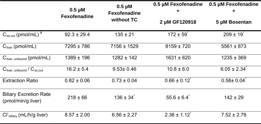
1.c. Determine the canalicular transport protein(s) involved in the biliary excretion of fexofenadine using isolated perfused livers from Mrp2-deficient TR- rats in combination with “specific” transport protein inhibitors (GF120918 and bosentan).
1.d. Define the role of basolateral and canalicular transport proteins in the overall hepatic disposition of fexofenadine using isolated perfused livers from transport protein-deficient mice.
Aim #2. Examine the mechanisms of hepatic uptake and excretion of 99m
Technetium mebrofenin (99mTc-MEB) and 99mTechnetium sestamibi (99mTc-MIBI) in intact hepatocytes, and the influence of altered hepatic transport protein function, on the hepatobiliary disposition of 99mTc-MEB and 99mTc-MIBI (Chapter 4).
2.a. Characterize 99mTc-MEB and 99mTc-MIBI uptake in suspended rat and human hepatocytes in the absence and presence of “specific” inhibitors of basolateral transport proteins.
and in the presence of “specific” transport protein inhibitors (e.g.,
GF120918).
2.c. Characterize 99mTc-MEB and 99mTc-MIBI hepatobiliary disposition in sandwich-cultured human hepatocytes in the absence and presence of “specific” transport protein inhibitors (e.g., GF120918 and MK-571).
Aim #3. Identify mechanisms of hepatic disposition of sorafenib in human hepatocytes, and evaluate 99mTc-MEB and 99mTc-MIBI as transport protein probe substrates to determine if their disposition is sensitive to changes in hepatic transport protein function as a result of hepatocellular carcinoma and cirrhosis (Chapters 5-6).
3.a. Investigate the hepatobiliary disposition of sorafenib in human hepatocytes.
3.b. Compare the pharmacokinetics and hepatic exposure of 99mTc-MEB and 99mTc-MIBI in a patient with hepatocellular carcinoma and Child’s Pugh B
References
1. A. Haimeur, G. Conseil, R. G. Deeley, and S. P. Cole. The MRP-related and BCRP/ABCG2 multidrug resistance proteins: biology, substrate specificity and regulation. Curr Drug Metab5: 21-53 (2004).
2. Y. Kato, H. Suzuki, and Y. Sugiyama. Toxicological implications of hepatobiliary transporters. Toxicology181-182: 287-90 (2002). 3. K. Maedaand Y. Sugiyama. Impact of genetic polymorphisms of
transporters on the pharmacokinetic, pharmacodynamic and toxicological properties of anionic drugs. Drug Metab Pharmacokinet23: 223-35 (2008).
4. N. Mizuno, T. Niwa, Y. Yotsumoto, and Y. Sugiyama. Impact of drug transporter studies on drug discovery and development. Pharmacol Rev
55: 425-61 (2003).
5. Y. Shitara, H. Sato, and Y. Sugiyama. Evaluation of drug-drug interaction in the hepatobiliary and renal transport of drugs. Annu Rev Pharmacol Toxicol45: 689-723 (2005).
6. C. Q. Xia, J. J. Yang, and L. S. Gan. Breast cancer resistance protein in pharmacokinetics and drug-drug interactions. Expert Opin Drug Metab Toxicol1: 595-611 (2005).
7. T. W. Looand D. M. Clarke. Membrane topology of a cysteine-less mutant of human P-glycoprotein. J Biol Chem270: 843-8 (1995).
8. L. Schmittand R. Tampe. Structure and mechanism of ABC transporters.
Curr Opin Struct Biol12: 754-60 (2002).
9. F. Thiebaut, T. Tsuruo, H. Hamada, M. M. Gottesman, I. Pastan, and M. C. Willingham. Cellular localization of the multidrug-resistance gene product P-glycoprotein in normal human tissues. Proc Natl Acad Sci U S A
84: 7735-8 (1987).
10. G. A. Graf, L. Yu, W. P. Li, R. Gerard, P. L. Tuma, J. C. Cohen, and H. H. Hobbs. ABCG5 and ABCG8 are obligate heterodimers for protein
trafficking and biliary cholesterol excretion. J Biol Chem278: 48275-82 (2003).
11. P. L. Ee, S. Kamalakaran, D. Tonetti, X. He, D. D. Ross, and W. T. Beck. Identification of a novel estrogen response element in the breast cancer resistance protein (ABCG2) gene. Cancer Res64: 1247-51 (2004).
marker Bcrp/ABCG2 enhances hypoxic cell survival through interactions with heme. J Biol Chem279: 24218-25 (2004).
13. H. Wang, L. Zhou, A. Gupta, R. R. Vethanayagam, Y. Zhang, J. D. Unadkat, and Q. Mao. Regulation of BCRP/ABCG2 expression by
progesterone and 17beta-estradiol in human placental BeWo cells. Am J Physiol Endocrinol Metab290: E798-807 (2006).
14. L. A. Doyle, W. Yang, L. V. Abruzzo, T. Krogmann, Y. Gao, A. K. Rishi, and D. D. Ross. A multidrug resistance transporter from human MCF-7 breast cancer cells. Proc Natl Acad Sci U S A95: 15665-70 (1998).
15. P. A. Fetsch, A. Abati, T. Litman, K. Morisaki, Y. Honjo, K. Mittal, and S. E. Bates. Localization of the ABCG2 mitoxantrone resistance-associated protein in normal tissues. Cancer Lett235: 84-92 (2006).
16. S. Kawabata, M. Oka, K. Shiozawa, K. Tsukamoto, K. Nakatomi, H. Soda, M. Fukuda, Y. Ikegami, K. Sugahara, Y. Yamada, S. Kamihira, L. A.
Doyle, D. D. Ross, and S. Kohno. Breast cancer resistance protein directly confers SN-38 resistance of lung cancer cells. Biochem Biophys Res Commun280: 1216-23 (2001).
17. M. Maliepaard, M. A. van Gastelen, L. A. de Jong, D. Pluim, R. C. van Waardenburg, M. C. Ruevekamp-Helmers, B. G. Floot, and J. H.
Schellens. Overexpression of the BCRP/MXR/ABCP gene in a topotecan-selected ovarian tumor cell line. Cancer Res59: 4559-63 (1999).
18. M. Nakagawa, E. Schneider, K. H. Dixon, J. Horton, K. Kelley, C. Morrow, and K. H. Cowan. Reduced intracellular drug accumulation in the absence of P-glycoprotein (mdr1) overexpression in mitoxantrone-resistant human MCF-7 breast cancer cells. Cancer Res52: 6175-81 (1992).
19. M. J. Zamek-Gliszczynski, K. Nezasa, X. Tian, J. C. Kalvass, N. J. Patel, T. J. Raub, and K. L. Brouwer. The important role of Bcrp (Abcg2) in the biliary excretion of sulfate and glucuronide metabolites of acetaminophen, 4-methylumbelliferone, and harmol in mice. Mol Pharmacol70: 2127-33 (2006).
20. H. Burger, H. van Tol, A. W. Boersma, M. Brok, E. A. Wiemer, G. Stoter, and K. Nooter. Imatinib mesylate (STI571) is a substrate for the breast cancer resistance protein (BCRP)/ABCG2 drug pump. Blood104: 2940-2 (2004).
induced by the epidermal growth factor receptor inhibitor Iressa (ZD1839, Gefitinib). Cancer Res65: 1770-7 (2005).
22. C. Erlichman, S. A. Boerner, C. G. Hallgren, R. Spieker, X. Y. Wang, C. D. James, G. L. Scheffer, M. Maliepaard, D. D. Ross, K. C. Bible, and S. H. Kaufmann. The HER tyrosine kinase inhibitor CI1033 enhances
cytotoxicity of 7-ethyl-10-hydroxycamptothecin and topotecan by inhibiting breast cancer resistance protein-mediated drug efflux. Cancer Res61: 739-48 (2001).
23. T. Ando, H. Kusuhara, G. Merino, A. I. Alvarez, A. H. Schinkel, and Y. Sugiyama. Involvement of breast cancer resistance protein (ABCG2) in the biliary excretion mechanism of fluoroquinolones. Drug Metab Dispos
35: 1873-9 (2007).
24. G. D. Kruh, H. Zeng, P. A. Rea, G. Liu, Z. S. Chen, K. Lee, and M. G. Belinsky. MRP subfamily transporters and resistance to anticancer agents.
J Bioenerg Biomembr33: 493-501 (2001).
25. M. Buchler, J. Konig, M. Brom, J. Kartenbeck, H. Spring, T. Horie, and D. Keppler. cDNA cloning of the hepatocyte canalicular isoform of the
multidrug resistance protein, cMrp, reveals a novel conjugate export pump deficient in hyperbilirubinemic mutant rats. J Biol Chem271: 15091-8 (1996).
26. D. Kepplerand J. Kartenbeck. The canalicular conjugate export pump encoded by the cmrp/cmoat gene. Prog Liver Dis14: 55-67 (1996). 27. T. P. Schaub, J. Kartenbeck, J. Konig, H. Spring, J. Dorsam, G. Staehler,
S. Storkel, W. F. Thon, and D. Keppler. Expression of the MRP2 gene-encoded conjugate export pump in human kidney proximal tubules and in renal cell carcinoma. J Am Soc Nephrol10: 1159-69 (1999).
28. G. E. Sandusky, K. S. Mintze, S. E. Pratt, and A. H. Dantzig. Expression of multidrug resistance-associated protein 2 (MRP2) in normal human tissues and carcinomas using tissue microarrays. Histopathology41: 65-74 (2002).
29. H. E. Meyer zu Schwabedissen, G. Jedlitschky, M. Gratz, S. Haenisch, K. Linnemann, C. Fusch, I. Cascorbi, and H. K. Kroemer. Variable
expression of MRP2 (ABCC2) in human placenta: influence of gestational age and cellular differentiation. Drug Metab Dispos33: 896-904 (2005). 30. Y. Cui, J. Konig, J. K. Buchholz, H. Spring, I. Leier, and D. Keppler. Drug


![Table 2.2. Accumulation, BEI and in vitro Cl biliary of [ 3 H]Taurocholate, [ 3 H]Digoxin and Fexofenadine in human SCH](https://thumb-us.123doks.com/thumbv2/123dok_us/8324484.2207074/119.918.99.832.277.756/table-accumulation-vitro-biliary-taurocholate-digoxin-fexofenadine-human.webp)