Complement Activation Contributes to Severe Acute
Respiratory Syndrome Coronavirus Pathogenesis
Lisa E. Gralinski,aTimothy P. Sheahan,aThomas E. Morrison,bVineet D. Menachery,a,cKara Jensen,aSarah R. Leist,a Alan Whitmore,dMark T. Heise,dRalph S. Barica
aDepartment of Epidemiology, University of North Carolina, Chapel Hill, North Carolina, USA
bDepartment of Immunology and Microbiology, University of Colorado School of Medicine, Aurora, Colorado,
USA
cDepartment of Microbiology and Immunology, University of Texas Medical Branch, Galveston, Texas, USA
dDepartment of Genetics, University of North Carolina, Chapel Hill, North Carolina, USA
Received11 August 2018Accepted15 August 2018Published9 October 2018 CitationGralinski LE, Sheahan TP, Morrison TE, Menachery VD, Jensen K, Leist SR, Whitmore A, Heise MT, Baric RS. 2018. Complement activation contributes to severe acute respiratory syndrome coronavirus pathogenesis. mBio 9:e01753-18.https://doi .org/10.1128/mBio.01753-18.
EditorKanta Subbarao, NIAID, NIH Copyright© 2018 Gralinski et al. This is an open-access article distributed under the terms of theCreative Commons Attribution 4.0 International license.
Address correspondence to Ralph S. Baric, [email protected].
This article is a direct contribution from a Fellow of the American Academy of Microbiology. Solicited external reviewers: Luis Enjuanes, Centro Nacional de Biotecnologia, CNB-CSIC; Stacey Schultz-Cherry, St. Jude Children's Research Hospital.
ABSTRACT Acute respiratory distress syndrome(ARDS) is immune-driven patholo-giesthatareobservedinseverecasesofsevereacuterespiratorysyndrome corona-virus (SARS-CoV) infection.SARS-CoVemerged in2002 to2003and ledtoaglobal outbreak of SARS. Aswith the outcome of human infection, intranasal infectionof C57BL/6J mice with mouse-adapted SARS-CoV results in high-titer virus replication withinthe lung,induction of inflammatorycytokines andchemokines,and immune cell infiltration within the lung. Using this model, we investigated the role of the complement systemduringSARS-CoVinfection.We observedactivationofthe com-plementcascadeinthelungasearlyasday 1followingSARS-CoVinfection.Totest whether this activation contributed to protective or pathologic outcomes, we uti-lizedmicedeficientinC3(C3–/–),thecentralcomponentofthecomplement system.
Relative to C57BL/6J control mice, SARS-CoV-infected C3–/– mice exhibited
signifi-cantly less weight loss and less respiratory dysfunction despite equivalent viral loads in the lung. Significantly fewer neutrophils and inflammatory monocytes were present in the lungs of C3–/– mice than in C56BL/6J controls, and
subse-quent studies revealed reduced lung pathology and lower cytokine and chemo-kine levels in both the lungs and the sera of C3–/–mice than in controls. These
studies identify the complementsystem as an important host mediator of SARS-CoV-induced disease and suggest that complement activation regulates a sys-temic proinflammatory response to SARS-CoV infection. Furthermore, these data suggest that SARS-CoV-mediated disease is largely immune driven and that in-hibiting complement signalingafter SARS-CoV infection might function as an ef-fective immunetherapeutic.
IMPORTANCE The complement system is a critical part of host defense to many bacterial, viral,and fungal infections. It works alongside pattern recognition recep-torsto stimulatehostdefensesystemsinadvance of activationof theadaptive im-mune response.Inthis study, wedirectlytest theroleof complement inSARS-CoV pathogenesis using a mouse model and show that respiratory disease is signifi-cantly reduced in the absence of complement even though viral load is un-changed. Complement-deficient mice have reduced neutrophilia in their lungs and reduced systemic inflammation, consistent with the observation that SARS-CoV pathogenesis is an immune-driven disease. These data suggest that inhibi-tion of complement signaling might be an effective treatment option following coronavirusinfection.
S
evere acute respiratory syndrome coronavirus (SARS-CoV) emerged in 2002 and 2003 from coronaviruses circulating in animal markets in China (1). Emergence of thisnovelvirusledtoaglobaloutbreakofrespiratorydisease,withover8,000human cases and 10% mortality (2, 3). In 2012, a new, related zoonotic coronavirus was identifiedintheMiddleEast,designatedMiddleEastrespiratorysyndromecoronavirus (MERS-CoV),causingsevererespiratorydiseasewithgreaterthan35%mortality(www.who.int/emergencies/mers-cov/en) (4).BothSARS-CoVandMERS-CoVcausearangeof
diseasefromasymptomaticcasestosevereacuterespiratorydistresssyndrome(ARDS) andrespiratoryfailure(5).Notably,metagenomicsandsyntheticvirusrecovery strate-gies have since revealedtheexistence of large pools of preepidemic SARS-like bat coronaviruseswhichreplicateinprimaryhumanairwayepithelial cells.Theseviruses arepoisedforemergencebecausetheybothefficientlyusehumanACE2entry recep-torsandresist existingvaccinesandimmunotherapeutics(6,7).Duetotheongoing threatandcontinuedemergenceofnew,highlypathogeniccoronavirusesfromanimal reservoirs,athoroughunderstandingofthehost-virusinteractionsthatdriveSARS-CoV pathogenesis will aid thepublic health response to current andfuture coronavirus outbreaks(8).
TheimportanceofcomplementinSARS-CoVpathogenesisiscontroversial.Previous studieshaveinvestigatedtheroleofknownpolymorphismsinthemannose-binding lectin(MBL)andMBL-associatedserineprotese-2(MASP2)genesinSARS-CoVinfection outcomefollowingthe2003outbreakbutwithconflictingresults.Oneretrospective analysisshowedthatpeoplewithlowordeficientserumMBLlevelsweremorelikely tobecomeinfectedwithSARS-CoV(9)thanthosewithhighMBLlevels,suggestingthat MBL and complement activation play a role in protecting the host from infection. However,asecondstudyfoundnoassociationbetweenMBLhaplotypeandSARS-CoV infectionstatus(10).Additionally,itwasshownMBLcanbindtotheSARS-CoVSpike proteininvitrobysomegroups(11)butnotbyothers(12).Examinationoftheroleof thedownstream complement geneMASP2foundno associationbetweengenotype andSARSsusceptibility(13).Together,theresultsleaveageneraluncertaintyaboutthe roleofcomplementinresponsetoSARS-CoVinfection.
Despitetheexistingbodyofliterature,theroleofcomplementinSARS-CoV patho-genesishasneverbeendirectlyassessedinvivo.Thecomplementsystemisanancient arm of theinnateimmune response comprisedof multiple proteinswhosereactive cascadeofcleavageproductscancoordinatetheinflammatoryresponseatthesitesof infectionandcanbedirectlyantimicrobial.Consistingofmorethan30solubleandcell surface-associated proteins,complement is amajor componentof innateimmunity thatfunctionstorecognizeandeliminateinvading pathogens(14).Activation ofthe complement system occurs through multiple mechanisms that include three well-describedpathways,theclassical,lectin,andalternativecomplementactivation path-ways(15),andresultsinproteolyticprocessingofvariouscomponentsofthe comple-mentsystem,includingC3,C4,andC5.ProteolyticprocessingofC3generatesanarray ofcleavageproductsthatareinvolvedinamplificationofcomplementactivitythrough formation ofC3andC5 convertases,opsonizationof pathogens,andattractionand activationofleukocytesofboththeinnateandadaptivearmsoftheimmuneresponse. Severalstudies,includingarecentstudyshowingthatcomplementblockaderesultsin reduceddiseaseinaMERS-CoVhumanDPP4transgenic(hDPP4-Tg)mousemodel(16), haveelucidatedprotectiveandpathogenicrolesforthecomplementsystemfollowing infectionbyavarietyofviralpathogens(17).Furthermore,thecomplementsystemhas well-describedrolesinotherpulmonarydiseases(18),especiallyafterinfluenzavirus andrespiratorysyncytialvirusinfection(19–21).
Inthisstudy,weassessedtheroleofthecomplementsysteminthepathogenesis of SARS-CoVinfection.Buildingfromasystemsbiologyanalysisthatsuggestedthat complement wasmodulatedduringSARS-CoVinfection,weconfirmedthat comple-mentwasactivateduponSARS-CoVchallenge.MicedeficientinC3(C3–/–),thecentral
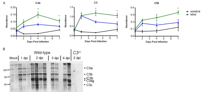
FIG 1 Omics characterization of complement pathway expression and activation. (A) Protein abundance at 1, 2, 4, and 7 days postinfection relative to that in mock-treated samples. Samples were taken from total lung homogenates, and error bars indicate standard errors of the means (SEM). Each point indicates the mean of results for 5 mice at a given time. (B) C3 protein cleavage is observed in the lung by Western blotting as early as 24 h following SARS-CoV MA15 infection of C57BL/6J mice. Numbers at the left are molecular weights (in thousands).
levelsofinflammatorycytokines/chemokinesinthelungandperiphery.Importantly, thekineticsandmagnitudeofvirusreplication inC3–/–andwild-typemicewerethe
same, showing that complement does not play a role in controlling virus replica-tion. We observed complement deposition inthelungs of SARS-CoV-infected mice, suggestingthat complement activationresults inimmune-mediateddamage tothe lung. Additionally, serum activation indicates that complement-mediated systemic inflammationmaydrivethepathogenicresponsetoSARS-CoVinfection.Together,the resultsindicate thatcomplement playsacriticalroleinSARS-CoVpathogenesisand that inhibition of the complement pathway might be an effective therapeutic to coronavirus-mediateddisease.
RESULTS
ComplementisactivatedinSARS-CoVMA15-infectedmice. Whileworkbyother
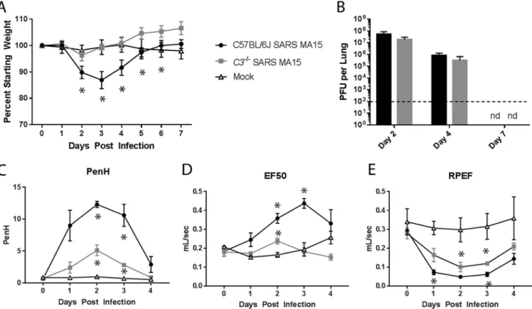
FIG 2 Characterization of C3 knockout mice. (A) Weight loss of SARS-CoV MA15-infected C57BL/6J mice,C3–/–mice, or mock-infected mice were measured over
time. (B) Lung titers of SARS-CoV MA15-infected C57BL/6J or C3–/–mice at 2, 4, and 7 days postinfection. nd, not determined. (A and B) Six to 8 mice were used
through day 4, and 3 to 4 mice were used for days 5 to 7. The respiratory function of SARS-CoV MA15-infected C57BL/6J andC3–/–mice and mock-infected
mice was measured using a Buxco whole-body plethysmography system for Penh, a measure of calculated airway resistance (C), EF50, midbreath expiratory flow (D), and RPEF, the rate of peak expiratory flow (E).*,P⬍0.05 between mock-infected mice and a given condition. (C to E) Three mice were used for each infection group, and two mock-infected mice were used.
MultiplecomplementpathwayscontributetoSARS-CoVMA15-induced
patho-genesis.To testtheimportanceofthecomplement signalingpathwayinSARS-CoV
pathogenesis, we infected C3–/– mice andC57BL/6J controls with SARS-CoV MA15.
Controlmiceexhibitedapproximately15%transientweightloss,withpeakweightloss atday3postinfection(Fig.2A).Incontrast,theC3–/–miceweresignificantlyprotected
frominfection,withnosignificantweightlossevidentatanytimepoint.Surprisingly, viraltitersinthelungweresimilarinC3–/–andC57BL/6Jcontrols(Fig.2B),indicating
thatthelackofdiseaseinC3–/–miceisuncoupledfromviralreplicationefficiencyand
that complementsignaling isnot necessary forSARS-CoVMA15clearance fromthe lung. WefurthermeasuredSARS-CoVMA15-induceddiseasebyassessingrespiratory functionusingwhole-bodyplethysmographyfollowinginfectionofC57BL/6JandC3–/–
mice.Enhancedpause(Penh)isacalculatedmeasureofairwayresistancethatwehave associated with airway debris following SARS-CoV MA15 infection (24). The 50% exhalation force(EF50)measurestheexhalationforcemidbreath,whichincreasesas breathingbecomesmoredifficult.Finally,theratioofpeakexpiratoryflow(RPEF)isthe timetopeakexpiratoryflowandhasbeenassociatedwithwheezingfollowing infec-tion. Allthreemetrics havebeenshowntochangesignificantly followingSARS-CoV MA15infection,withPenhandEF50increasingfollowinginfectionandRPEF decreas-ing.Combined,thesemeasurementsshowthatSARS-CoVMA15-infectedanimalshave alteredexhalationpatternsintheirbreathing.C3–/–miceexhibitadecreasedchangein
PenhandEF50levelsfollowingSARS-CoVMA15infectionrelativetothoseofinfected C57BL/6Jcontrolmice;however,RPEFvaluesweresimilarbetweeninfectionconditions (Fig.2CtoE).Together,thesedataindicatethatdespitethelackofweightlossinC3–/–
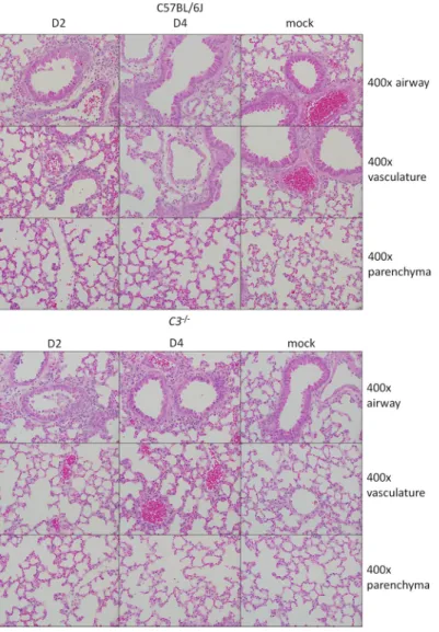
FIG3 Histological analysis of C57BL/6J and C3⫺/⫺lungs at 2 and 4 days postinfection. Representative
images show 400⫻ magnifications of the large airways (top row), vasculature (middle row), and parenchyma (bottom row) of the lung after SARS-CoV MA15 or mock infection of C57BL/6J or C3⫺/⫺
mice.
replicationorcompletelyabolishrespiratorydiseasefollowingSARS-CoVMA15 infec-tion.
In order to determine which arm of the complement pathway contributes to SARS-CoV MA15 pathogenesis,we infected knockoutmice lacking components up-streamofC3.C4-deficientmicelacktheabilitytosignalthroughboththeclassicaland lectinpathways,whilefB-deficientmicelacktheabilitytosignalthroughthealternative pathway.Bothmousestrainsshowedreducedweightlossrelativetothatofinfected controlmice(seeFig.S1inthesupplementalmaterial)at3dayspostinfection;however, neitherC4–/–norfB–/–micereproducedcompleteprotectionfromweightlossobserved
inC3–/–controls.Together,theresultssuggestthatmultiplearmsofthecomplement
pathwaymaybeactivatedandcontributetoSARS-CoV-mediateddiseasethroughC3 activation.
ReducedlungpathologyinC3–/–mice. AnalysisofSARS-CoVMA15-infectedlung
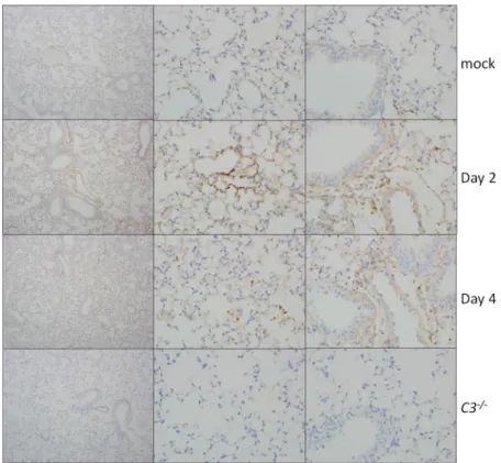
eosino-FIG4 Complement deposition staining. Complement deposition on lung tissue of C57BL/6J mice (top
three rows) was assessed by immunohistochemistry. Mice were examined at 2 and 4 dpi and mock infected or infected with SARS-CoV MA15. C3–/–mice (bottom row) showed no positive staining.
phils,at2dpithantheirwild-typecontrols,althoughthisrelationshipwasreversedlater ininfection.At4dpi,C57BL/6Jmicedisplayedpronouncedlungpathology,including inflammatorycellsinthelargeairwayandparenchyma,perivascularcuffing,thickening oftheinterstitialmembrane,andlowlevelsofintra-alveolaredema.Incontrast,C3–/–
miceshowedreducedscoresintheseareas,consistentwiththeimprovedrespiratory function observed in Fig. 2. Notably, the lung pathology results were not as pro-nounced as thecompleteabsence of weight loss,suggesting apossibledistinction between lung disease and overall pathogenesis. We also considered whether the decrease inSARS-CoV MA15 pathogenesis in C3–/– mice was due to reduced lung
damageintheabsenceof complementpathwaysignaling.Toinvestigatethis possi-bility,welookedforsignsofcomplementdepositiononSARS-CoVMA15lungtissue.At both2and4dpi,weobservedscatteredpositivestainingforcomplementinthelungs ofSARS-CoVMA15-infectedmice,suggestingthatlocaltissuedamagemight contrib-utetoSARS-CoVpathogenesis(Fig.4).Interestingly,stainingwasconsistentlyfoundin theparenchymaofthelungandnotinthelargeairways,whicharetheothermainsite ofSARS-CoVMA15replication.NopositivestainingwasobservedinthelungsofC3–/–
mice.
Diminishedinfiltrationofthelungsofaselectimmunepopulationofinfected
C3–/– mice. In order to identify and quantitate inflammatory cells in the lung, we
performedflowcytometryat4dpi.Inparallelwithhumansexhibitinglungpathology,
C3–/–miceexhibitedsignificantpulmonaryinfiltrationfollowingSARS-CoVMA15
infec-tion, butthis inflammationwasreducedrelativetothatobserved inwild-type mice. SARS-CoVMA15-infectedC57BL/6JandC3–/–micehadsimilartotalcellcountsaswell
assimilarpercentagesofCD45-positivecellsintheirlungs(datanotshown). Consis-tentlywith what wasobserved inhumanSARS-CoV patients(25), lymphopeniawas observedinthelungsofbothSARS-CoVMA15-infectedC57BL/6JandC3–/–micewith
FIG 5 Inflammatory cells of C57BL/6J mice andC3knockout mice. Flow cytometric analysis of inflammatory cells present in the lungs of SARS-CoV MA15-infected or mock-infected C57BL/6J orC3–/–mice at 4 days postinfection. (A) Lymphocytes; (B) myeloid-cell-derived cells (as defined by Misharin et al.
[68]); (C) CD4 T cell activation markers; (D) CD8 T cell activation markers; (E) CD11c–neutrophil activation markers.*,P⬍0.05. Error bars indicate SEM. Eight
mice were used for all infection groups, and 4 mice were used for all mock-infected groups. Neuts, neutrophils; DC, dendritic cells; MHCIIhi, a high fluorescence intensity for MHCII staining; MonoMac (inf), inflammatory monocyte-macrophages; Macs, macrophages.
significant differences wereobserved in levelsof Tcell activationbetween infected C57BL/6JandC3–/–mice;bothCD4andCD8TcellsinC3–/–miceexpressedmoreKi-67
(Fig.5CandD),anintracellularmarkerofproliferation,thanthoseinC57BL/6Jcontrols. AnalysisofmyeloidcellsinthelungshowedthatinfectedC56BL/6Jmicehad signifi-cantlyhigherlevelsof neutrophils,particularlynonactivated neutrophils,inthelung thaninfectedC3–/–mice(Fig.5BandE).Furthermore,inflammatorymonocytes,which
have previously been associated with increased SARS-CoVMA15 pathogenesis (26), weresignificantlyincreasedinthelungsofwild-typemicebutnotC3–/–mice(Fig.5B).
Finally,weobservedsignificantlymoredendriticcellsandalveolarmacrophagesinthe lungsof SARS-CoVMA15-infectedC3–/–mice thaninthelungsof infectedC57BL/6J
mice.Together,althoughC3–/–miceproducedarobustimmunecellinfiltration
follow-ingSARS-CoVinfection,theyhadsignificantreductionsinbothinflammatory mono-cytes and neutrophils relative to controls; both cell typesthat areassociated with SARS-CoVpathogenesis(27).Conversely,thepresenceofactivatedTcellsisassociated withrecoveryfollowinginfection(28).
indicating that the absence of C3 does not appear to significantly alter vascular permeability following infection with SARS-CoV (Fig. S2B).
FIG 6 Cytokine and chemokine abundance levels. Protein abundance in the lung was measured by Bioplex multiplex magnetic bead assay at days 2, 4, and
7 postinfection or in mock-infected mice. MIP-1a, MIP-1b, and monocyte chemoattractant protein (MCP) had similar concentrations in the lungs of C57BL/6J andC3–/–mice, with peak abundance at 2 days postinfection (A), while G-CSF, IL-6, TNF, and IL-1a expression was highest in C57BL/6J mice at 2 dpi (B).*,P⬍
0.05;‡,P⬍0.1. Error bars indicate SEM, and 3 to 4 mice were tested for each condition.
CytokineandchemokinelevelsaresignificantlydecreasedinthelungsofC3–/–
mice.TofurtherinvestigatetheinflammatoryresponsetoSARS-CoVMA15infection,
wemeasuredcytokineandchemokineproteinlevelsinthelunginthepresenceand absenceofcomplementsignaling.Multipleproteinexpressionpatternswereobserved inresponsetoinfection.MIP1a,MIP1b,andMCP1areallhighlyexpressedinthelung followingSARS-CoVMA15infectionofbothC57BL/6JandC3–/–mice(Fig.6A),
indicat-ingthatsomeinflammatorysignalingremainsintactintheabsenceofC3.Granulocyte colony-stimulating factor (G-CSF), interleukin 6 (IL-6), tumor necrosis factor alpha (TNF-␣),andIL-1acomprisedagroup ofcytokines andchemokines thatweremore highlyproducedinthelungsofC57BL/6JmicethaninC3–/–mice(Fig.6B),allpeaking
at 2 days postinfection.Notably, thesecytokines allhave arole inthe production, recruitment,ordifferentiationofneutrophils,consistentwiththeflowcytometryresults inFig.5B.WiththeexceptionofRANTES,allinflammatorycytokinesandchemokines weremeasuredatthehighestlevelsat2dpi,indicatingthatthehostimmuneresponse istriggeredquicklyfollowinginfectionwithSARS-CoVMA15.Together,theseresults indicate thattheabsenceofcomplement hasanimpact onthemagnitude ofsome cytokinesandchemokinesinthelung;however,robustproductioncanoccurineither thepresenceortheabsenceofC3.
SARS-CoVMA15inducessystemiccomplementactivation. Theabsenceof
com-plementsignalingresultedinreducedSARS-CoVMA15pathogenesis,asmeasuredby weight loss and a partial reduction of respiratory dysfunction, pathology, immune infiltration,and cytokineresponsesin thelung. We hypothesized thatsystemic dis-ease coupled with no changein viral titer might also drive important elements of complement-mediateddisease.Therefore,weexaminedserafromwild-typeandC3–/–
FIG 7 Systemic response to SARS-CoV MA15 infection. (A) C3 protein cleavage products are observed by Western blotting in the serum following infection. Molecular weights are noted at the left (in thousands). rMA15, recombinant MA15. (B) MCP-1 and RANTES levels are similarly elevated following infection in C57BL/6J andC3–/–mice. (C) G-CSF, KC, and IL-5 all have significantly higher expression in the sera of C57BL/6J mice than in those of C3–/–mice. IL-6 expression
was suggestive of differences. (D) Complement deposition staining in the kidneys of C57BL/6J mice.*,P⬍0.05;‡,P⬍0.06. Error bars indicate SEM, and 3 to 4 mice were used for each condition.
(keratinocyte chemoattractant or CXCL1) were presentin significantly higher abun-danceinthelungsofC57BL/6JmicethaninthoseofC3knockoutmice(Fig.7C).We furtherexaminedthepossibilitythatSARS-CoVMA15infectionleadstocomplement depositionoutsidethelungandfoundnosignsofincreasedcomplementstainingin thekidney(Fig.7D).Althoughnocomplementdepositionwasseen,thepresenceof bothactivatedcomplementandinflammatorycytokinesintheseralikelycontributesto asystemicinflammatoryresponsethat drivesSARS-CoVMA15-mediatedweight loss followinginfection.
DISCUSSION
infections (31) aswell as some influenzavirus and flavivirusinfections (22, 32,33). Furthermore,viruses,includingherpesviruses,poxviruses,astroviruses,flaviviruses,and retroviruses,encodegenestohelpthemevadedetectionbythecomplementsystem (17),strongevidencethatcomplementisimportantinthehostantiviralresponse.The host factors that drive protective (22) or pathogenic (34) complement-associated responsesinviralinfectionarenotwellunderstood.Ofparticularconcern,the anaphy-latoxinsC3a,C4a,andC5aareproducedduringactivationofthecomplementsignaling cascade;theyhave potentproinflammatoryproperties andcan triggerinflammatory cell recruitment and neutrophil activation (35). C3a and C5a blockade has been proposed asatreatmentforacute lunginjury(36), andanti-C5a antibodyhasbeen shown to protect mice from infection with influenzavirus (34) and, more recently, MERS-CoV (16).Complementrecognitionisimportantforthecontrolof paramyxovi-ruses(37),denguevirus(38),andhumanTlymphotrophicvirustype1(HTLV-1)(39), andmanymoreviruseshavedevelopedmeansofevadingdetectionbythe comple-ment system (17). In contrast,the data presented here, inconjunction with recent findingsforRossRivervirus(40,41),influenzavirus(34),andwell-established autoim-munedisease(42), demonstratethatcomplementsystemactivationcanalsoleadto exacerbateddisease.
Previousreportsclearlyestablishedtheabilityofmannose-bindinglectin(MBL)to bindtotheSARS-CoVspikeprotein(11),dependentonanN-linkedglycosylationsite; however,theroleofcomplementsignalinginSARS-CoVpathogenesiswasunclear(9, 10,13, 43).In this study,wedemonstratethat thecomplement systemisactivated followingSARS-CoVMA15infection(Fig.1B).However,wedidnotobserveanychange inviraltiterinC3–/–mice(Fig.2B),indicatinganimportantdifferencebetweeninvivo
andinvitrostudiesandtheuseofviralpseudoparticles.Theabsenceofcomplement signalingresultedinprotection fromSARS-CoVMA15-inducedweightloss,asshown through theuseof both C3-deficientmice(Fig. 2A)andactivation pathway-specific knockout mice (Fig. S1). Respiratory function in C3 knockout mice was improved relativetothatofcontrolmice,althoughsignificantchangesinPenhandRPEFwerestill observed,indicatingthattheeliminationofcomplementsignalingdidnotcompletely removetheeffectsofSARS-CoVMA15infection.Whileanalysisofthecellular inflam-matory response to SARS-CoVMA15 infectionrevealedmodest changesin histopa-thologyandoverallinflammatorycellrecruitmenttothelungs,significantdifferences were observed in pathogenic inflammatory monocyte and neutrophil populations, indicatingthatcomplementsignalingcontributestothebroaderimmuneresponseto infection. Immunohistochemical stainingrevealedthat SARS-CoVMA15 infection in-duced complement deposition in the lung (Fig. 4), similar to that associated with pathogenesisinRossRivervirus-infectedmice(41)andsomeinfluenzavirusinfections (34),anditislikelythatcomplementdepositioncontributestopulmonarydiseaseand inflammatorycellrecruitment.
The cytokines and chemokines IL-5, IL-6, KC (CXCL1), and G-CSF have higher abundancesinthelungsofSARS-CoVMA15-infectedwild-typemicethaninC3–/–mice
and are all known to promote neutrophil recruitment. Indeed, significantly more neutrophils were observed inthe lungs of SARS-CoV MA15-infected C56BL/6J mice than in the C3–/– mice (Fig. 5B). Interestingly, while there were fewer neutrophils
present,theneutrophilsfoundinthelungsofC3–/–miceinfectedwithSARS-CoVMA15
suggestthatanonpulmonarycausemightalsocontributetothelackofweightlossin
C3–/–mice.
Importantly, our data demonstrate that SARS-CoV MA15 infection activates the complement systemsystematicallyaswellasinthelung(Fig.7A and1B).Wild-type C57BL/6Jmiceexhibitedanincreasedabundanceofserumcytokinesandchemokines in response to SARS-CoV MA15 infection (Fig. 7C) in comparisonto C3–/– mice. In
particular,thepyrogeniccytokineIL-6(47)ispresentathigherabundanceinthelungs andseraofC57BL/6JmicethaninthoseofC3–/–mice.IL-1␣andTNF-␣arealsomore
abundantinthelungsofC57BL/6Jmice,suggestingthatwild-typebutnotC3–/–mice
developafeverresponsetoinfectionthatcontributestoweight lossandrespiratory dysfunction phenotypes. While the precise mechanism of complement activation followingSARS-CoVMA15infectionisstillunclear,itislikelythroughrecognitionofthe viralspikeglycoproteinandpartiallymediatedbyMBL(seeFig.S1inthesupplemental material).
Theanaphylatoxinsproducedbytheactivatedcomplementpathway,C4a,C3a,and C5a,haveimportantimmunostimulatoryrolesinvascularpermeabilityand inflamma-tory cellrecruitment (35, 48). C3a andC5a inparticular arenotedfor their rolesin causingmastcell degranulation,initiatingacytokinestorm,promotingvascular per-meability, and contributing to acute lung injury (36, 49, 50). Furthermore, a C5a antibody blockadewas recently shownto protect in amodel of highly susceptible MERS-CoV mice(16).Although therearenoreportsofmastcellactivationfollowing SARS-CoVorMERS-CoVinfection,activationhasbeenobservedbothinvitroandinvivo
following infection with influenza virus (51) andmay occur following othersevere respiratoryinfections,includingcoronaviruses.Mastcellsreleasecytokines,including IL-6, IL-1, and TNF-␣, consistent with the inflammatory profile observed following SARS-CoV MA15 infection. While this work cannot definitively conclude that the complement anaphylatoxins andmastcell activation contribute toSARS-CoV MA15 pathogenesis,thedataareconsistentwiththis possibility,andtheconceptwarrants furtherinvestigation.
Complement pathway activationisahallmark of bacterialinfection,and genetic deficienciesinthecomplement pathwayresultinenhancedsusceptibilityto Strepto-coccuspneumoniae,Neisseriameningitidis,andHaemophilusinfluenzaeinfections(52), aswellastosepsis.Interestingly,ithasalsobeenshownthatSARS-CoVMA15infection stimulates TLR4 (53, 54), which is classicallyknown as the lipopolysaccharide (LPS) receptor(55,56)andimportantforrecognitionofmanybacterialinfections.Combined, these datasuggest that hostrecognition of SARS-CoV MA15infection mayactivate similar pathwaysrecognizingabacterialinfection,leadingto immunesignaling cas-cades that cause systemic disease and enhance viral pathogenesis. While systemic activationofthecomplementpathwaymaybeusefulduringabacterialinfection,itis lesssoduringalocalizedacuteviralinfection,such asSARS-CoV.Furthermore, com-plementactivation,inconjunctionwiththepresenceofneutrophils,isknowntocause increasedvascularpermeability,aconditionthatisalsoobservedfollowingSARS-CoV infection(57–59)andwasassociatedwithpooroutcome.Baselinecomplement acti-vationalsoincreaseswithage(60,61),consistentwithincreasedSARS-CoVmorbidity andmortalityinagedpopulations.Finally,ithaspreviouslybeenreportedthatserum C5a levels are predictive of ARDS development (62) and that, in the absence of complement, animalsareprotectedfrom bacteriallyinduced “shocklung” (63), data consistent with thepathogenic rolethat wehave found forcomplement following SARS-CoVMA15infection.
Inthiswork,wedemonstratethatSARS-CoVMA15infectionactivatesthe comple-mentpathwayandthatcomplementsignalingcontributestodiseasefollowing infec-tion.Thisdiseaseislikelymediatedbycomplementproteindepositioninthelungas wellassystemiccomplementactivationandinflammation.Notably,theabsenceofC3
aretreatedwitheitheraC3areceptor(C3aR)antagonistorantibodiestoC5a(16,34). AsimilartreatmentmightbeeffectiveinmitigatingSARS-CoVMA15-induceddisease, asSARS,MERS, andinfluenzahavecommondiseasemanifestations,including devel-opment of acute lung injury. Given the large array of zoonotic strains poised for cross-speciestransmission,broad-basedinhibitorsofemergingcoronavirusinfections areahighpriority(64).Pinpointingtheprecisearmsofthecomplementpathwaythat contributetoSARS-CoVwillhelpfurtheridentifytherapeutictargetswhileminimizing unnecessaryinhibitionoftheimmuneresponse.Thisworksuggeststhatinvestigation of anticomplement drugs for treatment of coronavirus infections is warranted and wouldpairwellwithdirectantiviraltherapeutics.
MATERIALSANDMETHODS
Virusesandcells.Stocks of recombinant mouse-adapted SARS-CoV (MA15) (65) were propagated
and their titers were determined in Vero E6 cells and stored as single-use aliquots at ⫺80°C as previously described (66). Tissue titers of MA15 were determined by plaque assay on Vero E6 cells as previously described (66, 67), with a limit of detection of 100 PFU. All experiments using live virus were performed in a class II biological safety cabinet in a certified biosafety level 3 laboratory with negative air pressure and redundant exhaust fans; personnel wore personal protective equipment, including Tyvek suits, hoods, and powered air-purifying respirators.
Mouseexperiments.C57BL/6J (stock number 000664) and C3–/–(stock number 003641) mice were
purchased from Jackson Laboratories. fB⫺/⫺mice were generously provided by Charles Jennette (UNC),
and C4–/–mice were provided by Mark Heise (UNC). All animal husbandry and experiments were
performed in accordance with all University of North Carolina at Chapel Hill Institutional Animal Care and Use Committee guidelines. Age-matched (10- to 11-week-old) female mice were anesthetized with a mixture of ketamine-xylazine and intranasally inoculated with 50 l of phosphate-buffered saline (PBS) or 105PFU of SARS-CoV MA15 diluted in PBS. Mice were monitored for disease signs and weighed at 24-h
intervals.
Microarrayandproteomicsanalysis.Microarray and proteomics analyses were performed on a
time course and according to a dose-response study published by Gralinski et al. by following the same methods (23). The proteomics data (experiment SM001) are publically available through the PNNL (http://omics.pnl.gov) Web portal. Briefly the mean intensity (abundance) for each protein was then graphed as an average (5 mice for each infection, 3 mice for mock infection) for each group at each time point. Missing or absent values were not scored; however, if no value was observed in any of the samples at a time point, the sample was registered with a single 0, representing “not detected.”
Histologicalanalysis.At the times indicated in the figures, mice were euthanized using an overdose
of isoflurane, and lung tissue was fixed in 10% formalin. Tissues were embedded in paraffin, and 5-m sections were prepared. To determine the extent of inflammation and tissue pathology, tissues were stained with hematoxylin and eosin and scored in a blind manner from 0 (no sign of phenotype) to 3 (widespread and severe phenotype).
Plateletcounts.For bronchoalveolar lavage (BAL), immediately following euthanasia, 1 ml of PBS
was injected into the lung through the trachea by using a 22-gauge Exel Safelet catheter tip (Fisher). This fluid was then drawn back out and used for subsequent analysis. Two hundred fifty microliters of BAL fluid was used for absolute counting of gross cell types using an Abaxis VetScan HM5 analyzer.
Complement deposition staining. Lung sections from SARS-CoV MA15-, Ross River virus-, or
mock-infected mice were stained for the presence of C3 by the Animal Histopathology and Laboratory Medicine Core at the University of North Carolina. SARS-CoV MA15 lung samples were tested from 20-week-old mice at 1, 2, 4, and 7 days after infection with 105PFU of virus. Staining was performed using
a goat anti-mouse C3 primary antibody (MP Biomedicals).
Immunoblot analysis.Mice were perfused with PBS, and then lung tissue was dissected and
homogenized in lysis buffer (50 mM Tris [pH 8.0], 150 mM NaCl, 1% Nonidet P-40, 0.5% deoxycholate, and 0.1% sodium dodecyl sulfate [SDS] supplemented with Complete protease inhibitor cocktail [Roche]). Total protein concentrations were determined by using the Coomassie Plus assay kit (Pierce). Dilutions of serum or 20- to 30-g aliquots of protein were diluted in an equal volume of 2⫻ SDS sample buffer, and SDS-polyacrylamide gel electrophoresis was performed. Proteins were transferred onto polyvi-nylidene fluoride membranes (Bio-Rad). Membranes were blocked in 1⫻ PBS–5% milk– 0.1% Tween 20 and incubated with goat anti-mouse C3 antibody (1:1,000; Cappel) overnight at 4°C. Membranes were washed in PBS– 0.1% Tween 20 and incubated with rabbit anti-goat-horseradish peroxidase (1:10,000; Sigma) for 1 h at room temperature. After a washing step, proteins were visualized by enhanced chemiluminescence (Amersham) according to the manufacturer’s instructions.
Flowcytometry.Following euthanasia at 4 days postinfection, mice were perfused with 10 ml of PBS
Cy5.5 (clone 145-2C11; eBioscience), CD4-BUV737 (clone RMA4-5; BD), CD8-phycoerythrin (PE) (clone 53-6.7; BD), CD19-BV650 (clone 6D5; BioLegend), CD25-BV510 (clone PC61; BD), CD44-BV786 (clone IM7; BD), CD62L-BUV395 (clone MEL-14; BD), Ki-67–fluorescein isothiocyanate (FITC) (clone SolA15; eBioscience), CD69-PECF594 (clone H1.2F3; BD), NK1.1-PE Cy7 (clone PK136; eBioscience), PD-1-BV605 (clone J43; BD), CD45-LCA–APC-R700 (clone 30-F11; BD), CD11b-BV785 (clone M1/70; BioLegend), CD11c-PECF594 (clone HL3; BD), MHC II-APC (clone M5/114.15.2; eBioscience), Ly6G-APC-Fire780 (clone 1A8; BioLegend), Ly6C-BV605 (clone AL-21; BD), SiglecF-BV650 (clone E50-2440; BD), CD80-BUV737 (clone 16-10A1; BD), CD86-BV421 (clone GL1; BD), and CD103-PerCP-eFluor710 (clone 2E7; eBioscience). After being stained, cells were fixed in 2% paraformaldehyde overnight and then stored in PBS until acquisition within 24 h. A minimum of 100,000 events were collected using an LSRII cytometer (Becton, Dickinson), and analysis was completed using FlowJo software version 10 (TreeStar). All samples were first evaluated through subsequent gates for (i) mononuclear cells, (ii) doublet exclusion, (iii) dead-cell exclusion based on uptake of a fixable live/dead cell discriminator (Invitrogen), and CD45-LCA expression before downstream analyses. Reported cell frequencies were normalized to the percentage of total CD45-LCA⫹events, where appropriate.
Whole-body plethysmography.Respiratory function was measured using whole-body
plethysmog-raphy as described by Menachery et al. (24). Briefly, mice were loaded into individual chambers and allowed to acclimate for 30 min before a 5-min measurement window. Measurements were recorded every 2 s for a total of 150 measurements per time point per mouse.
Cytokine and chemokine protein analysis. The small center lung lobe of each mouse was
homogenized in 1 ml of PBS and briefly centrifuged to remove debris. Fifty microliters of homogenate was used to measure cytokine and chemokine protein abundance using a Bio-Plex Pro mouse cytokine 23-plex assay (Bio-Rad) according to the manufacturer’s instructions.
Statistical analyses.Percent starting body weights, viral titers, and inflammatory cell numbers were
evaluated for statistically significant differences by the Mann-Whitney test or Student’s ttest using GraphPad Prism software.
Accession number(s).The microarray data were previously deposited in the GEO database under
accession number GSE33266 (23).
SUPPLEMENTAL MATERIAL
Supplemental material for this article may be found athttps://doi.org/10.1128/mBio .01753-18.
FIG S1, TIF file, 0.4 MB.
FIG S2, TIF file, 0.9 MB.
TABLE S1, DOCX file, 0.01 MB.
ACKNOWLEDGMENTS
Research was supported by grants from the NIAID of the NIH (AI100625 to R.S.B. and M.T.H., AI106772 to R.S.B., AI109761 to R.S.B., and K99AG049092 to V.D.M.). Animal histo-pathology was performed by the Animal Histohisto-pathology and Laboratory Medicine Core at the University of North Carolina, which is supported in part by an NCI Center Core Support Grant (5P30CA016086-41) to the UNC Lineberger Comprehensive Cancer Center. The UNC Flow Cytometry Core Facility is supported in part by Cancer Center Core Support Grant P30 CA016086 to the UNC Lineberger Comprehensive Cancer Center. The funders had no role in study design, data collection and interpretation, or the decision to submit the work for publication.
The content is solely the responsibility of the authors and does not necessarily represent the official views of the NIH.
REFERENCES
1. Chinese SARS Molecular Epidemiology Consortium. 2004. Molecular evolu-tion of the SARS coronavirus during the course of the SARS epidemic in China. Science 303:1666 –1669.https://doi.org/10.1126/science.1092002. 2. Rota PA, Oberste MS, Monroe SS, Nix WA, Campagnoli R, Icenogle JP,
Peñaranda S, Bankamp B, Maher K, Chen MH, Tong S, Tamin A, Lowe L, Frace M, DeRisi JL, Chen Q, Wang D, Erdman DD, Peret TC, Burns C, Ksiazek TG, Rollin PE, Sanchez A, Liffick S, Holloway B, Limor J, McCaustland K, Olsen-Rasmussen M, Fouchier R, Günther S, Osterhaus AD, Drosten C, Pallansch MA, Anderson LJ, Bellini WJ. 2003. Characterization of a novel coronavirus associated with severe acute respiratory syndrome. Science 300:1394 –1999.https://doi.org/10.1126/science.1085952.
3. Christian MD, Poutanen SM, Loutfy MR, Muller MP, Low DE. 2004. Severe acute respiratory syndrome. Clin Infect Dis 38:1420 –1427.https://doi
.org/10.1086/420743.
4. Zaki AM, van Boheemen S, Bestebroer TM, Osterhaus AD, Fouchier RA. 2012. Isolation of a novel coronavirus from a man with pneumonia in Saudi Arabia. N Engl J Med 367:1814 –1820.https://doi.org/10.1056/
NEJMoa1211721.
5. Hui DS, Memish ZA, Zumla A. 2014. Severe acute respiratory syndrome vs. the Middle East respiratory syndrome. Curr Opin Pulm Med 20: 233–241.https://doi.org/10.1097/MCP.0000000000000046.
6. Menachery VD, Yount BL, Jr, Debbink K, Agnihothram S, Gralinski LE, Plante JA, Graham RL, Scobey T, Ge XY, Donaldson EF, Randell SH, Lanzavecchia A, Marasco WA, Shi ZL, Baric RS. 2015. A SARS-like cluster of circulating bat coronaviruses shows potential for human emergence. Nat Med 21:1508 –1513.https://doi.org/10.1038/nm.3985.
Zhang SY, Wang LF, Daszak P, Shi ZL. 2013. Isolation and characterization of a bat SARS-like coronavirus that uses the ACE2 receptor. Nature 503:535–538.https://doi.org/10.1038/nature12711.
8. Cheng VC, Lau SK, Woo PC, Yuen KY. 2007. Severe acute respiratory syn-drome coronavirus as an agent of emerging and reemerging infection. Clin Microbiol Rev 20:660 – 694.https://doi.org/10.1128/CMR.00023-07. 9. Ip WK, Chan KH, Law HK, Tso GH, Kong EK, Wong WH, To YF, Yung RW,
Chow EY, Au KL, Chan EY, Lim W, Jensenius JC, Turner MW, Peiris JS, Lau YL. 2005. Mannose-binding lectin in severe acute respiratory syndrome coronavirus infection. J Infect Dis 191:1697–1704.https://doi.org/10.1086/ 429631.
10. Yuan FF, Tanner J, Chan PK, Biffin S, Dyer WB, Geczy AF, Tang JW, Hui DS, Sung JJ, Sullivan JS. 2005. Influence of Fc␥RIIA and MBL polymorphisms on severe acute respiratory syndrome. Tissue Antigens 66:291–296.
https://doi.org/10.1111/j.1399-0039.2005.00476.x.
11. Zhou Y, Lu K, Pfefferle S, Bertram S, Glowacka I, Drosten C, Pohlmann S, Simmons G. 2010. A single asparagine-linked glycosylation site of the severe acute respiratory syndrome coronavirus spike glycoprotein facil-itates inhibition by mannose-binding lectin through multiple mecha-nisms. J Virol 84:8753– 8764.https://doi.org/10.1128/JVI.00554-10. 12. Leth-Larsen R, Zhong F, Chow VT, Holmskov U, Lu J. 2007. The SARS
coronavirus spike glycoprotein is selectively recognized by lung surfactant protein D and activates macrophages. Immunobiology 212: 201–211.https://doi.org/10.1016/j.imbio.2006.12.001.
13. Wang Y, Yan J, Shi Y, Li P, Liu C, Ma Q, Yang R, Wang X, Zhu L, Yang X, Cao C. 2009. Lack of association between polymorphisms of MASP2 and susceptibility to SARS coronavirus infection. BMC Infect Dis 9:51.https://
doi.org/10.1186/1471-2334-9-51.
14. Mathern DR, Heeger PS. 2015. Molecules great and small: the comple-ment system. Clin J Am Soc Nephrol 10:1636 –1650.https://doi.org/10
.2215/CJN.06230614.
15. Ricklin D, Hajishengallis G, Yang K, Lambris JD. 2010. Complement: a key system for immune surveillance and homeostasis. Nat Immunol 11: 785–797.https://doi.org/10.1038/ni.1923.
16. Jiang Y, Zhao G, Song N, Li P, Chen Y, Guo Y, Li J, Du L, Jiang S, Guo R, Sun S, Zhou Y. 2018. Blockade of the C5a-C5aR axis alleviates lung damage in hDPP4-transgenic mice infected with MERS-CoV. Emerg Mi-crobes Infect 7:77.https://doi.org/10.1038/s41426-018-0063-8. 17. Stoermer KA, Morrison TE. 2011. Complement and viral pathogenesis.
Virology 411:362–373.https://doi.org/10.1016/j.virol.2010.12.045. 18. Sarma VJ, Huber-Lang M, Ward PA. 2006. Complement in lung disease.
Autoimmunity 39:387–394.https://doi.org/10.1080/08916930600739456. 19. Chang WC, White MR, Moyo P, McClear S, Thiel S, Hartshorn KL,
Taka-hashi K. 2010. Lack of the pattern recognition molecule mannose-binding lectin increases susceptibility to influenza A virus infection. BMC Immunol 11:64.https://doi.org/10.1186/1471-2172-11-64.
20. Thielens NM, Tacnet-Delorme P, Arlaud GJ. 2002. Interaction of C1q and mannan-binding lectin with viruses. Immunobiology 205:563–574.
https://doi.org/10.1078/0171-2985-00155.
21. Bera MM, Lu B, Martin TR, Cui S, Rhein LM, Gerard C, Gerard NP. 2011. Th17 cytokines are critical for respiratory syncytial virus-associated airway hyper-reponsiveness through regulation by complement C3a and tachykinins. J Immunol 187:4245– 4255.https://doi.org/10.4049/jimmunol.1101789. 22. O’Brien KB, Morrison TE, Dundore DY, Heise MT, Schultz-Cherry S. 2011.
A protective role for complement C3 protein during pandemic 2009 H1N1 and H5N1 influenza A virus infection. PLoS One 6:e17377.https://
doi.org/10.1371/journal.pone.0017377.
23. Gralinski LE, Bankhead A, III, Jeng S, Menachery VD, Proll S, Belisle SE, Matzke M, Webb-Robertson BJ, Luna ML, Shukla AK, Ferris MT, Bolles M, Chang J, Aicher L, Waters KM, Smith RD, Metz TO, Law GL, Katze MG, McWeeney S, Baric RS. 2013. Mechanisms of severe acute respiratory syndrome coronavirus-induced acute lung injury. mBio 4:e00271-13.
https://doi.org/10.1128/mBio.00271-13.
24. Menachery VD, Gralinski LE, Baric RS, Ferris MT. 2015. New metrics for evaluating viral respiratory pathogenesis. PLoS One 10:e0131451.
https://doi.org/10.1371/journal.pone.0131451.
25. Lee N, Hui D, Wu A, Chan P, Cameron P, Joynt GM, Ahuja A, Yung MY, Leung CB, To KF, Lui SF, Szeto CC, Chung S, Sung JJ. 2003. A major outbreak of severe acute respiratory syndrome in Hong Kong. N Engl J Med 348:1986 –1994.https://doi.org/10.1056/NEJMoa030685. 26. Channappanavar R, Fehr AR, Vijay R, Mack M, Zhao J, Meyerholz DK,
Perlman S. 2016. Dysregulated type I interferon and inflammatory monocyte-macrophage responses cause lethal pneumonia in
SARS-CoV-infected mice. Cell Host Microbe 19:181–193.https://doi.org/10.1016/j
.chom.2016.01.007.
27. van den Brand JM, Haagmans BL, van Riel D, Osterhaus AD, Kuiken T. 2014. The pathology and pathogenesis of experimental severe acute respiratory syndrome and influenza in animal models. J Comp Pathol 151:83–112.https://doi.org/10.1016/j.jcpa.2014.01.004.
28. Zhao J, Zhao J, Perlman S. 2010. T cell responses are required for protection from clinical disease and for virus clearance in SARS-CoV-infected mice. J Virol 84:9318 –9932.https://doi.org/10.1128/JVI.01049-10.
29. Nesargikar PN, Spiller B, Chavez R. 2012. The complement system: history, pathways, cascade and inhibitors. Eur J Microbiol Immunol 2:103–111.https://doi.org/10.1556/EuJMI.2.2012.2.2.
30. Merle NS, Church SE, Fremeaux-Bacchi V, Roumenina LT. 2015. Comple-ment system part I—molecular mechanisms of activation and regula-tion. Front Immunol 6:262.https://doi.org/10.3389/fimmu.2015.00262. 31. Berends ET, Kuipers A, Ravesloot MM, Urbanus RT, Rooijakkers SH. 2014.
Bacteria under stress by complement and coagulation. FEMS Microbiol Rev 38:1146 –1171.https://doi.org/10.1111/1574-6976.12080.
32. Fuchs A, Lin TY, Beasley DW, Stover CM, Schwaeble WJ, Pierson TC, Diamond MS. 2010. Direct complement restriction of flavivirus infection requires glycan recognition by mannose-binding lectin. Cell Host Mi-crobe 8:186 –195.https://doi.org/10.1016/j.chom.2010.07.007. 33. Mehlhop E, Diamond MS. 2006. Protective immune responses against
West Nile virus are primed by distinct complement activation pathways. J Exp Med 203:1371–1381.https://doi.org/10.1084/jem.20052388. 34. Sun S, Zhao G, Liu C, Wu X, Guo Y, Yu H, Song H, Du L, Jiang S, Guo R,
Tomlinson S, Zhou Y. 2013. Inhibition of complement activation allevi-ates acute lung injury induced by highly pathogenic avian influenza H5N1 virus infection. Am J Respir Cell Mol Biol 49:221–230.https://doi
.org/10.1165/rcmb.2012-0428OC.
35. Bosmann M, Ward PA. 2012. Role of C3, C5 and anaphylatoxin receptors in acute lung injury and in sepsis. Adv Exp Med Biol 946:147–159.
https://doi.org/10.1007/978-1-4614-0106-3_9.
36. Wang R, Xiao H, Guo R, Li Y, Shen B. 2015. The role of C5a in acute lung injury induced by highly pathogenic viral infections. Emerg Microbes Infect 4:e28.https://doi.org/10.1038/emi.2015.28.
37. Johnson JB, Capraro GA, Parks GD. 2008. Differential mechanisms of complement-mediated neutralization of the closely related paramyxo-viruses simian virus 5 and mumps virus. Virology 376:112–123.https:// doi.org/10.1016/j.virol.2008.03.022.
38. Avirutnan P, Hauhart RE, Marovich MA, Garred P, Atkinson JP, Diamond MS. 2011. Complement-mediated neutralization of dengue virus re-quires mannose-binding lectin. mBio 2:e00276-11. https://doi.org/10
.1128/mBio.00276-11.
39. Ikeda F, Haraguchi Y, Jinno A, Iino Y, Morishita Y, Shiraki H, Hoshino H. 1998. Human complement component C1q inhibits the infectivity of cell-free HTLV-I. J Immunol 161:5712–5719.
40. Morrison TE, Fraser RJ, Smith PN, Mahalingam S, Heise MT. 2007. Com-plement contributes to inflammatory tissue destruction in a mouse model of Ross River virus-induced disease. J Virol 81:5132–5143.https:// doi.org/10.1128/JVI.02799-06.
41. Gunn BM, Morrison TE, Whitmore AC, Blevins LK, Hueston L, Fraser RJ, Herrero LJ, Ramirez R, Smith PN, Mahalingam S, Heise MT. 2012. Man-nose binding lectin is required for alphavirus-induced arthritis/myositis. PLoS Pathog 8:e1002586.https://doi.org/10.1371/journal.ppat.1002586. 42. Chen M, Daha MR, Kallenberg CG. 2010. The complement system in systemic autoimmune disease. J Autoimmun 34:J276 –J286.https://doi .org/10.1016/j.jaut.2009.11.014.
43. Zhang H, Zhou G, Zhi L, Yang H, Zhai Y, Dong X, Zhang X, Gao X, Zhu Y, He F. 2005. Association between mannose-binding lectin gene polymor-phisms and susceptibility to severe acute respiratory coronavirus infec-tion. J Infect Dis 192:1355–1361.https://doi.org/10.1086/491479. 44. Abi Abdallah DS, Egan CE, Butcher BA, Denkers EY. 2011. Mouse
neu-trophils are professional antigen-presenting cells programmed to in-struct Th1 and Th17 T-cell differentiation. Int Immunol 23:317–326.
https://doi.org/10.1093/intimm/dxr007.
45. Tsui PT, Kwok ML, Yuen H, Lai ST. 2003. Severe acute respiratory syndrome: clinical outcome and prognostic correlates. Emerg Infect Dis 9:1064 –1069.https://doi.org/10.3201/eid0909.030362.
46. Haick AK, Rzepka JP, Brandon E, Balemba OB, Miura TA. 2014. Neutro-phils are needed for an effective immune response against pulmonary rat coronavirus infection, but also contribute to pathology. J Gen Virol 95:578 –590.https://doi.org/10.1099/vir.0.061986-0.
of immunity: the immune system feels the heat. Nat Rev Immunol 15:335–349.https://doi.org/10.1038/nri3843.
48. Gorski JP, Hugli TE, Muller-Eberhard HJ. 1979. C4a: the third anaphyla-toxin of the human complement system. Proc Natl Acad Sci U S A 76:5299 –5302.https://doi.org/10.1073/pnas.76.10.5299.
49. Guo RF, Ward PA. 2005. Role of C5a in inflammatory responses. Annu Rev Immunol 23:821– 852.https://doi.org/10.1146/annurev.immunol.23
.021704.115835.
50. Peng Q, Li K, Sacks SH, Zhou W. 2009. The role of anaphylatoxins C3a and C5a in regulating innate and adaptive immune responses. Inflamm Allergy Drug Targets 8:236 –246.https://doi.org/10.2174/187152809788 681038.
51. Hu Y, Jin Y, Han D, Zhang G, Cao S, Xie J, Xue J, Li Y, Meng D, Fan X, Sun LQ, Wang M. 2012. Mast cell-induced lung injury in mice infected with H5N1 influenza virus. J Virol 86:3347–3356.https://doi.org/10.1128/JVI .06053-11.
52. Ram S, Lewis LA, Rice PA. 2010. Infections of people with complement deficiencies and patients who have undergone splenectomy. Clin Mi-crobiol Rev 23:740 –780.https://doi.org/10.1128/CMR.00048-09. 53. Gralinski LE, Menachery VD, Morgan AP, Totura AL, Beall A, Kocher J,
Plante J, Harrison-Shostak DC, Schafer A, Pardo-Manuel de Villena F, Ferris MT, Baric RS. 2017. Allelic variation in the Toll-like receptor adaptor protein Ticam2 contributes to SARS-coronavirus pathogenesis in mice. G3 (Bethesda) 7:1653–1663.https://doi.org/10.1534/g3.117.041434. 54. Totura AL, Whitmore A, Agnihothram S, Schafer A, Katze MG, Heise MT,
Baric RS. 2015. Toll-like receptor 3 signaling via TRIF contributes to a protective innate immune response to severe acute respiratory syn-drome coronavirus infection. mBio 6:e00638-15.https://doi.org/10.1128/
mBio.00638-15.
55. Poltorak A, He X, Smirnova I, Liu MY, Van Huffel C, Du X, Birdwell D, Alejos E, Silva M, Galanos C, Freudenberg M, Ricciardi-Castagnoli P, Layton B, Beutler B. 1998. Defective LPS signaling in C3H/HeJ and C57BL/10ScCr mice: mutations in Tlr4 gene. Science 282:2085–2088.
https://doi.org/10.1126/science.282.5396.2085.
56. Hoshino K, Takeuchi O, Kawai T, Sanjo H, Ogawa T, Takeda Y, Takeda K, Akira S. 1999. Cutting edge: Toll-like receptor 4 (TLR4)-deficient mice are hyporesponsive to lipopolysaccharide: evidence for TLR4 as the lps gene product. J Immunol 162:3749 –3752.
57. Gralinski LE, Ferris MT, Aylor DL, Whitmore AC, Green R, Frieman MB, Deming D, Menachery VD, Miller DR, Buus RJ, Bell TA, Churchill GA, Threadgill DW, Katze MG, McMillan L, Valdar W, Heise MT, Pardo-Manuel de Villena F, Baric RS. 2015. Genome wide identification of SARS-CoV susceptibility loci using the collaborative cross. PLoS Genet 11:e1005504.
https://doi.org/10.1371/journal.pgen.1005504.
58. Fett C, DeDiego ML, Regla-Nava JA, Enjuanes L, Perlman S. 2013. Com-plete protection against severe acute respiratory syndrome
coronavirus-mediated lethal respiratory disease in aged mice by immunization with a mouse-adapted virus lacking E protein. J Virol 87:6551– 6559.https:// doi.org/10.1128/JVI.00087-13.
59. Franks TJ, Chong PY, Chui P, Galvin JR, Lourens RM, Reid AH, Selbs E, Mcevoy CPL, Hayden CDL, Fukuoka J, Taubenberger JK, Travis WD. 2003. Lung pathology of severe acute respiratory syndrome (SARS): a study of 8 autopsy cases from Singapore. Hum Pathol 34:743–748.https://doi
.org/10.1016/S0046-8177(03)00367-8.
60. Naito AT, Sumida T, Nomura S, Liu ML, Higo T, Nakagawa A, Okada K, Sakai T, Hashimoto A, Hara Y, Shimizu I, Zhu W, Toko H, Katada A, Akazawa H, Oka T, Lee JK, Minamino T, Nagai T, Walsh K, Kikuchi A, Matsumoto M, Botto M, Shiojima I, Komuro I. 2012. Complement C1q activates canonical wnt signaling and promotes aging-related pheno-types. Cell 149:1298 –1313.https://doi.org/10.1016/j.cell.2012.03.047. 61. McGeer EG, Klegeris A, McGeer PL. 2005. Inflammation, the complement
system and the diseases of aging. Neurobiol Aging 26:94 –97.https://
doi.org/10.1016/j.neurobiolaging.2005.08.008.
62. Hammerschmidt DE, Weaver LJ, Hudson LD, Craddock PR, Jacob HS. 1980. Association of complement activation and elevated plasma-C5a with adult respiratory distress syndrome. Pathophysiological relevance and possible prognostic value. Lancet i:947–949.
63. Hosea S, Brown E, Hammer C, Frank M. 1980. Role of complement activation in a model of adult respiratory distress syndrome. J Clin Invest 66:375–382.https://doi.org/10.1172/JCI109866.
64. Sheahan TP, Sims AC, Graham RL, Menachery VD, Gralinski LE, Case JB, Leist SR, Pyrc K, Feng JY, Trantcheva I, Bannister R, Park Y, Babusis D, Clarke MO, Mackman RL, Spahn JE, Palmiotti CA, Siegel D, Ray AS, Cihlar T, Jordan R, Denison MR, Baric RS. 2017. Broad-spectrum antiviral GS-5734 inhibits both epidemic and zoonotic coronaviruses. Sci Transl Med 9:eaal3653.https://doi.org/10.1126/scitranslmed.aal3653.
65. Roberts A, Deming D, Paddock C, Cheng A, Yount B, Vogel L, Herman BD, Sheahan T, Heise M, Genrich GL, Zaki SR, Baric R, Subbarao K. 2007. A mouse adapted SARS coronavirus causes disease and mortality in BALB/c mice. PLoS Pathog 3:e5.https://doi.org/10.1371/journal.ppat.0030005. 66. Yount B, Curtis K, Fritz E, Hensley L, Jahrling P, Prentice E, Denison M,
Geisbert T, Baric R. 2003. Reverse genetics with a full length infectious cDNA of the severe acute respiratory syndrome coronavirus. Proc Natl Acad Sci U S A 100:12995–13000.https://doi.org/10.1073/pnas.1735582100. 67. Sheahan T, Rockx B, Donaldson E, Sims A, Pickles R, Corti D, Baric R. 2008.
Mechanisms of zoonotic severe acute respiratory syndrome coronavirus host range expansion in human airway epithelium. J Virol 82:2274 –2285. https://doi.org/10.1128/JVI.02041-07.
68. Misharin AV, Morales-Nebreda L, Mutlu GM, Budinger GR, Perlman H. 2013. Flow cytometric analysis of macrophages and dendritic cell sub-sets in the mouse lung. Am J Respir Cell Mol Biol 49:503–510.https://