MODULATION OF ADENO-ASSOCIATED VIRUS TRANSDUCTION BY THE PROMYELOCYTIC LEUKEMIA PROTEIN, ARSENIC TRIOXIDE, AND PROTEASOME
INHIBITORS
Angela Marie Mitchell
A dissertation submitted to the faculty at the University of North Carolina at Chapel Hill in partial fulfillment of the requirements for the degree of Doctor of Philosophy in the Department
of Microbiology and Immunology in the School of Medicine.
Chapel Hill 2013
©2013
ABSTRACT
Angela Marie Mitchell: Modulation of adeno-associated virus transduction by the promyelocytic leukemia protein, arsenic trioxide, and proteasome inhibitors
(Under the direction of R. Jude Samulski)
Adeno-associated virus (AAV) has been developed as a gene therapy vector and has been utilized in over 100 clinical trials, which demonstrate increasing efficacy. However, the efficacy of systemic applications is often hampered by low transgene expression at lower doses or loss of transgene expression over time at higher doses. Therefore, mechanisms are needed to increase rAAV transduction efficiency without increasing viral dose.
The promyelocytic leukemic protein (PML) is a known cell-intrinsic antiviral factor, which has not been examined in the context of rAAV. Using PML knockout mice, we determined that PML inhibits rAAV transduction up to 50-fold in a serotype-independent
manner and at several doses. Mechanistically, this transduction inhibition occurred at the level of second-strand DNA synthesis and not at earlier transduction steps. We further demonstrated human PML inhibited rAAV transduction, mostly through the actions of PML isoform II. This effect was extended to rAAV and wild type AAV production and replication. These data
demonstrate PML inhibits rAAV transduction and suggest that PML may be an important target for efforts to enhance rAAV transduction.
Moreover, various cell stressors enhance rAAV transduction through diverse
increase in rAAV transduction in vitro, in cell lines from several cell type and species origins. This transduction increase was due to reaction oxygen species dependent stabilization of rAAV virions at the perinuclear region. As2O3 increased transduction in vivo with several rAAV
serotypes. Therefore, As2O3 treatment and the dependent mechanisms are promising avenues to
enhancing rAAV transduction.
ACKNOWLEDGEMENTS
First, I would like to thank my mentor, Jude Samulski, for his help and support during my graduate career. His help in training me to be an independent scientist has been invaluable. I would also like to thank my committee members, Mark Heise, Aravind Asokan, Tal Kafri, and Lishan Su, for their time and help throughout my time at UNC. They have been very open and available for questions and I greatly value their support. In addition, I would like to thank all of the current and former members of the Samulski lab for their suggestions, support, and
friendship. In particular, I would like to thank Karen Hogan, without whom the lab could not run. I would also like to thank Chengwen Li who is always willing to look at data and offer
suggestions and Matthew Hirsch for his insightful analyses. In addition, I would like to thank Sarah Nicolson and Jayme Warischalk who have made their journeys through the Samulski lab with me and have been available for support through all the trials of graduate school. The Samulski lab is part of the larger family of the UNC Gene Therapy Center and I would like to thank everyone in the GTC for the support, help, and fun. Finally, I would like to thank all of my family: my parents, grandparents, aunts, uncles, cousins, and my cousins’ children. I cannot thank them enough for their love and support.
Flow Cytometry Core, UNC Small Animal Imagining Facility, UNC Microscopy Services Laboratory, and UNC Animal Clinical Chemistry Core.
TABLE OF CONTENTS
LIST OF FIGURES ...x
LIST OF ABBRIVIATIONS ... xii
CHAPTER I. Introduction ...1
AAV Biology ...1
rAAV Vector Biology ...4
Clinical Successes with rAAV ...6
Systemic Transduction ...10
Enhancement of rAAV Transduction ...13
Dissertation Objectives ...21
II. The promyelocytic leukemia protein is a cell-intrinsic defense inhibiting parvovirus DNA replication ...27
Summary ...27
Introduction ...28
Materials and Methods ...30
Results ...38
III. Arsenic trioxide stabilizes accumulations of adeno-associated virus virions at the perinuclear region, increasing transduction
in vitro and in vivo ...64
Summary ...64
Introduction ...65
Materials and Methods ...69
Results ...73
Discussion ...80
IV. Mechanistic insights into the enhancement of adeno-associated virus transduction by proteasome inhibitors ...98
Summary ...98
Introduction ...98
Results and Discussion ...100
Conclusions ...105
V. Conclusions and Future Directions ...111
Summary of Findings ...112
Future Perspectives for the Role of PML in Parvovirus Biology and rAAV Gene Therapy ...112
Future Perspectives Relating to As2O3 Enhancement of rAAV Transduction ....115
Future Perspectives for the Mechanism of rAAV Transduction Enhancement Following Proteasome Inhibitor Treatment ...117
Concluding Remarks ...118
LIST OF FIGURES
Figure 1.1: AAV2 genome organization ...24
Figure 1.2: The transduction pathway of AAV ...25
Figure 1.3: AAV genome replication factors and pathway ...26
Figure 2.1: PML knockout enhances rAAV2 transduction at several vector doses in both male and female mice ...50
Figure 2.2: Supplemental data related to Figure 2.1 ...52
Figure 2.3: Enhancement of rAAV transduction by PML knockout is conserved among several serotypes ...54
Figure 2.4: rAAV transduction enhancement is organ specific and correlates With PML expression ...55
Figure 2.5: PML expression is not increased by rAAV2 transduction ...56
Figure 2.6: PML does not inhibit rAAV cell entry, nuclear localization, or uncoating ...57
Figure 2.7: PML inhibits rAAV second-strand DNA synthesis ...58
Figure 2.8: Human PML, especially isoform II, inhibits rAAV transduction and second-strand synthesis ...59
Figure 2.9: Supplemental data related to Figure 2.8 ...60
Figure 2.10: Production and replication of rAAV and AAV2 are inhibited by PMLII ...61
Figure 2.11: PMLII inhibits the infection of wildtype AAV2 ...62
Figure 2.12: Alignment of PMLII and PMLIII protein sequences ...63
Figure 3.1: HEK-293 cells transduced by rAAV2 after As2O3 treatment ...89
Figure 3.3: Mechanistic insights regarding rAAV2 transduction after As2O3
treatment ...92
Figure 3.4: Subcellular localization of rAAV2-Cy5 virions after As2O3 treatment ...93
Figure 3.5: Role of ROS in the rAAV2 transduction effects of As2O3...94
Figure 3.6: rAAV2 transduction effects of As2O3 in vivo ...95
Figure 3.7: Transduction from several serotypes of rAAV after As2O3 treatment ...96
Figure 3.8: Model for effect of arsenic trioxide on AAV transduction ...97
Figure 4.1: Carfilzomib enhances rAAV2 transduction ...106
Figure 4.2: Serine and cysteine protease inhibition does not enhance rAAV transduction ...107
Figure 4.3: Bortezomib and carfilzomib act on rAAV2 transduction through the same mechanism ...108
Figure 4.4: Bortezomib is more efficient at enhancing rAAV transduction in vivo than carfilzomib ...109
Figure 5.1: rAAV transduction model demonstrating the findings of this dissertation ...119
LIST OF ABBREVIATIONS
AAP Assembly activating protein AAV Adeno-associated virus
AAV2 AAV serotype 2 (other serotypes denoted in similar manner)
Ad Adenovirus
Ad-DBP Ad DNA binding protein ALT Alanine amino transferase As2O3 Arsenic trioxide
AST Aspartate aminotransferase DHE Dihydroethidium
EBV Epstein Barr virus F.IX Factor IX
HCMV Human cytomegalovirus
HFF hTERT Human foreskin fibroblast immortalized with telomerase HIV Human immunodeficiency virus
HSV Herpes simplex virus i.p. intraperitoneal
LCA Leber’s congenital amaurosis LPL Lipoprotein lipase
MVM Minute virus of mice NAC N-acetyl-L-cysteine
PCNA Proliferating cell nuclear antigen PI Proteasome inhibitor
PML Promyelocytic leukemia protein
PMLI PML isoform I (other isoforms denoted in similar manner) PML-/- PML knockout mouse
PML+/+ Wild-type mouse
PMSF Phenylmethane sulfonylfluoride rAAV Recombinant AAV vector RFC Replication factor C ROS Reactive oxygen species RPA Replication protein A
RPE65 Retinal pigment epithelium-specific protein 65 kDa scAAV self-complementary AAV
ssAAV single-stranded AAV TR Terminal repeat TRIM Tripartite motif
Vg Vector genome
CHAPTER 1
Introduction
AAV Biology
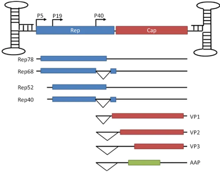
Adeno-associated virus (AAV) is a small single-stranded DNA virus of the parvovirus family. AAV was first discovered as a contaminant of adenovirus (Ad) stocks (1) and later determined to be dependent upon Ad for replication, leading to its classification as a dependovirus. This classification in the dependovirus genus separates AAV from the other members of the parvovirus subfamily, the autonomous parvovirus and erythrovirus genera (2). Although the majority of the population is seropositive for AAV (3), to date, no pathogenicity has been linked to AAV (4). AAV’s 4.7 kb genome encodes two genes, Rep and Cap, flanked by inverted terminal repeats (TR) (Fig. 1.1), which are the only viral elements required in cis for genome packaging (5). The Rep gene encodes four non-structural proteins, Rep78, Rep68, Rep52, and Rep40, using two promoters and alternative splicing. These proteins function to control viral transcription, nick the genome to allow completion of genome replication, integrate the genome in a site-specific manner, and package the genome into the capsid (6, 7).
start sites and alternative splicing (2, 8). The capsid proteins share their C-terminal domain, the VP3 common region, and differ in their N-terminal domains. The unique regions of VP1 and VP2 are denoted as VP1u and VP1/2 common region, respectively, and have motifs including punitive nuclear localization signals and a phospholipase domain (9, 10). AAP is responsible for targeting of the capsid proteins to the nucleolus and assembling them into the T=1 icosahedral capsid (8). VP1, VP2, and VP3 assemble in a ratio of 1:1:10 and thus capsids contain five copies of VP1, five copies of VP2, and fifty copies of VP3 surrounding the positive- or negative-sense viral genome (11).
on chromosome 19 (20, 21). The AAV genomes persist until replication is activated by the presence of a helper virus.
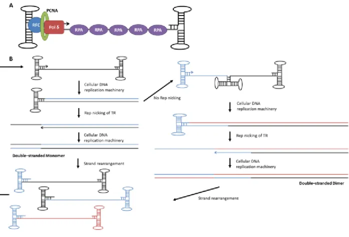
Although AAV was first discovered in association with Ad (1), several other viruses are also able to provide helper functions for AAV, including herpes simplex virus (HSV) (22), human papillomavirus (HPV) (23), and vaccinia (24). Furthermore, conditions of cellular stress, including treatment with hydroxyurea or UV light in the presence of oncogenes, have been demonstrated to substitute for AAV helper virus functions (25, 26). AAV helper viruses increase the efficiency of or are necessary for a number of steps in AAV life cycle. The helper viruses increase the efficiency of nuclear transport of AAV (27), facilitate second-strand DNA synthesis (16), encourage genome circularization (28), release Rep repression of the viral p5 promoter (29), increase the efficiency of mRNA splicing, transport, and translation (30-32), allow genome replication (33, 34), and provide a means of cell escape (35). Therefore, in the presence of a helper virus, AAV genomes are replicated by cellular DNA replication machinery such as DNA polymerase δ, proliferating cell nuclear antigen (PCNA), replication factor C (RFC), and
presumably in the nucleolus, with the aid of Rep and AAP (8) and the virions are released from the cell by helper virus cell lysis, completing AAV’s lifecycle.
Although the majority of AAV biology has been generated using AAV2, many different serotypes of AAV have been isolated from the tissues of a broad range of animal species and show between 49% and 99% identity in capsid amino acid sequence (38). These serotypes can have markedly different tissue tropisms; for instance, AAV1 is largely muscle-tropic, while AAV2 is liver-tropic and AAV9 is systemic (39). Capsid structures are available for many of the serotypes (11, 40-46), allowing the influence of various capsid regions on the steps of the viral lifecycle to be elucidated.
rAAV Vector Biology
To produce an AAV virus-like particle or vector (rAAV), the viral genes can be
completely removed from the vector genome and supplied in trans with the TRs being the only viral elements required in cis (5). A transgene cassette is then substituted for the viral genes and the vector is produced through a triple transfection method with an Ad helper plasmid
minimizing the risk of wild-type virus production (52). Through this method, the same transgene cassette can be packaged into the capsids of various AAV serotypes, allowing comparisons between the serotypes. In addition, self-complementary rAAV genomes can be produced which contain a mutation in the Rep nicking element in one TR, producing an rAAV genome which can self-anneal avoiding the rate-limiting step of second-strand DNA synthesis and increasing
transduction kinetics and efficiency (Fig. 1.2) (53, 54). These features make rAAV highly amenable as a gene therapy vector.
rAAV is thought to undergo the same transduction steps and intracellular trafficking as wild-type AAV in the absence of a helper virus through the step of second-strand DNA
synthesis. However, due to the lack of Rep protein in rAAV transduction, rAAV cannot integrate in a site-specific manner and so only low-level (<0.5%), illegitimate integration occurs (55-57). The vast majority of genomes persist episomally as head-to-tail concatemers (58). The
56). Therefore, this low level of illegitimate integration has not prevented the utilization of rAAV to delivery genes for clinical gene therapy applications.
Clinical Successes with rAAV
Given AAV’s many advantages as a gene delivery vector, over 100 clinical trials to date are utilizing or have utilized rAAV for gene therapy purposes (http://www.abedia.com/wiley). The results of these trials strongly demonstrate that rAAV-mediated gene delivery is a very safe method for delivering transgenes in the clinic (4). In addition, recent clinical trials, especially in restricted transduction sites, have begun to demonstrate successes in reaching their efficacy goals. Of the trials conducted, several stand out including trials leading to the licensing of the first commercial gene therapy product in Europe (60), trials for the treatment of retinal disease (61), trials for the treatment of CNS diseases (62), and trials for congestive heart failure (63). Two of these applications are discussed in more detail below.
Glybera®, a commercial gene therapy product. Glybera®, gene therapy product marketed to treat lipoprotein lipase deficiency (LPLD), was approved for commercial use in Europe in November of 2012 and is seeking approval in other markets. LPLD, caused by loss of function mutations in the lipoprotein lipase (LPL) gene or its functional partners, is an orphan disease affecting approximately one in five hundred thousand to one in one million people, although certain geographical regions such as eastern Canada exhibit higher rates of disease (60). LPL is produced by muscle cells and adipocytes and secreted into circulation where it binds to the luminal surface of blood vessels (60). After a meal, LPL is responsible for clearing
triglycerides from chylomicrons, whereas LPL affects triglycerides in very low-density
cleared from the plasma, leading to plasma triglyceride levels 10 to 100-fold higher than normal. These high levels of triglycerides lead to complications including acute and chronic pancreatitis (60). As the serum half-life of LPL is very short prohibiting enzyme replacement therapy, the only treatment available for LPLD prior to Glybera® was dietary management to enact an extremely low fat diet, which was often not effective.
The lack of effective treatments for LPLD led to the development of AAV-mediated gene addition approaches to target LPLD resulting from mutations in LPL. Early LPLD gene therapy studies in animal models were conducted with Ad vectors and demonstrated proof-of-principle that delivery of LPL could led to decreases in serum triglyceride levels and disease symptoms; however, the highly inflammatory response to Ad vectors led to short-term transgene expression and immune responses directed against the transgene (64, 65). Therefore, further studies were conducted with rAAV1, which is a muscle-tropic serotype of AAV. These studies also utilized a naturally occurring gain of function mutant of LPL, LPLS447X, which is present in approximately 20% of the human population and results in low plasma triglyceride levels and reduced risk of cardiovascular disease (66). Preclinical studies in LPL deficient mice demonstrated that
intramuscular injection of rAAV1- LPLS447X resulted in decreased plasma triglyceride levels and disease symptoms lasting more than one year, suggesting that this approach led to long-term disease improvements (67). Further preclinical studies in a feline LPLD model also demonstrate improvements although the treatment was limited by an antibody response to the human LPL transgene expressed (68). Given these promising preclinical results, clinical trials were initiated to determine whether the treatment would be safe and effective in humans.
using 40 to 60 simultaneous intramuscular injections (69). Most importantly, this study resulted in no serious adverse events. In addition, it demonstrated a significant but transient decrease in plasma triglyceride levels, thought to be limited by an immune response. A larger dose escalation study conducted with 14 patients and utilizing immunosuppression for 12 weeks after treatment demonstrated significant decreases in plasma triglyceride levels that were, however, still
transient, suggesting that the short-term nature of the decrease was not due to an immune
response (70). Nevertheless, signs of clinical improvement, including a decrease in incidences of pancreatitis, changes in the patients’ tolerance for certain food, and changes in the profile of lipids present in the blood out to two years following treatment, as well as long-term muscle expression of the LPLS447X transgene, suggested that plasma triglyceride levels might not be an appropriate biomarker of successful treatment and that treatment with rAAV1- LPLS447X had the capacity to improve patient outcomes. Therefore, a third clinical study was conducted looking for the effects of treatment with rAAV1- LPLS447X on abdominal pain, pancreatitis, and chylomicron plasma clearance.
This third clinical trial enrolled five patients and demonstrated that rAAV1- LPLS447X treatment resulted in an improvement of chylomicron metabolism (71). In addition, the patients reported increased energy, increased ability to eat, decreased abdominal pain, and decreased incidence of pancreatitis through two years post-treatment. A retrospective study of 22 of the 27 patients treated previously determined that treatment resulted in decreased incidence of
commercial use in Europe, representing a great success for both AAV-mediated gene delivery and gene therapy in general.
Retinal targeted AAV gene therapy. Thus far, mutations in approximately 200 genes have been linked to inherited retinal diseases, including Bardet-Biedl syndrome, chorioretinal atrophy, cone dystrophy, cone-rod dystrophy, congenital stationary night blindness, Leber’s congenital amaurosis (LCA), macular degeneration, and retinitis pigmentosa
(https://sph.uth.edu/retnet/home.htm). These retinal diseases are an especially attractive target for AAV-mediated gene therapy. The small site of delivery and restricted numbers of cells to
transduce allow very small doses of rAAV to be effective in expressing transgenes (72). Moreover, the site of transduction is reached by relatively easy surgery and the specific cells transduced can be tailored by subretinal or intravitreal injection to target different retinal cell layers (73). In addition, subretinal injections lead to immune privilege, avoiding immune responses to the transgene or the vector (74). Furthermore, various rAAV serotypes are capable of targeting different cell types in the retina allowing customization of delivery (75). Finally, non-invasive technologies, such as tomography and electroretinography, allow efficacy
outcomes to be easily evaluated (72). For all of these reasons, AAV-mediated gene therapy for retinal disease is a promising avenue of investigation.
delivery (72, 76-78). LCA, an early onset form of retinal degeneration, accounts for about 5% of retinal disease and is the leading cause of childhood blindness (75). Mutations leading to this disease have been identified in 14 genes to date (https://sph.uth.edu/retnet/home.htm); however, mutations to RPE65 account for approximately 6 to 16% of LCA cases (75). RPE65 is an enzyme responsible for converting the all-trans-retinal formed when photoreceptors signal to 11-cis-retinal, allowing photoreceptors to signal again (79). Three clinical trials delivering RPE65 using an rAAV2 vector were initiated based on very promising small and large animal models and reported their results in 2008 (80-82). All of these trials demonstrated the safety of the treatment and the two trials utilizing strong promoters demonstrated improvements in various measures of visual function including nystagmus, visual fields, dark-adapted perimetry, and mobility at low luminance (81, 82). In total, these trials treated 30 patients and have now reported lasting improvements through three years post-treatment (78). In addition, a follow up study treated the contralateral eye in three patients, demonstrating safety and efficacy with re-administration via the subretinal injection (83). Based in part on these results, eight clinical trials utilizing rAAV to treat retinal diseases have been initiated since 2008 for indications including age-related macular degeneration, choroderaemia, and a phase III trial for LCA
(http://www.abedia.com/wiley), exemplifying the promise of rAAV-mediated gene therapy.
Systemic Transduction
efficacy of these approaches. These findings are exemplified by the results garnered from trials to treat hemophilia B, one of the earliest targets of rAAV-mediated gene therapy. Hemophilia B is an X-linked monogenetic disease caused by loss of function mutations to the factor IX (F.IX) gene affecting approximately 1 in 25 000 males (84). Hemophilia B can be characterized by the percentage of normal F.IX activity possessed by the patient (84). Patients with mild disease (5% to 30% activity) usually only have bleeding episodes in response to major trauma or surgery and are often diagnosed as adults. Patients with moderate disease (1% to 5% activity) generally have bleeding episodes after injury and relatively few spontaneous episodes. The majority of
hemophilia B patients have severe disease (less than 1% activity) and experience frequent spontaneous bleeding into muscle tissue and joints, leading to long-term tissue damage.
Hemophilia B is generally treated with injections of exogenous F.IX; however, this treatment is extremely expensive and often not feasible for those in developing countries (84). In addition, 2% to 4% of patients treated with exogenous F.IX develop inhibitory antibodies, necessitating more complicated treatments (84). For these reasons, hemophilia B is considered a good target for rAAV-mediated gene therapy.
Given hemophilia B’s strength as a target for gene therapy, four clinical trials have utilized rAAV to deliver F.IX to the liver (85-88), while one trial used rAAV to delivery F.IX to the muscle (89, 90). In most of these trials, no serious adverse events related to the vector were reported. Here, we will focus on the liver directed gene therapy approaches. The early clinical trials utilized rAAV2 as this was the first serotype developed and approved for clinical use. The results of a dose escalation trial treating 7 patients with severe hemophilia at a range of
rAAV2-F.IX doses from 8×10¹⁰ vector genomes (vg)/kg to 2×10¹² vg/kg were published in 2006 (86). At
rAAV2-F.IX, suggesting that the dose was not high enough to allow for successful transduction. At the high dose, F.IX levels initially reached moderate or even mild hemophilia levels in the first 5 weeks post-transduction; however, after this time, the levels of F.IX decreased, returning to baseline by 10 weeks post-transduction. This decrease in transduction was thought to be the result of an immune response and correlated with a small increase in liver enzyme levels in one patient. Although the trial did not reach efficacy goals, the results demonstrated the safety of the treatment and encouraged further investigations.
A later clinical trial tried to improve these results by altering the vector used to deliver the F.IX gene (88). Specifically, the trial utilized a rAAV8 vector, which has much higher transduction efficiency in the liver than rAAV2, a self-complementary genome that avoids the rate-limiting step of second-strand DNA synthesis, and a codon-optimized version of the F.IX gene. The trial treated six patients with severe hemophilia with doses of scAAV2/8-F.IX ranging from 2×10¹¹ vector genomes (vg)/kg to 2×10¹² vg/kg. The patients in the low and moderate dose
for other systemic gene therapy applications utilizing rAAV as well. Furthermore, it is clear that even the clinical successes with rAAV would benefit from higher transgene expression in order to improve the level of efficacy observed. Given all of these data, strategies are needed to increase the level of transgene expression without increasing vector dose.
Enhancement of rAAV Transduction
As clinical studies with rAAV have repeatedly demonstrated the need for increased transgene expression from rAAV vectors, several approaches have been undertaken in order to increase the efficiency of rAAV transduction. These approaches include random mutagenesis and directed evolution approaches to identify vectors that have high transduction potential in specific targets, understanding limiting steps in AAV biology and altering rAAV to avoid these barriers, and pharmacologically altering the environment of the cell in order to improve the efficiency of transduction. These approaches will be discussed in more detail below.
specific alterations, this approach has been applied both in vitro and in vivo to isolate capsids that are efficient in transducing certain cell types (95, 96), evading neutralizing antibody responses (92), targeting tissues of interest and detargeting other tissues (94), and increasing efficiency for specific disease models (94). Three examples of successful directed evolution approaches are detailed below.
The first rAAV directed evolution approach utilizing libraries that re-assort Cap genes from different AAV serotypes attempted to isolate rAAV capsids that could infect a hamster melanoma cell line, CS1, generally refractory to rAAV transduction (96). The authors created a library of several million clones and allowed five rounds of replication on CS1 cells in the presence of Ad, resulting in the recovery of a single clone named AAV1829. This clone was a chimera of AAV serotypes consisting of the N-terminus of AAV1, followed by a short region of AAV8, a long section of AAV2, and the C-terminus of AAV9 and shared heparan binding ability with AAV2. This clone demonstrated increased transduction compared to the parental serotypes on CS1 cells and several murine melanoma cell lines and equal transduction to rAAV2 on human melanoma cell lines. When tested in vivo, this clone demonstrated lower transduction of the liver and muscle and a switch in brain transduction from neuronal tropism to neural progenitor cell tropism. These results demonstrated that a library of AAV clones from several AAV serotypes can be generated and used to isolate clones that perform better than the parental serotype.
Another study, demonstrating the breadth of libraries applicable to directed evolution, selected for AAV clones that could successfully transduce primary human astrocytes, as these cells are important targets for gene therapy but are generally poorly transduced by rAAV (95). Three libraries were generated in this study and then combined before four rounds of selection on the primary astrocytes: AAV2 Cap with random mutagenesis, a shuffled capsid library as described above, and a peptide display library based on AAV2. This approach isolated several clones containing point mutations to AAV2 Cap, as well as several chimeric clones that were more efficient at transducing astrocytes than rAAV2. When tested in vivo, the isolated clones demonstrated a 3 to 5-fold higher percentage of astrocytes transduced than rAAV2. In addition, with sub-retinal injection, one of the clones demonstrated increased tropism for Müller glia. Interestingly, despite the large number of clones in the initial libraries, an attempt to select the libraries separately through five rounds of selection resulted in no improvements over rAAV2’s transduction. These results demonstrate that directed evolution approaches are capable of identifying rAAV clones that transduce specific cell types more efficiently than the parental rAAV serotypes; however, they also suggest that the results of the directed evolution can vary widely from trial to trial and that beneficial clones can sometimes be lost during cycling.
The approach of directed evolution has been taken farther and applied to in vivo selection experiments. Specifically, a chimeric library was used to select for clones which could cross the blood brain barrier in areas of seizure induced damage and transduce the damaged areas in order to create a vector selective for the damaged tissue (94). Since coinfection with Ad is not
to transduce cells successfully. Consequently, a variety of isolated clones were screened for efficient transduction. Two clones were isolated that exhibited the desired pattern of transduction of the damaged regions of the brain following seizures. In addition, these clones exhibited
decreased transduction of many peripheral organs compared to the parental serotypes, suggesting that the biopanning experiment not only placed positive pressure on transduction of neurons in the damaged areas of the brain but also exhibited negative selection for transduction of
peripheral organs. Therefore, given the lack of replication during the cycling, this in vivo selection approach can be very useful for isolating tissue specific clones as long as care is taken to screen multiple clones. Furthermore, clones isolated in this type of in vivo selection
experiment are likely to be specific for the specific disease model utilized and may not be capable of being generalized to other models.
Engineering of rAAV to alter or avoid limiting steps in transduction. An alternative approach to the library-based directed evolution is to understand specific steps in the rAAV transduction pathway that are inefficient and alter rAAV to increase its efficiency at these steps or avoid these steps entirely. For this approach, rAAV’s transduction pathway needs to be understood and then strategies devised to alter specific steps. Two examples of this strategy being successfully employed are the development of self-complementary rAAV and the transfer of receptor binding domains between rAAV serotypes. The generation of self-complementary AAV began with the observation that rAAV2 transduction was significantly enhanced by co-infection with Ad (16). This was an interesting observation given that many of the helper
functions described for Ad at the time, such as activation of viral promoters or promotion of viral mRNAs splicing (29-32), were not applicable to the transduction of rAAV. The authors
determined that the effect of Ad was mediated by the Ad E4Orf6 protein and observed increased double-stranded rAAV genomes despite equal amounts of uncoated DNA, suggesting that Ad enhanced rAAV second-strand DNA synthesis (16). As this enhancement of second-strand synthesis led to transduction levels orders of magnitude higher, the authors concluded that second-strand DNA synthesis is the rate-limiting step in rAAV transduction. This conclusion was further collaborated by in vivo experiments demonstrating that rAAV DNA was present in the nucleus of a high percentage of hepatocytes, most of which demonstrate no transgene expression, and that this expression could be rescued by co-infection with Ad (98).
of genomes packed should be single-stranded dimers consisting of a two copies of the transgene in opposite orientations with TRs present on both ends as well as in between the transgene cassettes. When released from the capsid, this self-complementary molecule can fold back on itself to form a double-stranded hairpin molecule. In fact, when these particle were isolated based on their density, they demonstrated 20-fold higher transduction than a comparable transgene cassette packaged in single-stranded form and a reprieve from reliance on DNA synthesis for transgene expression. In addition, when the vectors were utilized in vivo, the authors
demonstrated both increased kinetics of transgene expression and increased steady state levels of transduction. These results demonstrated that self-complementary vectors had greatly increased transgene expression through avoidance of the rate-limiting step in rAAV transduction of second-strand DNA synthesis. The self-complementary rAAV technology was then further improved by deleting the D element of one TR, and so removing the Rep nicking stem, forcing a self-complementary molecule to be packaged (53). This mutation led to a much higher
percentage of self-complementary genomes packaged and greater ease in self-complementary AAV production. Self-complementary vectors allowed for transduction in the mouse liver, brain, and muscle much higher than that of single-stranded rAAV. Therefore, the development of self-complementary rAAV is a compelling example of knowledge of rAAV biology leading to improvements in rAAV that circumvent limiting steps in transduction.
Another example of advances in the understanding of rAAV biology leading to altered vectors with differential transduction is the transfer of primary receptor binding footprints
binding is not essential for transduction, and strong liver transduction, while rAAV1 does not bind heparan sulfate and has weak liver transduction. Swapping of each amino acid between rAAV1 and rAAV6 followed by heparan affinity column experiments demonstrated that altering single amino acid, 531 (rAAV1 E531K, rAAV6 K531E), could both remove heparan binding ability from rAAV6 and confer heparan binding ability to rAAV1 (99). In addition, the authors demonstrated that a comparable mutation in a different serotype of AAV, rAAV8 E533K, could also confer heparan binding. The heparan binding mutant of rAAV1 demonstrated greatly increased transduction in both a human-derived liver cell line and in the liver in vivo. These data demonstrate that understanding of the receptors utilized by AAV and their binding location on the AAV capsid can lead to alterations and enhancement of transduction.
biology can lead to improvements in the efficiency of transduction. Therefore, elucidation of rAAV biology is a useful strategy leading to improvements in rAAV transduction.
Pharmacological approaches to enhance rAAV transduction and elucidate AAV
biology. A third approach to enhancing rAAV transduction is to utilize pharmacological agents to alter the cellular milieu and so enhance rAAV transduction. Many pharmacological agents, specifically chemotherapeutics that cause cellular stress, have been observed to enhance rAAV transduction in a serotype independent manner. These stressors include DNA damaging agents, such as radiation (UV, gamma, X-ray), tritiated thymidine, and cisplatinum (16, 103), DNA synthesis inhibitors, such as aphidicolin and hydroxyurea (16, 104), topoisomerase inhibitors, such as etoposide and camptothecin (104), and proteasomal inhibitors, such as MG132, LLnL, and bortezomib (105-109). Some pharmacological treatments have also been derived that affect rAAV in a serotype-specific manner. For instance, treatment of cells with neuraminidase to remove sialic acid moieties results in inhibited rAAV1 transduction, which utilizes sialic acid as its primary receptor, has no effect on rAAV2 transduction, which utilizes heparan sulfate as its primary receptor, and results in enhanced rAAV9 transduction, which utilizes galactose as its primary receptor (100). Therefore, while some treatments target universal steps in rAAV transduction, others target specific transduction steps that differ between serotypes.
In addition to simply enhancing rAAV transduction, the changes in the transduction pathway of rAAV in the presence of these pharmacological agents can also lead to advances in the understanding of rAAV biology. For instance, a study examining the effects of hydroxyurea and proteasome inhibitors, as well as several siRNA treatments, demonstrated the importance of trafficking of rAAV capsids between the nucleolus and the nucleoplasm for successful
MG132, resulted in increased nucleolar accumulation of rAAV virions, whereas treatment with hydroxyurea resulted in increased localization of virions to the nucleoplasm. The drugs both enhance transduction and co-treatment with the two drugs results in a synergistic increase in transduction. These data led the authors to hypothesize that virions entering the nucleus stably accumulate in the nucleolus and then move to the nucleoplasm where uncoating of the genome can occur. This hypothesis, derived from changes occurring due to pharmacological treatments, can now be further investigated to elucidate the details of the nuclear steps in rAAV
transduction. Therefore, pharmacological agents are useful tools not only for enhancing rAAV transduction, but also for revealing rAAV biology.
Dissertation Objectives
In summary, rAAV is an important gene delivery vector for clinical applications and has recently demonstrated success in reaching efficacy goals, especially in restricted sites such as the retina; however, low levels of transgene expression and loss of transgene expression over time have hampered many clinical systemic gene delivery efforts. These results led to the need to enhance rAAV transduction without increasing vector dose. The goal of this dissertation is to identify methods by which rAAV transduction, specifically in vivo transduction, can be increased. Towards this aim, we have undertaken several approaches: we have identified a cellular restriction factor that inhibits rAAV transduction, we have identified a chemotherapeutic approved for use in humans that enhances rAAV transduction, and we have explored the
mechanisms by which a known class of pharmacological agents enhances rAAV transduction. Thus, of the approaches for enhancing rAAV transduction discussed above, we pursued gaining greater knowledge of the limiting steps in rAAV transduction as well as utilizing
affect rAAV biology. The results of these approaches are detailed in Chapters 2, 3, and 4, while the final chapter will discuss the implications and future directions of these discoveries in the broader context of rAAV biology and applications towards clinical gene therapy.
Specifically, Chapter 2 will answer the question does the promyelocytic leukemia protein (PML) inhibit rAAV transduction. PML is a tripartite motif protein that has been demonstrated to play many cellular roles including as a cell-intrinsic antiviral defense factor. PML can inhibit both RNA and DNA viruses through many varied mechanisms (110-114) and the importance of PML can be demonstrated by the wide variety of viruses that encode factors that block or modify PML activities (115, 116). Despite the wide range of viruses inhibited by PML, including AAV’s traditional helper viruses HSV and Ad (115, 116), the functional role of PML in the transduction and replication of parvoviruses has not been examined, although a possible role is suggested by the partial temporal colocalization of PML with the replication centers of both AAV (117) and minute virus of mice (MVM) (118). In this chapter, we utilized PML knockout mice, as well as knockdown and overexpression studies in human cells, to determine that PML inhibits rAAV second-strand synthesis leading to up to 50-fold inhibition of transduction in vivo. We also determined that human PML isoform II was mainly responsible for this effect and that the effect could be extended both to the production of rAAV and to the replication of wild-type AAV. These results will have implications for the enhancement of rAAV transduction and possibly for the replication other parvoviruses.
Chapter 3 will answer the question does arsenic trioxide (As2O3) enhance the
transduction of rAAV. As discussed above, many cellular stressors including chemotherapeutic agents have been demonstrated to enhance rAAV transduction (16, 103-105). We utilized As2O3,
(119) and is under evaluation for the treatment of many other types of leukemia (120, 121). This compound affects many different cellular pathways including inducing reactive oxygen species formation, inhibiting BCL-2 and NFκB activation, decreasing mitochondrial membrane
potential, changing histone acetylation patterns and, at high doses and with long treatments, degrading PML and inducing cell-intrinsic apoptosis (122). We determined that As2O3 treatment
caused a dose-dependent increase in rAAV transduction in a number of human and non-human cell lines through stabilization of perinuclear accumulations of rAAV virions. In addition, we determined that this effect was mediated by reactive oxygen species and that As2O3 treatment led
to up to 10-fold enhancement of rAAV transduction in vivo. These results will have implications for the pharmacological enhancement of rAAV transduction.
In chapter 4, we will answer the question of whether proteasome inhibition is sufficient for the enhancement of rAAV transduction and examine the mechanisms of proteasome inhibitor effects on rAAV transduction. Although many groups have demonstrated that proteasome
Figure 1.3: AAV genome replication factors and pathway.(A) Several cellular replication factors have been demonstrated to be involved in AAV DNA replication, which requires only leading strand DNA synthesis. These factors include DNA polymerase δ, PCNA, which
CHAPTER 2
The promyelocytic leukemia protein is a cell-intrinsic defense inhibiting parvovirus DNA replication1
Summary
The promyelocytic leukemia protein (PML) is a viral restriction factor inhibiting
processes from uncoating to transcription to cell survival. Here, we investigated PML’s effect on adeno-associated virus (AAV), a parvovirus used for gene delivery. Although dependovirus (AAV) and autonomous parvovirus (MVM) replication centers can colocalize with PML, PML’s functional effect on parvoviruses is unknown. We determined PML knockout enhances rAAV2 transduction up to 56-fold at a range of vector doses in both male and female mice. PML inhibited several rAAV serotypes, suggesting a conserved mechanism, and organ specificity correlated with PML expression. Mechanistically, PML inhibited rAAV second-strand DNA synthesis, precluding inhibition of self-complementary rAAV. We confirmed the effect of human PML on rAAV transduction through knockdown experiments and linked the highest level of inhibition to PML isoform II. PMLII overexpression resulted in inhibition of second-strand synthesis, vector production, and genome replication. Moreover, PMLII inhibited wild-type AAV2 production and infectivity. Our data demonstrate PMLII inhibits AAV second-strand
1Adapted for this dissertation from: Mitchell A.M., Hirsch M.L, Li C., and R.J. Samulski.
synthesis and replication, processes necessary for all parvoviruses, suggesting implications for both general parvovirus replication and AAV-mediated gene delivery.
Introduction
Adeno-associated virus (AAV) is a helper-dependent member of the Parvoviridae family, which, in addition to AAV, contains other viruses of clinical and veterinary importance such as B19 parvovirus, human bocavirus, and canine parvovirus. AAV consists of an icosahedral capsid surrounding a single-stranded DNA genome encoding two genes, Rep and Cap, and has been developed as a gene delivery vector for gene therapy applications. For use as a vector or virus-like particle (rAAV), the viral genes can be removed and replaced with a transgene cassette, the terminal repeats being the viral only elements required in cis (5). Although clinical
rAAV-mediated gene therapy has demonstrated increasing success in reaching efficacy goals, especially in restricted sites such as the eye (124), low transgene expression or loss of expression over time have repeatedly compromised the efficacy in other clinical trials (86, 88). Therefore, efforts to increase the efficiency of rAAV transduction without increasing vector dose are imperative.
same transduction steps as wild-type AAV through second-stand synthesis but cannot proceed with replication, making AAV a good model for studying the initial transduction of parvoviruses without the later steps of replication occluding the results.
The AAV helper viruses Ad and HSV share the ability to modify or degrade the
To avoid PML antiviral activity, many viruses encode PML modifying proteins. For instance, Ad E4Orf3 binds directly to PMLII and causes its rearrangement from spherical PML bodies to track-like structures (116). Furthermore, HSV encodes ICP0, an E3 ligase, that causes proteasomal degradation of PML (115). In a natural AAV infection, the PML modifying
properties helper viruses may protect AAV from any potential inhibitory effects of PML.
Moreover, not only have AAV replication centers been shown to colocalize with PML track-like structures in the presence of Ad (117), but the replication centers of an autonomous parvovirus, minute virus of mice (MVM), have also been demonstrated to colocalize with PML bodies during specific times in its replication cycle (118). However, the functional consequences of PML on parvovirus transduction and replication have not been examined. Therefore, we asked whether, in the absence of a helper virus, PML is capable of inhibiting rAAV transduction. To address this question, we utilized PML knockout mice, siRNA-mediated knockdown in human cells, and overexpression of PML isoforms. We demonstrated PML inhibits the transduction of rAAV both in vivo and in human cells in culture. This inhibition is due to the prevention of second-stand synthesis and the majority of inhibition can be traced to PML isoform II. PMLII can also inhibit the production of both rAAV and wild-type AAV and the infection of wild-type AAV. These data may lead to strategies for enhancing the efficiency of rAAV mediated gene therapy. In addition, these data may have implications for the other members of the parvovirus family.
Materials and Methods
penicillin, and 100 g/ml streptomycin at 37°C with 5% CO2. Adult mouse tail fibroblasts
cultured in the media above supplemented with 1X MEM non-essential amino acids. AAV vectors were produced through cesium gradient purification as has been described (52). Self-complementary rAAV and corresponding control vectors were purified to yield pure virus as has been described (132). Wild-type AAV was produced as for rAAV except that the plasmid
encoding AAV’s genes and the transgene cassette plasmid were replaced with pSSV9. Virus and vectors were titered by qPCR (52).
Isolation of adult mouse tail fibroblasts. Adult tail snip fibroblasts were isolated using the protocol on the ENCODE Database entitled “Establishment and Propagation of Adult Mouse Fibroblast Cultures” (http://genome.ucsc.edu/ENCODE/protocols/cell/ mouse/Fibroblast_Stam_ protocol.pdf). Briefly, tail snips were clipped into Hank’s Balanced Salt Solution (HBSS, Life Technologies) and minced with a razor blade. The tissue was then digested with collagenase Type XI-S at a final concentration of 1000 U/ml in HBSS for 30 minutes at 37°C. Tissue was washed once with HBSS, resuspended in 0.05% trypsin-EDTA (Life Technologies), and incubated at 37°C for 20 minutes. The tissue was then resuspended in complete growth media, pipetted to dissociate cells, and seeded in 35-mm plates with tissue clumps under glass
coverslips. Media was changed every four days. When cells were subcultured, the cells were detached with 0.25% trypsin-EDTA (Life Technologies) and were seeded ratios of 1:2 to 1:4.
Animals and in vivo transduction assays. All animal experiments were conducted in accordance with the policies of the Institutional Animal Care and Use Committee at the University of North Carolina at Chapel Hill. Wild-type 129/SV and PML knockout
129/SV-PMLtm1Pppmice (133) were a kind gift from Dr. Pier Paolo Pandolfi (Beth Israel Deaconess
by retro-orbital injection. Transduction from luciferase carrying vectors was assayed by live luciferase imaging (132). Transduction from GFP vectors was determined using a GFP ELISA kit (Cell Biolabs, Inc.). Briefly, livers were harvested at 7 days post-transduction and minced. A sample (approximately 50 mg) was lysed with RIPA buffer, homogenized with a Tissue-Tearor (BioSpec Products), and total protein concentration was determined using the Bio-Rad Protein Assay and BioRad SmartSpec Plus spectrophotometer. Equal amounts of protein were used to proceed with the GFP ELISA as per manufacturer’s directions. A Bio-Rad iMark plate reader was used to determine absorbance and pg GFP per mg total protein was calculated.
Biodistribution experiments and PML expression analysis. Mice were treated with rAAV as for transduction experiments and the specified organs were harvested and frozen on day 14 post-transduction. The organs were minced and small samples were taken for luciferase assay and vector genome quantification. Luciferase samples were lysed in 2X passive lysis buffer (Promega) and homogenized with a TissueLyser (Qiagen) for 5 min at 40 Hz. The lysate was cleared by centrifugation and luciferase activity was assayed as per the manufacturer’s
qPCR protocols and primers. To determine PML expression levels from cDNA made from mouse tissues and from human cells, qPCR assays were designed using the Universal Probe Library Assay Design Center (Roche). The amplicon was designed to be in the 5’ region shared between PML isoforms. For mouse tissues, the primers and probe for PML were as follows: 5’-AGAGGAACCCTCCGAAGACT-3’, 5’-ATTCCTCCTGTATGGCTTGC-3’, and Mouse Universal Probe Library probe 76 (Roche). The primers and probe for mouse GAPDH were as follows: 5’-GGGTTCCTATAAATACGGACTGC-3’, 5’-CCATTTTGTCTACGGGA CGA-3’, and Mouse Universal Probe Library probe 52 (Roche). For human cells, the primers and probe for PML were as follows: 5’-TTCTGCTCCAACCCCAAC-3’, 5’-CGCTGATGTCGC
ACTTGA-3’, and Human Universal Probe Library probe 5 (Roche). The primers and probe for human GAPDH were as follows: 5’-ATCACTGCCACCCAGAAGACT-3’, 5’-ACACGGAAGG CCATGCCA-3’, and Mouse Universal Probe Library probe 34 (Roche). The qPCR reactions were run with LightCycler 480 Probes Master mix (Roche) on the following program in the Roche LightCycler 480: 95°C 10 minutes, [95°C 10 s, 60°C 30 s, 72°C 1 s (acquisition)] 45 cycles, 40°C 30 minutes. Standard curves for each primer set were used to determine efficiency and calculate relative expression.
(Life Technologies) added to DNase treated samples to act as a carrier. We determined the purity of isolated nuclei to be greater than 99.5% using the EnzChek acid phosphatase assay kit (Life Technologies).
siRNA and in vitro transduction assays. We used SMARTpool On-TARGETplus PML siRNA (Thermo Scientific) to knockdown human PML and On-TARGETplus Non-targeting Control Pool siRNA (Thermo Scientific as a negative control. HuH7 cells were seeded and transfected with DharmaFECT as per the manufacturer’s instructions with slight modification. HuH7 cells were transfected with 25 nM siRNA 48 hours and 24 hours prior to transduction. At time of transduction, the media was changed to contain the indicated dose of rAAV and cells were incubated for 24 hours. Cells were harvested with 1X passive lysis buffer (Promega) and luciferase and protein assays were performed as above. Mouse fibroblasts were seeded 16 hours prior to transduction and the media was changed to contain rAAV at the indicated dose at the time of transduction. Transduction was assayed as for siRNA experiments 48 hours post-transduction.
digested with AgeI (New England Biolabs) and a 4.2 kb band containing the plasmid backbone without the EGFP gene was gel purified (Qiagen). For retrieval of the PMLII DNA, the pEGFP-C3-PMLII plasmid was PCR amplified with the primers 5’-AAACCGGTCCATGGAGCCTGC ACC-3’ and 5’-CCCTTCTCTTGTAACCTTGGAATTC GC-3’ and Illustria Hot Start Mix RTG (GE Healthcare). The PCR program was as follows: 95°C for 5 minutes, [95°C for 30 s, 60°C for 30 s, 72°C for 120 s] 30 cycles, 72°C for 5 minutes, 4°C ∞. The ends of the PCR product were blunted with Klenow polymerase, PCR purified, and then digested with AgeI. The 2.6 kb PCR product was then gel purified. The fragments were ligated with T4 DNA ligase, transformed into electrocompetent DH10B (Life Technologies), and colonies were picked based on sequence.
assayed for transduction by luciferase assay. Luciferase values from transduction were
normalized to those from transfection. For the self-complementary rAAV experiment, HeLa cells were seeded and transfected with pTR-CMV-PMLII or pTR-CMV-Luciferase and carrier DNA and transduced with indicated doses of single stranded or self-complementary rAAV2-CMV-EGFP 24 hours post-transfection. Transduction was assayed at 24 hours by flow cytometry (132).
loaded on 12% Mini-PROTEAN TGX precast polyacrylamide gels (Bio-Rad) and
electrophoresed at 225V for 35 minutes in Tris/Glycine/SDS buffer (Bio-Rad). Proteins were transferred to a nitrocellulose membrane using an iBlot (Life Technologies) on P3 for 4 minutes. AAV capsid proteins were detected with B1 antibody, rep proteins with IF11 antibody, and actin with Abcam ab8226.
Replication and infection assays. For rAAV DNA replication assays, HEK293 cells were seeded at a 1:4 density into 10 cm plates 24 hours prior to transfection with PMLII or control plasmid. For transfection, 6 µg of pEGFP-C3 or pEGFP-PMLII were combined with 300 µl DMEM without FBS or antibiotics and 40 µl PEI Max (Polysciences, Inc) and incubated for 10 minutes at room temperature. The entire volume was added to the plate and cells were incubated for 6 hours. The media was then changed to remove the transfection reagents. Three days following transfection of the PML overexpression plasmid, or an eGFP expression plasmid, an additional transfection was performed to investigate rAAV replication. Three plasmids were used in this PEI transfection: i) the adenoviral helper plasmid XX680 (10ug), pXR2 (3ug) which supplies Rep2 and Cap2, and an AAV vector plasmid (3ug) pITR2-CBA-luc. Three days
following the second transfection, HIRT DNA was isolated using modified protocol (135) and digested overnight with DpnI (New England Biolabs). Samples were separated on an alkaline gel, transferred to a nitrocellulose membrane (Amersham XL) and hybridization was performed with the product of a random primed labeling reaction (Roche) using the packaged transgenic DNA sequence as template (which also served as our size standard; (52)). Blots were exposed to film and DNA quantitation of the replication products was performed using ImageJ.
pEGFP-C3-PMLII and 3 μg pXX680 or salmon sperm DNA in 300 µl OptiMEM and 40 µl PEI Max. Transfection cocktails were incubated 10 minutes at room temperature and the total volume was added to the plates. Media was changed to remove transfection reagents 6 hours
post-transfection. At 24 hours post-transfection, the cells were concurrently seeded into 24-well plates at 1×10⁵ cells/well and infected with the indicated dose of AAV2. Total DNA was harvested 48 hours post-transduction by DNeasy Blood and Tissue Kit and numbers of viral genomes per cell were analyzed by qPCR.
Data Analysis. We determined the statistical significance of all data using the non-parametric Kruskal-Wallis test and considered a p-value of less than 0.05 to be significant.
Results
PML knockout enhances rAAV2 transduction in vivo. During wild-type AAV’s natural lifecycle, replicating AAV may be sheltered from the effect of PML by the PML modifying activity of its helper viruses. We hypothesized that PML may inhibit rAAV
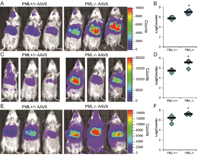
transduction in environments where helper viruses are not present. To test this hypothesis, we transduced wild-type (PML+/+) and PML knockout (129/SV-PMLtm1Ppp, PML-/-) mice with a range of rAAV2-luciferase doses and examined transduction by live imaging. Although we observed very low levels of transduction with 1×10¹⁰ vector genome (vg)/mouse, we observed expression of luciferase in the area of the liver at day 11 post-transduction in PML-/- mice whereas no expression was observable in PML+/+ mice (Fig. 2.1A & Fig. 2.2A). At a five-fold higher dose of virus (5×10¹⁰ vg/mouse), we observed transduction at the site of injection in PML+/+ mice, although liver expression was still low; however, liver and injection site
dose of AAV, similar transduction levels were observed in PML+/+ mice as with 5×10¹⁰ vg/mouse; however, PML-/- mice demonstrated increasing levels of liver and injection site transduction (Fig. 2.1C). At a high dose of rAAV2 (5×10¹¹ vg/mouse), we observed high levels of transduction in all mice, but more transduction in PML-/- mice (Fig. 2.1D). By quantifying the light output, we created doses curves for transduction (Fig. 2.2B) and observed significant enhancement of transduction in PML-/- mice as compared to PML+/+ mice at all of the tested doses in either total transduction (Fig. 2.1E) or transduction in the area of the liver (Fig. 2.1F), demonstrating that knockout of PML can enhance rAAV2 transduction and suggesting that PML can inhibit rAAV2 transduction.
Effect of PML knockout is conserved among several rAAV serotypes. After determining that PML inhibited rAAV2 transduction in vivo, we examined the transduction of various serotypes of rAAV in PML-/- mice to determine whether the transduction pathway affected by PML is specific to rAAV2 or is conserved among rAAV serotypes. Therefore, we transduced PML+/+ and PML-/- mice with 1×10¹¹ vg/mouse rAAV6, rAAV8, or rAAV9 and assayed transduction by live luciferase imaging. At 7 days post-transduction, we observed enhanced rAAV6 transduction in the area of the liver in PML-/- mice (Fig. 2.3A) and quantified this enhancement at 15.0-fold (Fig. 2.3B). In addition, we observed higher transduction from rAAV8 (Fig. 2.3C) and quantitated this increase at 47.0-fold at 3 days post-transduction (Fig. 2.3D). With rAAV9, we observed increased transduction in PML-/- mice at day 7
post-transduction (Fig. 2.3E) and quantified this difference at 5.6-fold (Fig. 2.3F). Thus, these data demonstrate PML knockout can enhance transduction of a number of rAAV serotypes,
suggesting PML inhibits a process in AAV transduction conserved between serotypes. PML knockout enhances rAAV transduction in a manner correlating to PML
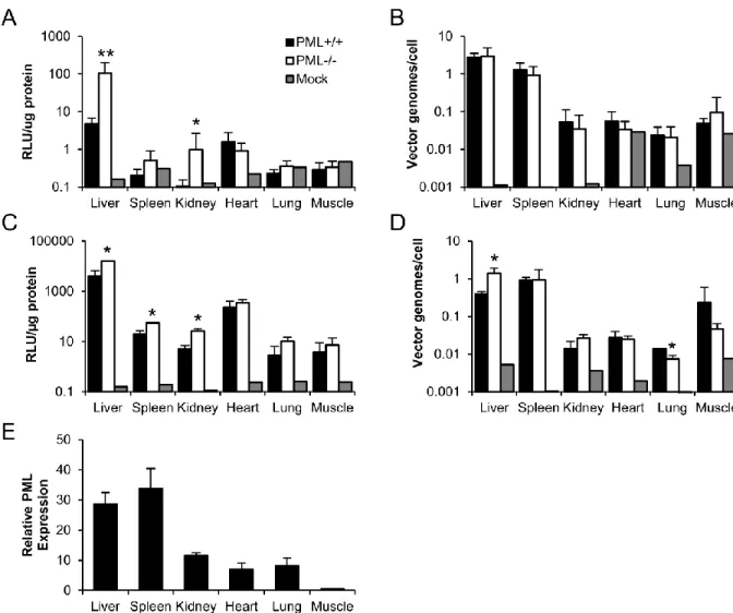
Therefore, we investigated the effect of PML knockout on the biodistribution of rAAV9, a systemic vector.
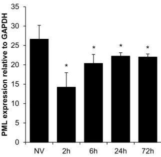
As with rAAV2, we transduced PML-/- and PML+/+ mice with rAAV9 and harvested tissues at 14 days post-transduction. Similarly to rAAV2, we observed significant increases in luciferase activity in the liver (3.9-fold), spleen (2.8-fold), and kidney (5.2-fold), and no significant increases in heart, lung, and muscle (Fig. 2.4C). In addition, the only significant changes in vector genome copy number observed were in the liver where copy number was 3.5-fold higher in PML-/- mice and the lung where copy number was 2-fold lower in PML-/- mice (Fig. 2.4D). To investigate the organ specificity further, we examined the expression of PML in the PML+/+ mice by qRT-PCR. We observed the highest levels of PML expression in the liver and spleen, intermediate levels of expression in the kidney, low levels of expression in the heart and lung, and very low levels of expression in the muscle (Fig. 2.4E). Interestingly, we observed no increase in PML expression in the liver following rAAV2 transduction (Fig. 2.5), suggesting rAAV transduction does not induce PML transcription. As the levels of PML expression appear to correlate with the increases in rAAV9 transduction we observed (Fig. 2.4C), this suggests the organ specificity of rAAV transduction enhancement observed with PML knockout occurs based on varying PML expression levels. Furthermore, our genome copy number data (Fig. 2.4B & 2.4D) suggest changes in rAAV genome number are not necessary for PML’s effect on rAAV transduction.
rAAV transduction that PML knockout affects, we transduced PML-/- and PML+/+ mice with rAAV2 and harvested liver tissue at several times post-transduction. We first asked whether the difference occurred in cell entry by determining the vector genome copy number at 1 day and 7 days post-transduction. We observed no difference in copy number between PML+/+ and PML -/-mice at either time point (Fig. 2.6A), suggesting that the effect of PML occurs post-entry. This agrees with our biodistribution data, demonstrating no difference in rAAV2 vector genome copy number 14 days post-transduction (Fig. 2.4B). We next performed nuclear fractionation to determine levels of nuclear entry at these time points. At both 1 day and 7 days, we observed equal numbers of nuclear vector genomes in PML+/+ and PML-/- mice (Fig. 2.6B), suggesting PML acts after rAAV2 nuclear entry. The next step in rAAV transduction after nuclear entry is uncoating of the vector genome; therefore, we assayed the numbers of uncoated genomes by performing a DNase protection assay on our nuclear fractions. We observed no difference in the numbers of unprotected (uncoated) genomes between PML+/+ and PML-/- mice (Fig. 2.6C), suggesting PML acts after this transduction step on either second-strand DNA synthesis or transcription.
To access whether PML knockout affects second-strand DNA synthesis, we transduced PML+/+ and PML-/- mice with self-complementary and single-stranded rAAV8-EGFP and determined transduction in harvested liver tissue at 7 days post-transduction.
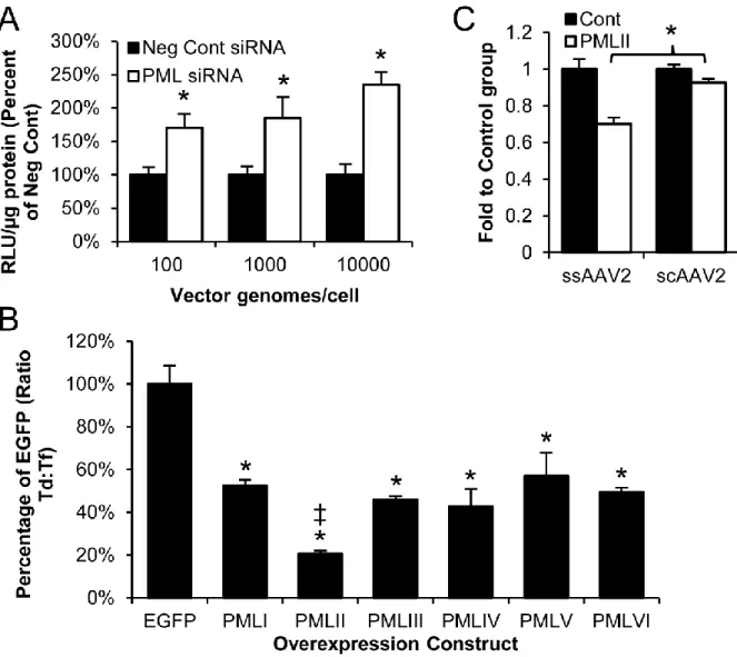
substantiate this conclusion, we treated fibroblasts from the PML+/+ and PML-/- mice with a wide range of rAAV2 doses to determine whether there is a difference in the lower transduction threshold. We hypothesized that transduction increases with PML knockout might be greater at lower vector doses if PML affects second-strand synthesis as annealing of genomes could not compensate for lack of second-strand synthesis. In fact, we observed transduction in the 100 vg/cell group for PML-/- cells but did not observe transduction in the PML+/+ cells until the 500 vg/cell group (Fig. 2.7B), demonstrating a lower threshold for transduction in PML-/- cells. These data also agree with our in vivo dosing data showing similar thresholding at our lowest rAAV2 dose (Fig. 2.1A). The differences in the threshold for transduction support our mechanism of PML inhibition of rAAV second-strand DNA synthesis. Taken together, our results demonstrate PML knockout enhances second-strand DNA synthesis of rAAV vectors, suggesting PML can inhibit this transduction step.
minor isoforms, all of which share their N-terminal domains and differ in their C-terminal domains (126). Of the major isoforms, isoforms I through VI are nuclear (130) and could possibly mediate the effect of PML on rAAV2 second-strand synthesis. We acquired plasmids expressing EGFP-tagged versions of these nuclear isoforms (137), expressed them in HeLa cells (Fig. 2.9B), and examined the effect of these isoforms on rAAV2 transduction. We determine that expression of five of the six isoforms (PMLI, PMLIII, PMLIV, PMLV, and PMLVI)
resulted in an approximately 2-fold decrease in transduction, while expression of PMLII resulted in a 4.9-fold decrease in transduction (Fig. 2.8B). To confirm our in vivo mechanism, we then examined whether PMLII could inhibit self-complementary rAAV2 transduction. In fact, although we observed a significant decrease in the number of cells transduced with single-stranded rAAV2 after PMLII overexpression, we observed significantly less inhibition of self-complementary rAAV2 transduction (Fig 2.8C), confirming human PML isoform II is
responsible for the inhibition of rAAV second-strand synthesis.
PMLII overexpression inhibits rAAV2 and wildtype AAV2 production and
replication. All of our data thus far address the role of PML in rAAV transduction; however,
given our second-strand synthesis mechanism we set out to determine whether PML plays a role in rAAV genome replication and virus production. We began by examining the effect of PMLII overexpression on virus production by encoding PMLII as a transgene for rAAV vectors. We determine the presence of the PMLII transgene resulted in a 6.8-fold decrease in the yield of vector (Fig. 2.10A). As the effect of PML on rAAV transduction was through second-stand synthesis, we hypothesized that PMLII inhibits rAAV production on the level of genome replication. Therefore, we performed a replication assay to determine whether the level of
fact, we observed slightly lower levels of the replicative monomer and dimer form of the vector genome in PMLII expressing cells (Fig. 2.10B), which quantified at 73% of the control value (Fig. 2.10C). We then examined whether PML could also inhibit the production of wild-type AAV by producing wild-type AAV2 from an infectious clone with or without PMLII
overexpression and assaying the levels of virus produced. As with rAAV2, we observed a 6.5-fold decrease in wild-type AAV2 when PMLII was overexpressed (Fig. 2.10D), demonstrating PML also inhibits wild-type AAV2. We further investigated this production effect with both wild-type and recombinant AAV by examining levels of the rep and capsid proteins. We
observed greatly reduced levels of all three capsid proteins, as well as the four Rep proteins, with PMLII overexpression (Fig. 2.10E), although the degree of decrease varied. These data
Discussion
In this study, we have examined the functional role of PML in the transduction of AAV and in AAV production and infection, in order to determine whether PML can inhibit AAV. We determined PML inhibits rAAV transduction in vivo in a manner that correlates with PML expression level and that is conserved among serotypes. Utilizing subcellular fractionation as well as self-complementary rAAV, we demonstrated that the inhibition of rAAV transduction by PML appeared to occur at the level of conversion of the single-stranded vector genome to a functional double-stranded form. In addition, we established that human PML, especially PMLII, could also inhibit rAAV2 transduction, production, and wild-type AAV2 infection. To our knowledge, these data represent the first time a functional role for PML in the transduction or replication of a parvovirus has been described.
Four pieces of data contribute to our conclusion that PML inhibits rAAV second-strand DNA synthesis: (1) Equal the numbers of vector genomes completed the pre-second-strand synthesis transduction steps of cell entry, nuclear entry, or uncoating in vivo in PML+/+ and PML
-/- mice. (2) Neither PML knockout in vivo nor PML knockdown in human cells had an effect on
Furthermore, although PML affects the replication of many viruses through sequestration of viral components (111, 114) or prevention of transcription (110, 113), we are not aware of any other studies demonstrating a PML effect specifically on genome replication. AAV genome replication relies on cellular replication machinery including DNA polymerase δ, replication factor C, proliferating cell nuclear antigen, and replication protein A (36, 37), although second-strand synthesis has not been directly examined. In the future, it will be interesting to investigate whether any of these factors are involved in PML’s effect on rAAV second-strand synthesis. Moreover, as the same replication factors are involved in MVM DNA replication (138) and PML overexpression greatly inhibited AAV2 replication, it will be interesting to determine whether PML plays a role on the second-strand synthesis and DNA replication of other parvoviruses.
In addition to investigating the role of PML in rAAV transduction in vivo, we examined the effect of human PML on AAV in order to eliminate the possibility that the PML effects were specific to mouse and determined human PML could also inhibit rAAV transduction.
Furthermore, given PML acted at a nuclear step in transduction, we examined the six major nuclear human PML isoforms and elucidated their effect on rAAV. We determined
hypotheses is correct. Nevertheless, our data clearly demonstrate that PMLII, and specifically its unique region, is important for rAAV transduction inhibition.
The unique region of PMLII consists of the majority of exon 7b and spans from amino acid 571 to 824, the C-terminus (Fig. 2.12). Interestingly, while PMLI and PMLII are the most abundant PML isoforms, the majority of known PMLII functions are interactions with AAV’s helper viruses Ad and HSV. Specifically, Ad E4Orf3 binds within amino acids 645 to 674 of PMLII and rearranges it to form tract-like structures (116). In addition, the conserved region 3 on Ad E1A-13S interacts with PMLII and may enhance viral and cellular transcription (139).
Furthermore, HSV has two distinct mechanisms to decrease PMLII levels, ICP27 induced alternant splicing (140) and degradation of PML by ICP0 (115). In fact, PMLII expression decreases the replication of ICP0 null HSV (141). From these studies, it is clear that PMLII plays an important role in DNA virus replication that is still being elucidated. Further studies with AAV may clarify the role of PML in both DNA virus replication and other cellular processes.