www.wjpr.net Vol 8, Issue 11, 2019. 1466
FORMULATION AND EVALUATION OF ETHOSOMAL GEL OF
DICLOFENAC SODIUM AND CAPSAICIN
Komal Gupta*
M-Pharm Scholar, IIMT College of Pharmacy plot no. 20-A near Lg, Crossing, Knowledge
Park III, Noida, Uttar Pradesh, 201308.
ABSTRACT
Ethosomes are ethanolic liposomes. Ethosomes are drug delivery
carriers that are noninvasive and causes the drugs to transfer to the
deep layers of skin and/or the systemic circulation. These are
composed mainly of phospholipids such as phosphatidylcholine (PC),
phosphatidyl serine (PS) and phosphatidic acid, high concentration of
ethanol and water. The aim of the study was to improve the
transdermal penetration of diclofenac sodium, poorly soluble drug. The
purpose of the work was to formulate the vesicular carrier of
combination of Diclofenac Sodium; an anti-inflammatory drug and
Capsaicin; a powerful counterirritant in the form of ethosomes using
phospholipids. The prepared ethosomes were characterized for
vesicular size distribution, vesicular shape and surface morphology, percent entrapment
efficiency, drug excipient compatibility study and zeta potential. Topical gels were prepared
by cold method using carbopol 934 and ethosomal suspensions were incorporated into
carbopol gel. The ethosomal gels were evaluated for pH, drug content, viscosity and in vitro
drug permeation studies. The release profile was evaluated using release studies of different
developed ethosomal formulations and comparison with available brands in markets. Results
revealed that ethosome formulations can act as drug reservoir in the skin and extend the
pharmacologic effects of diclofenac sodium.
KEYWORDS: ethosomes, ethanol, vesicular, entrapment efficiency, pharmacologic,
viscosity.
1. INTRODUCTION
Topical delivery can defined as the application of a drug containing formulation to the skin to
Volume 8, Issue 11, 1466-1481. Research Article ISSN 2277– 7105
Article Received on 19 August 2019,
Revised on 09 Sept. 2019, Accepted on 30 Sept. 2019,
DOI: 10.20959/wjpr201911-16001
*Corresponding Author Komal Gupta
directly treat cutaneous disorder or general disease with the intent of possessing the
therapeutic or other properties of the drug to the skin surface or within the skin. Topical
preparations are frequently applied for the functioning at the site of their action with respect
to penetration of drug into the underlying skin layers or mucous membranes. The wide range
of topical products constitute semisolid preparations that comprise of ointment, cream, and
gel.[1]
Basic Principle of permeation
In the initial step, drugs molecules along the hair follicles or sweat ducts enter the skin and
then absorption is done by the follicular epithelium and sebaceous glands. When a constant
state has been achieved, diffusion by stratum corneum becomes the dominant pathway.
Drug Permeation incorporates the underlying steps
A) Sorption by stratum corneum.
B) Penetration of the drug through viable epidermis.
C) Drug uptake in the dermal papillary layer by capillary network.
Gels
Gels are system of semisolid preparation in which a liquid phase is contrived within a natural
or synthetic gums polymeric matrix of three dimensional in which a vast degree of cross
linking has been produced.[2] The USP defines gels as a semisolid system consisting of dispersion produced by either inorganic particles of small size or organic molecules that are
of larger size incorporated and penetrated by liquid.[3]
ETHOSOMES
“Ethosomes are ethanolic liposomes”.[4]
Ethosomes are drug delivery carriers that are noninvasive and causes the drugs to transfer to
the deep layers of skin and/or the systemic circulation. These are vesicles that are soft,
malleable and are designed for increasing the delivery of pharmaceutical agents. The vesicles
are greatly known for their application in cellular communication and transportation of
particle for no of years. Vesicle permit the controlling of rate of drug release for an extended
time, by protecting the drug from immune response or other systems and thus has the
potential to deliver just the right quantity of drug and maintain that concentration constant for
larger periods of time. The major advancement in research of vesicle include finding of a
phospholipids such as phosphatidylcholine (PC), phosphatidyl serine (PS) and phosphatidic
acid, high concentration of ethanol and water.[7] The use of lipid vesicles in delivery systems for curing the skin disorders has influenced enhanced attention. Further, it is mostly
concluded that classic liposomes are of no value as transdermal drug delivery carriers
because they do not deeply penetrate the skin, but rather remain limited to the superficial
layer of the stratum corneum.[8,9] Only uniqually designed vesicles were proven to allow drug through transdermal delivery.[10] Ethanol is known as an efficient permeation enhancer.[11]
2. EXPERIMENTAL WORK
A) Preformulation Studies
Pre-formulation studies on the obtained sample of drug were performed for identification and
compatibility studies.
i. Organoleptic properties: The organoleptic properties like general appearance i.e. nature,
color, odour etc. were evaluated by visual observations
ii. Melting point: Melting point was determined with the help of melting point apparatus by
the capillary method.
iii. Determination of absorption maxima (λ max) of drug, mixture of both drugs (i.e.
combination of Diclofenac sodium and Capsaicin) by UV/VIS spectrophotometer UV
scan of drugs: UV scans was done to know the λ max for preparation of standard curves
in Acetonitrile (ACN): Water: Buffer (0.05% v/v Ortho phosphoric acid buffer): 75:10:15,
ethanol and distilled water. For this purpose, Stock solutions of drug samples were
prepared in three solvents. ACN: water: buffer was used as solvent for both drugs.
Preparation of standard curve of drugs using UV/VIS spectrophotometer: For determination
of content of drug, standard plot of drug was prepared in ethanol and in distilled water, in a
suitable concentration range. These are analyzed spectrophotometrically at 276 nm for
diclofenac sodium and at 280 nm for capsaicin using UV/VIS spectrophotometer.[12]
iv. Solubility: Solubility of drug was determined in various solvents such as water, methanol,
ethanol, chloroform, dichloromethane, DMSO, 0.1 N HCl and phosphate buffer pH 6.8.
v. Partition Coefficient: The partition coefficient of a drug is usually referred to as the
oil/water equilibrium partition coefficient and is therefore a measure of the drug lipid
solubility. Partition coefficient of Diclofenac Sodium and Capsaicin were determined at
37±5℃ with 10 ml of octanol and saturate it with 10 ml of water by shaking with
drug about 10 mg of was incorporated into this solution and was shaken on wrist action
mechanical stirrer. Two layers were separated through separating funnel and filtered
through whatmann filter paper, and the amount of drug solubilized was determined by
measuring the absorbance at 276 nm for Diclofenac Sodium and 280 nm for Capsaicin
against reagent blank through double beam UV/VIS spectrophotometer in both solutions.
vi. Drug Excipient compatibility studies: The possible interaction between drugs and
excipients were studied by FTIR spectroscopy.[13]
B) Preparation of Ethosomes
Ethosomes were prepared by cold method using phospholipids, ethanol and drugs
subsequently it was incorporated into topical gel which was prepared by using Carbopol-934.
Soya phosphatidyl choline (SPC) was dissolved along with the drug in ethanol in a beaker.
The beaker was sealed or covered with aluminum foil. Magnetic stirrer was used to stir the
solution at 1500 rpm and Milli pore water was added at a constant rate of 1 ml/min at room
temperature through a syringe pump. After addition of water stirring was continued for
additional 30 min. The preparation was stored at 4º C. Ethosome prepared by the above
procedure were subjected to sonication at 4℃ causing probe sonicator in 3 cycles of 5
minutes with 5 minutes rest between the cycles. The optimization of formulations was carried
out by varying SPC and alcohol concentration from 1-3% and 20–40%, respectively.
Ethosomal formulation was composed from drug, lipids and ethanol. Composition of
ethosomal formulations and drug ratio were as shown in below table 2.1.
Table 2.1: Composition of ethosomal formulations and drug ratio
S.N Name of Drug Conc. Of drug Lipid Drug: lipid ratio
1. Diclofenac Sodium 1 % w/w Soya lecithin 1:3
2. Capsaicin 0.05 % w/w Soya lecithin 1:60
C) Evaluation of Ethosomes[14]
The prepared ethosomes of anti-inflammatory drugs; Diclofenac Sodium and Capsaicin were
evaluated for following parameters.
i. Vesicular shape and surface morphology: Vesicular shape of the ethosomal
preparations were investigated by using a transmission electron microscope (TEM) with
an accelerating voltage of 80KV
ii. Determination of percentage entrapment efficiency: Percent entrapment efficiency of
ultracentrifugation technique.
Entrapment efficiency was calculated by using the formula.
% EE = Qt−Qs × 100
Qt
Where % EE is the Percent entrapment efficiency.
Qt is the theoretical amount of drugs (either Diclofenac sodium or Capsaicin) that was added
and Qs is the amount of drugs detected only in supernatant.[15]
iii. Zeta potential determination: Zeta potential is defined as the difference in potential
between the surface of the tightly bound layer and the electro-neutral region of the solution.
Zeta potential of vesicles was determined using Zeta sizer at 25±1℃.[16]
D) Preparation of carbopol gel base[17]
Carbopol gel was prepared using Carbopol 934 as a gelling agent in 1% w/w concentration
with distilled water using mechanical stirrer. Accurately weighed amount of Carbopol 934
was taken, dispersed in distilled water with mild stirring and allowed to swell for 24 hr. to
[image:5.595.150.435.452.564.2]obtain 1% gel. Other excipients were also added with continuous stirring.
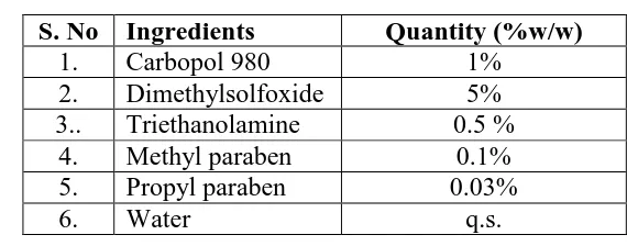
Table 2.2: Composition of carbopol gel formulations.
S. No Ingredients Quantity (%w/w)
1. Carbopol 980 1%
2. Dimethylsolfoxide 5%
3.. Triethanolamine 0.5 %
4. Methyl paraben 0.1%
5. Propyl paraben 0.03%
6. Water q.s.
E) Incorporation of Ethosomal Suspension into Gel
Ethosomal suspension (10ml) containing DS (100 mg) and CAP (5 mg) was slowly added in
the carbopol gel with continuous stirring at 700rpm in a closed vessel and maintained at
temperature 30℃ until homogeneous in order to achieve ethosomal gel. The pH was then
adjusted to neutral using triethanolamine and stirred slowly till a gel was obtained.[18]
F)Evaluation of Ethosomal Gel[19]
The prepared ethosomal gels were evaluated for following parameters:
pH: The pH measurement of formulations was carried out using a pH meter by dipping
Rheological studies: The rheological analysis of prepared gels was measured using
Brookfield Viscometer.
Drug content: Gel formulations (100 mg) was dissolved in phosphate buffer (pH 6.8)
and filtered and the volume was made to 100 ml with phosphate buffer. The resultant
solution was suitably diluted with phosphate buffer and absorbance was measured at 281
nm for both drugs; Diclofenac Sodium and Capsaicin.[20]
In-vitro drug permeation studies: In-vitro drug release study of Diclofenac Sodium and
Capsaicin from ethosomal gel formulation was studied using a Franz glass diffusion cell.
3. RESULT AND DISCUSSION
A) Preformulation studies
i. Organoleptic properties of drug: Diclofenac Sodium was found to be a white to slightly
yellowish crystalline powder, slightly hygroscopic and odorless. Capsaicin was found to
be pure dark red solid, highly volatile, pungent odor and burning taste detectable.
ii. Melting point: The obtained sample of drug of diclofenac sodium and capsaicin were
found to be in the range of 275-277℃ and 62-65℃ respectively. It complies with the
standard thus indicating the purity of the drug sample.
iii. Determination of absorption maxima (λ max) of drug by UV spectrophotometer. UV
[image:6.595.93.501.479.687.2]scans of Diclofenac Sodium.
Table 3.1: UV scans of Diclofenac sodium.
Sr. No. Wavelength Abs.
1 340 0.001
2 276 1.741
3 220.80 1.719
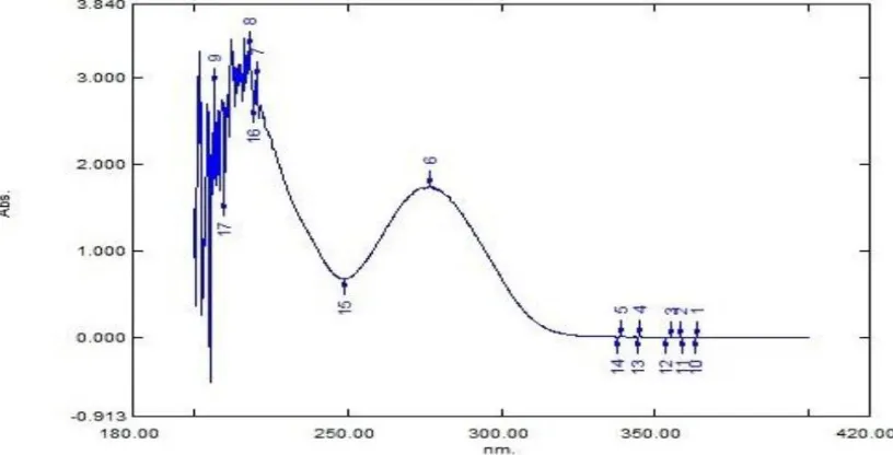
UV scans of Capsaicin
An absorption maximum (λ max) of drug was determined by UV/VIS spectrophotometer. UV
[image:7.595.108.484.242.446.2]spectra of drug in Ethanol and ACN: Water: Buffer showed maxima absorbance at 280 nm.
Fig 3.2: UV scan of Capsaicin in ACN: Water: Buffer.
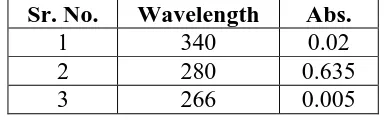
Table 3.2: UV scans of capsaicin.
Sr. No. Wavelength Abs.
1 340 0.02
2 280 0.635
3 266 0.005
iv. Solubility: Diclofenac Sodium was found to be freely soluble in methanol, soluble in
ethanol (95%), soluble in water, phosphate buffer (pH 7.2), slightly soluble in glacial acetic
acid and acetone, practically insoluble in chloroform, ether and toluene. Capsaicin was found
to be freely soluble in alcohol; soluble in ether, chloroform, benzene and DMSO; slightly
soluble in HCL, petroleum; insoluble in water.
v. Partition coefficient: The partition coefficient of Diclofenac sodium was found to be
13.60 and partition coefficient of Capsaicin was found to be 4.73. The partition coefficient
[image:7.595.201.395.511.571.2]lipophilic drugs.
vi. Drug Excipients compatibility study: The possible interaction between drugs and
excipients were studied by FTIR spectroscopy.
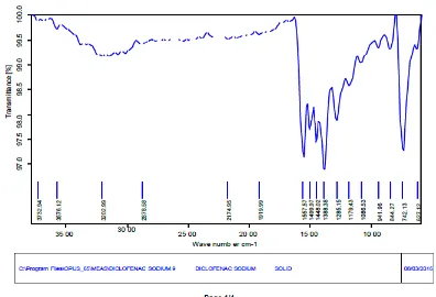
[image:8.595.87.483.180.450.2]IR Spectrum of Diclofenac Sodium
Fig 3.3: IR spectrum of Diclofenac Sodium.
Structure
In this IR spectrum, pure Diclofenac Sodium showed major peaks at 3202, 742, 1558, 1448
Table 3.3: IR interpretation of Diclofenac sodium.
Functional group Wave number (cm-1)
N-H stretch 3202cm-1
C-Cl stretch 742 cm-1
C=O stretch 1558cm-1
C=C stretch 1448cm-1
C-H stretch 2878cm-1
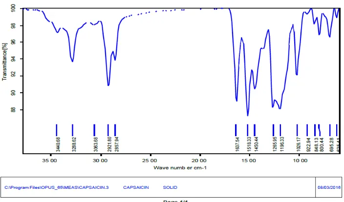
IR Spectrum of Capsaicin
Fig 3.4: IR spectrum of Capsaicin.
Structure
In this IR spectrum, pure Capsaicin showed major peaks at 3440, 2921, 1637, 1518 and 1026
[image:9.595.123.475.214.421.2]cm-1. The following peaks were observed which may be due to presence of different functional groups of Capsaicin.
Table 3.4: IR interpretation of capsaicin.
Functional group Wave number (cm-1)
N-H stretch 3440cm-1
C=O stretch 1637cm-1
C-O stretch 1026cm-1
C=C stretch 1518cm-1
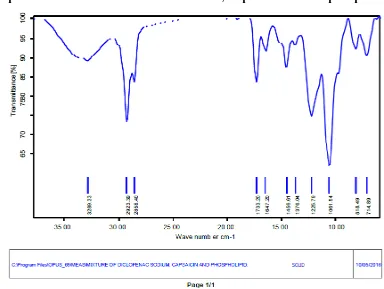
FTIR Spectrum of Mixture of Diclofenac Sodium, Capsaicin and Phospholipid
Fig 3.5: IR Spectrum Diclofenac Sodium, Capsaicin and Phospholipid
In this IR spectrum, Diclofenac Sodium, Capsaicin and Phospholipid showed major peaks at
3289, 2923, 2858, 1733, 1647, 1456, 1061, 818 and 714 cm-1. The following peaks were observed which may be due to presence of different functional groups of mixture of drugs
[image:10.595.167.442.531.650.2]and phospholipid.
Table 3.5: IR interpretation of Diclofenac Sodium, Capsaicin and Phospholipid.
Functional group Wave number (cm-1)
N-H stretch 3289 cm-1
C-H stretch 2923 cm-1 and 2858 cm-1
C=C stretch 1456 cm-1
C-O stretch 1061 cm-1
N-H rocking 818 cm-1
C=O stretch 1733 cm-1 and 1647 cm-1
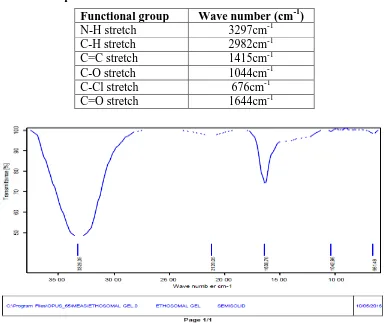
Fig 3.6: IR spectrum of Ethosomal formulation.
In this IR spectrum, Formulation Ethosome containing drugs showed major peaks at 3297,
2982, 1644, 1415, 1044 and 676 cm-1. The following peaks are observed which may be due to presence of different groups of drugs and phospholipid.
Table 3.6: IR interpretation of Ethosomal formulation.
Functional group Wave number (cm-1)
N-H stretch 3297cm-1
C-H stretch 2982cm-1
C=C stretch 1415cm-1
C-O stretch 1044cm-1
C-Cl stretch 676cm-1
C=O stretch 1644cm-1
[image:11.595.103.489.421.744.2]In this IR spectrum, Formulation of Ethosomal Gel which was containing drugs showed
[image:12.595.171.426.166.243.2]major peaks at 3328, 1638, 1044 and 661 cm-1. The following peaks are observed which may be due to presence of different functional groups of drugs and excipients.
Table 3.7: IR interpretation of ethosomal gel.
Functional group Wave number (cm-1)
N-H stretch 3328cm-1
C-O stretch 1044cm-1
C-Cl stretch 661cm-1
C=O stretch 1638cm-1
From IR data, it was observed that functionality of both drugs have remained unaffected,
including intensities of the peak. This suggests that during development of formulation,
excipients have not reacted with the drugs to give rise to reactant products. So there is no
interaction between them which is in favor to keep for formulation.
B) Characterization of Ethosomes
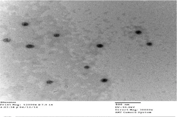
i. Vesicular Shape and Surface Morphology: Investigation of formulations by TEM
indicated spherical structure of vesicles with a smooth surface.
Fig 3.8: Transmission Electron Photomicrographs of Ethosomal formulation.
The prepared ethosomes were found to be spherical and discrete in shape. The vesicles size
range was found to be 50 to 58.8 nm.
ii. Percentage Entrapment Efficiency (%EE): The % entrapment efficiency of ethosomal
[image:12.595.148.449.426.624.2]respectively.
iii. Zeta Potential: Zeta potential of ethosomal formulations of Diclofenac Sodium and
Capsaicin was found to be -12.4 ± 2.85. It was observed that zeta potential of prepared
vesicles has sufficient charge to inhibit aggregation of vesicles and indicating stable vesicles.
C) Evaluation of Ethosomal Gel (EG)
pH
Table 3.8: pH of ethosomal gel.
S. No. Formulation code pH
1 EG-C 1% 6.73 ±0.30
2 EG-C 2% 5.23 ±0.57
3 Marketed formulation 6.8 ± 0.35
Drug Content: The drug content of prepared ethosomal gel of Diclofenac Sodium and
Capsaicin were found to be 96.20 ± 0.86 % and 94.25 ± 0.76 % respectively.
Viscosity: The viscosity of EG-C 1%, EG-C 2% and marketed formulation at 100 rpm of
spindle was found to be 16100 ± 310, 35400 ± 416 and 20400 ± 350. The viscosity of
formulation EG-C 1% was found to be optimum value.
In vitro drug permeation study: Result revealed that % cumulative drug permeated
from Diclofenac Sodium and Capsaicin ethosomal formulations were found to be 52.56 ±
0.54 and 64.76 ± 0.42 respectively. Drugs permeation from ethosomal gel was found to
be higher as compared to the carbopol gel as well as in comparison with marketed
formulation.
4. CONCLUSION
In this study ethosomal gel of Diclofenac Sodium, a NSAID and Capsaicin, a powerful
counterirritant was formulated and evaluated. Ethosomes of the drugs were prepared by cold
method using phospholipids, ethanol; subsequently this combination of ethosomal suspension
was incorporate into topical gel which is prepared by using Carbopol-934.
The prepared ethosomes were characterized for vesicles size, vesicle shape and surface
morphology, percentage entrapment efficiency by ultracentrifugation method, zeta potential
and drug-drug, drug-excipients compatibility studies by IR spectroscopy. The prepared
studies.
IR study concluded that no major interaction occurred between drug, phospholipids and carbopol-934 used in the present study.
Morphology of vesicles; the prepared ethosomes were spherical and discrete in shape.
Average particle size of the prepared ethosomal formulations was found to be 55.13 nm. Percentage entrapment efficiency of prepared ethosomal formulations of DS and CAP
were found to be 55.72 ± 0.44 % and 72.44 ± 0.98 % respectively.
The pH value of prepared ethosomal gels (EG-C1 %, EG-C2 % and marketed formulation
A) were found to be 6.73 ± 0.20, 5.23 ± 0.057 and 6.8 ± 0.35 respectively. Adjustment of
gel formulations pH nearby to skin pH will avoid skin irritation and other related
problems. The result showed pH of formulations EG-C1% was found to be in range of
skin pH.
Drug content of prepared ethosomal gel DS and CAP was found to be 96.20 ± 0.86 % and
94.25.76 ± 0.76 % respectively. Viscosities of ethosomal gel formulation (C 1%,
EG-C2% and marketed formulation A) were found to be 16100 ± 310, 35400 ± 416 and
20400 ± 350 c.p respectively at 100 rpm. The result showed that viscosity of formulation
EG-C 2% was higher than formulation EG-C 1%. The viscosity of formulation EG-C 1%
was found to optimum value.
The order of drug permeate was found to be:- Ethosomal gel formulation > marketed
formulation > carbopol gel in phosphate buffer solution (pH 6.8)
Result revealed that % cumulative drug permeate from ethosomal formulations loaded DS and CAP were found to be 52.56 ± 0.54 % and 64.76 ± 0.42 % respectively. Drugs permeate
(i.e. combination of Diclofenac Sodium and Capsaicin) from ethosomal gel was higher as
compared to the carbopol gel and marketed formulation of same drug.
From the above studies it was concluded that Ethosomal formulation is the best formulation
for improving permeability of Diclofenac Sodium and Capsaicin.
REFERENCES
1. C. Surber, F.A. Davis, Dermatological and transdermal formulation, Pharma info net,
2002; 119: 323-403.
2. G.S. Banker, C.T. Rhodes, Modern pharmaceutics, 2ndedn., Vol. 40, Marcel Dekker, inc.,
3. H.C. Ansel, N.G. Popovich, Allen V. Loyd, Pharmaceutical Dosage Forms and Drug
Delivery Systems, 8thEdn, B.I. Publications Pvt Ltd, 2005; 415-419.
4. Chandel, V. Patil, R. Goyal, H. Dhamija and B. Parashar, Ethosomes: A Novel Approach
towards Transdermal. IJPCS, 2012; 1(2): 563-569.
5. Manosroi, P. Jantrawut, N. Khositsuntiwong, W. Manosroi and J. Manosroi, Novel
Elastic Nanovesicles for Cosmeceutical and Pharmaceutical Applications, Chiang Mai. J.
Sci, 2009; 36(2): 168-178.
6. E. Touitou, N. Dayan, L. Bergelson, B. Godin and M. Eliaz, Ethosomes novel vesicular
carriers for enhanced delivery: characterization and skin penetration properties, J. Control
Release, 2000; 65: 403-418.
7. S. Patel, Ethosomes: A promising tool for transdermal delivery of drug, Pharma. Info.
Net, 2007; 5(3).
8. O. Braun-Falco, H.C. Kortung, H.I. Maibach (Eds.), Griesbach Conference: Liposome
Dermatics, Springer-Verlag, Ber lin, Heidelberg, 1992; 38-43.
9. E. Touitou, H.E. Junginger, N.D. Weiner, M. Mezei, Liposomes as carriers for topical
and transdermal delivery, J. Pharm. Sci, 1992; 9: 1189–1203.
10.G. Cevc, D. Gebauer, J. Stieber, A. Schatzlein, G. Blume, Ultraflexible vesicles,
transfersomes, have an extremely low pore penetration resistance and transport
therapeutic amount of insulin across the intact mammalian skin, Biochim. Biophys. Acta,
1998; 1368: 201–215.
11.Berner, P. Liu, Alcohols, in: E.W. Smith, H.I. Maibach (Eds.), Percutaneous Penetration
Enhancers, CRC Press, Boca Raton, Fl, 1995; 45–60.
12.S. Jain, A.K Tiwary, B. Sapra, N. K Jain, Formulation and Evaluation of ethosomes for
transdermal delivery of lamivudine, AAPS Pharm. Sci. Tech, 2007; 8(4): E1-E9.
13.E.R. Bendas, M.I. Tadros, Enhanced Transdermal Delivery of Salbutamol Sulfate via
Ethosomes, AAPS Pharm. Sci. Tech, 2007; 8(4): E1-E8.
14.A.K. Barupal, Suman Ramteke and Vandana Gupta, Preparation and characterization of
ethosomes for topical delivery of aceclofenacc, Ind. J. Pharm. Sci. 72(5) (2010) 582-586.
15.S.D. Maurya, Enhanced transdermal permeation of indinavir sulphate through stratum
corneum via novel Permeation enhancers: ethosomes, Der Pharmacia Lettre, 2010; 2(5):
208-220.
16.S.C. Chandran, A. Shirwaikar, M.R. Kuriakose and N.S. Sabna, Development and
evaluation of ethosomes for transdermal delivery of fluconazole, J. Chem. Bio. Phy. Sci,
17.P. Verma, K. Pathak, Nanosized ethanolic vesicles loaded with econazole nitrate for the
treatment of deep fungal infections through topical gel formulation, Nanomed:
Nanotechnol. Biol. Medic, 2011; 8(4): 489-496.
18.Sheer, M. Chauhan, Ethosomes as vesicular carrier for enhanced transdermal delivery of
ketoconazole – Formulation and Evaluation, J. Pharma. Cosmetol, 2011; 1(3): 1-14.
19.A.K. Garg, L.M. Negi, M. Chauhan, Gel containing ethosomal vesicles for transdermal
delivery of aceclofenac, Int. J. Pha. Pharma. Sci, 2010; 2(2): 102-108.z
20.V. Dave, D. Kumar, S. Lewis, S. Paliwal, Ethosome for enhanced transdermal drug