organic papers
o764
Nobuo Okabeet al. C7H6O5H2O DOI: 10.1107/S1600536801012041 Acta Cryst.(2001). E57, o764±o766 Acta Crystallographica Section EStructure Reports Online
ISSN 1600-5368
Gallic acid monohydrate
Nobuo Okabe,* Hasuyo Kyoyama and Misato Suzuki
Faculty of Pharmaceutical Sciences, Kinki University, Kokawae 3-4-1, Higashiosaka, Osaka 577-8502, Japan
Correspondence e-mail: [email protected]
Key indicators Single-crystal X-ray study
T= 296 K
Mean(C±C) = 0.005 AÊ
Rfactor = 0.049
wRfactor = 0.188
Data-to-parameter ratio = 15.0
For details of how these key indicators were automatically derived from the article, see http://journals.iucr.org/e.
#2001 International Union of Crystallography Printed in Great Britain ± all rights reserved
In the crystal structure of the title compound, 3,4,5-tri-hydroxybenzoic acid monohydrate, C7H6O5H2O, the gallic
acid molecule is essentially planar and has two intramolecular hydrogen bonds between hydroxyl groups. The H atoms of the three hydroxyl groups are oriented in the same direction around the ring, and form intra- and intermolecular hydrogen bonds. The crystal structure is stabilized by all available intermolecular hydrogen bonds, including also those involving the water molecule.
Comment
Gallic acid, 3,4,5-trihydroxybenzoic acid, is a naturally occur-ring plant phenol which has antitumor and anti-oxidative activity. It induces apotosis in the human myelogenous leukemic cell line (Sakaguchiet al., 1999; Satoh & Sakagami, 1997). Therefore the determination of its crystal structure is important for the structural clari®cation of its biological function. Recently the crystal structure was determined in a monohydrate form by Jiang et al. (2000). We have now determined its structure as a different monohydrated form, (I).
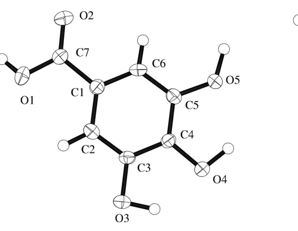
The molecular structure of (I) is essentially planar, as shown in Fig. 1. In the molecule, all the H atoms of the three hydroxyl groups are oriented in the same direction around the ring, forming two intramolecular hydrogen bonds between a pair of hydroxyl groups at positions 3 and 4, and at positions 4 and 5. The hydroxyl groups at positions 3 and 4 are also linked to the water-O atom and to the hydroxyl-O atom of a neighbouring molecule by a bifurcated hydrogen bond. This hydrogen-bonding scheme is different from that reported by Jianget al.
(2000), in which only one intramolecular hydrogen bond is present, and one of the three H atoms of the hydroxyl groups is oriented in the reverse direction to the others. Furthermore, a hydrogen bond is formed between the carboxyl groups in the
previous structure, but this interaction is not present in the structure reported here. All of the possible hydrogen bonds are present as either intra- or intermolecular interactions, as shown in Table 2. The two different crystal structures and hydrogen-bonding schemes observed for gallic acid mono-hydrate may have a role in the biological function of this compound.
Experimental
The crystal was obtained by slow evaporation of an ethanol solution.
Crystal data C7H6O5H2O
Mr= 188.13
Monoclinic,P2=n a= 14.15 (1) AÊ
b= 3.622 (9) AÊ
c= 15.028 (10) AÊ
= 97.52 (7)
V= 764 (1) AÊ3
Z= 4
Dx= 1.636 Mg mÿ3
MoKradiation Cell parameters from 18
re¯ections
= 10.0±14.3
= 0.15 mmÿ1
T= 296.2 K Needle, colorless 0.500.100.03 mm Data collection
Rigaku AFC-5Rdiffractometer
!±2scans
Absorption correction: none 2109 measured re¯ections 1766 independent re¯ections 764 re¯ections withI> 2(I)
Rint= 0.040
max= 27.5
h= 0!18
k=ÿ4!0
l=ÿ19!19 3 standard re¯ections
every 150 re¯ections intensity decay: 0.1% Re®nement
Re®nement onF2
R[F2> 2(F2)] = 0.049
wR(F2) = 0.188
S= 0.91 1766 re¯ections 118 parameters
H-atom parameters not re®ned
w= 1/[2(F
o2) + (0.1P)2]
whereP= (Fo2+ 2Fc2)/3
(/)max< 0.001 max= 0.31 e AÊÿ3 min=ÿ0.31 e AÊÿ3
Table 1
Selected geometric parameters (AÊ,).
O1ÐC7 1.317 (5)
O2ÐC7 1.203 (5)
O3ÐC3 1.373 (4)
O4ÐC4 1.370 (4)
O5ÐC5 1.372 (4)
C1ÐC2 1.396 (5)
C1ÐC6 1.380 (5)
C1ÐC7 1.487 (5)
C2ÐC3 1.376 (4)
C3ÐC4 1.391 (5)
C4ÐC5 1.390 (5)
C5ÐC6 1.377 (4)
C2ÐC1ÐC6 119.7 (3)
C2ÐC1ÐC7 122.2 (3)
C6ÐC1ÐC7 118.0 (3)
C1ÐC2ÐC3 119.3 (3)
O3ÐC3ÐC2 119.8 (3)
O3ÐC3ÐC4 119.2 (3)
C2ÐC3ÐC4 121.0 (3)
O4ÐC4ÐC3 117.4 (3)
O4ÐC4ÐC5 123.2 (3)
C3ÐC4ÐC5 119.3 (3)
O5ÐC5ÐC4 116.8 (3)
O5ÐC5ÐC6 124.0 (3)
C4ÐC5ÐC6 119.6 (3)
C1ÐC6ÐC5 121.0 (3)
O1ÐC7ÐO2 122.6 (3)
O1ÐC7ÐC1 113.5 (3)
O2ÐC7ÐC1 123.8 (3)
O1ÐC7ÐC1ÐC2 2.7 (5)
O1ÐC7ÐC1ÐC6 178.6 (3)
O2ÐC7ÐC1ÐC2 ÿ175.3 (4)
O2ÐC7ÐC1ÐC6 0.2 (6)
O3ÐC3ÐC2ÐC1 ÿ179.0 (3)
O3ÐC3ÐC4ÐO4 ÿ0.2 (5)
O3ÐC3ÐC4ÐC5 179.0 (3)
O4ÐC4ÐC3ÐC2 ÿ177.3 (3)
O4ÐC4ÐC5ÐO5 0.9 (5)
O4ÐC4ÐC5ÐC6 178.9 (4)
O5ÐC5ÐC4ÐC3 ÿ178.9 (3)
O5ÐC5ÐC6ÐC1 177.2 (3)
C1ÐC2ÐC3ÐC4 ÿ1.9 (6)
C1ÐC6ÐC5ÐC4 ÿ1.5 (6)
C2ÐC1ÐC6ÐC5 1.4 (6)
C2ÐC3ÐC4ÐC5 1.8 (6)
C3ÐC2ÐC1ÐC6 0.3 (6)
C3ÐC2ÐC1ÐC7 175.7 (4)
C3ÐC4ÐC5ÐC6 ÿ0.1 (6)
C5ÐC6ÐC1ÐC7 ÿ174.2 (4)
Table 2
Hydrogen-bonding geometry (AÊ,).
DÐH A DÐH H A D A DÐH A
O3ÐH3 O4 0.850 2.328 2.692 (8) 106.3
O4ÐH4 O5 0.945 2.306 2.752 (8) 108.9
O3ÐH3 O6i 0.850 1.949 2.770 (4) 161.6
O4ÐH4 O5ii 0.945 1.907 2.767 (8) 150.0
O5ÐH5 O2iii 0.851 1.878 2.729 (4) 177.7
O6ÐH7 O4ii 0.820 2.182 2.977 (4) 163.8
O6ÐH8 O3iv 0.812 1.941 2.747 (4) 170.9
O1ÐH1 O6iii 0.830 1.859 2.686 (4) 174.2
Symmetry codes: (i)1
2ÿx;yÿ1;21ÿz; (ii)12ÿx;y;12ÿz; (iii) 1ÿx;1ÿy;1ÿz; (iv) 1
2x;ÿy;zÿ12.
All H atoms were located from difference Fourier maps and were not re®ned.
Data collection: MSC /AFC Diffractometer Control Software
(Molecular Structure Corporation & Rigaku Corporation, 1999); cell re®nement:MSC/AFC Diffractometer Control Software; data reduc-tion: TEXSAN (Molecular Structure Corporation & Rigaku Corporation, 1999); program(s) used to solve structure:SIR88 (Burla
et al., 1989) andDIRDIF(Beurskenset al., 1992); program(s) used to re®ne structure:SHELXL97 (Sheldrick, 1997); molecular graphics:
ORTEPII (Johnson, 1976); software used to prepare material for publication:TEXSAN.
References
Beurskens, P. T., Admiraal, G., Beurskens, G., Bosman, W. P., Garcia-Granda, S., Gould, R. O., Smith, J. M. M. & Smykalla, C. (1992).DIRDIF.University of Nijmegen, The Netherlands.
Burla, M. C., Camalli, M., Cascarano, G., Giacovazzo, C., Spagna, R. & Viterbo, D. (1989).J. Appl. Cryst.22, 389±403.
Johnson, C. K. (1976).ORTEPII. Oak Ridge National Laboratory, Tennessee, USA.
Acta Cryst.(2001). E57, o764±o766 Nobuo Okabeet al. C7H6O5H2O
o765
organic papers
Figure 1
organic papers
o766
Nobuo Okabeet al. C7H6O5H2O Acta Cryst.(2001). E57, o764±o766 Molecular Structure Corporation & Rigaku Corporation (1999).MSC/AFCDiffractometer Control SoftwareandTEXSAN(Version 1.10). MSC, 3200 Research Forest Drive, The Woodlands, TX 77381, USA, and Rigaku, 3-9-12 Akishima, Tokyo, Japan.
Jiang, R.-W., Ming, D.-S., But, P. H. P. & Mak, T. C. W. (2000).Acta Cryst.C56, 594±595.
Satoh, K. & Sakagami, H. (1997).Anticancer Res.17, 2181±2184.
Sakaguchi, N., Inoue, M., Isuzugawa, K., Ogihara, Y. & Hosaka, K. (1999).
Biol. Pharm. Bull.22, 471±475.
supporting information
sup-1
Acta Cryst. (2001). E57, o764–o766
supporting information
Acta Cryst. (2001). E57, o764–o766 [doi:10.1107/S1600536801012041]
Gallic acid monohydrate
Nobuo Okabe, Hasuyo Kyoyama and Misato Suzuki
S1. Comment
Gallic acid, 3,4,5-trihydroxybenzoic acid, is a naturally occurring plant phenol which has antitumor and anti-oxidative
activity. It induces apotosis in the human myelogenenous leukemic cell line (Sakaguchi et al., 1999; Satoh & Sakagami,
1997). Therefore the determination of its crystal structure is important for the structural clarification of its biological
function. For this reason, we intended to analyse its crystal structure. Recently, this was determined in a monohydrate
form by Jiang et al. (2000). We have mow determined its structure as a different monohydrated form, (I).
The molecular structure of (I) is essentially planar as shown in Fig. 1. In the molecule, all the H atoms of the three
hydroxyl groups are oriented in the same direction around the ring, forming two intramolecular hydrogen bonds between
a pair of hydroxyl groups at positions 3 and 4, and at positions 4 and 5. The hydroxyl groups at positions 3 and 4 are also
linked to the water-O atom and to the hydroxyl-O atom of a neighbouring molecule by a bifurcated hydrogen bond. This
hydrogen-bonding scheme is different from that reported by Jiant et al. (2000), in which only one intramolecular
hydrogen bond is present, and one of the three H atoms of the hydroxyl groups is oriented in the reverse direction to the
others. Furthermore, a hydrogen bond is formed between the carboxyl groups in the previous structure, but this
interaction is not present in the structure reported here. All of the possible hydrogen bonds are present as either intra- or
intermolecular interactions, as shown in Table 2. The two different crystal structures and hydrogen-bonding schemes
observed for gallic acid monohydrate may have a role in the biological function of this compound.
S2. Experimental
The crystal was obtained by slow evaporation of an ethanol solution.
S3. Refinement
supporting information
sup-2
[image:5.610.133.438.95.332.2]Acta Cryst. (2001). E57, o764–o766 Figure 1
ORTEPII (Johnson, 1976) drawing of the title compound with the atomic numbering scheme. Ellipsoids for non-H atoms
correspond to 50% probability.
(I)
Crystal data
C7H6O5·H2O Mr = 188.13
Monoclinic, P2/n a = 14.15 (1) Å
b = 3.622 (9) Å
c = 15.028 (10) Å
β = 97.52 (7)°
V = 764 (1) Å3 Z = 4
F(000) = 392.0
Dx = 1.636 Mg m−3
Mo Kα radiation, λ = 0.7107 Å Cell parameters from 18 reflections
θ = 10.0–14.3°
µ = 0.15 mm−1 T = 296 K Needle, colorless 0.50 × 0.10 × 0.03 mm
Data collection
Rigaku AFC-5R diffractometer
ω–2θ scans
2109 measured reflections 1766 independent reflections 764 reflections with I > 2σ(I)
Rint = 0.040
θmax = 27.5°, θmin = 4° h = 0→18
k = −4→0
l = −19→19
3 standard reflections every 150 reflections intensity decay: 0.1%
Refinement
Refinement on F2 R[F2 > 2σ(F2)] = 0.049 wR(F2) = 0.188 S = 0.91
1766 reflections 118 parameters
supporting information
sup-3
Acta Cryst. (2001). E57, o764–o766 w = 1/[σ2(F
o2) + (0.1P)2] where P = (Fo2 + 2Fc2)/3 (Δ/σ)max < 0.001
Δρmax = 0.31 e Å−3 Δρmin = −0.31 e Å−3
Special details
Refinement. Refinement using reflections with F2 > -10.0 σ(F2). The weighted R-factor (wR) and goodness of fit (S) are based on F2. R-factor (gt) are based on F. The threshold expression of F2 > 2.0 σ(F2) is used only for calculating R-factor (gt).
Fractional atomic coordinates and isotropic or equivalent isotropic displacement parameters (Å2)
x y z Uiso*/Ueq
O1 0.3779 (2) 0.2043 (9) 0.7302 (2) 0.0449 (8) O2 0.4899 (2) 0.4049 (9) 0.6523 (2) 0.0447 (9) O3 0.1018 (2) −0.1394 (8) 0.5049 (2) 0.0338 (7) O4 0.1550 (2) 0.0075 (8) 0.3430 (2) 0.0334 (7) O5 0.3351 (2) 0.2679 (8) 0.3286 (2) 0.0336 (7) O6 0.5102 (2) 0.4879 (8) 0.1313 (2) 0.0370 (7) C1 0.3455 (2) 0.175 (1) 0.5732 (2) 0.0243 (8) C2 0.2535 (2) 0.044 (1) 0.5784 (2) 0.0253 (8) C3 0.1931 (2) −0.0162 (10) 0.5004 (2) 0.0230 (8) C4 0.2207 (2) 0.0606 (10) 0.4172 (2) 0.0228 (8) C5 0.3126 (2) 0.191 (1) 0.4124 (2) 0.0248 (8) C6 0.3740 (2) 0.243 (1) 0.4905 (2) 0.0264 (8) C7 0.4124 (3) 0.272 (1) 0.6544 (2) 0.0280 (9)
H1 0.4126 0.2858 0.7748 0.0464*
H2 0.2292 −0.0105 0.6347 0.0464*
H3 0.0777 −0.2436 0.4565 0.0464*
H4 0.1808 0.0710 0.2901 0.0464*
H5 0.3893 0.3730 0.3331 0.0473*
H6 0.4346 0.3306 0.4847 0.0464*
H7 0.4673 0.3675 0.1493 0.0556*
H8 0.5347 0.3636 0.0958 0.0556*
Atomic displacement parameters (Å2)
U11 U22 U33 U12 U13 U23
supporting information
sup-4
Acta Cryst. (2001). E57, o764–o766
C7 0.027 (2) 0.031 (2) 0.025 (2) −0.003 (2) 0.001 (1) −0.001 (2)
Geometric parameters (Å, º)
O1—C7 1.317 (5) O6—H8 0.812
O1—H1 0.830 C1—C2 1.396 (5)
O2—C7 1.203 (5) C1—C6 1.380 (5)
O3—C3 1.373 (4) C1—C7 1.487 (5)
O3—H3 0.850 C2—C3 1.376 (4)
O4—C4 1.370 (4) C2—H2 0.975
O4—H4 0.945 C3—C4 1.391 (5)
O5—C5 1.372 (4) C4—C5 1.390 (5)
O5—H5 0.851 C5—C6 1.377 (4)
O6—H7 0.820 C6—H6 0.929
O1···O6i 2.686 (4) O3···O6v 2.769 (4)
O1···O2ii 3.576 (5) O3···O3vi 3.039 (5)
O2···O5i 2.718 (4) O3···C3ii 3.432 (5)
O2···O6i 3.275 (9) O4···O5vii 2.772 (4)
O2···C6i 3.321 (5) O4···O6vii 2.977 (4)
O2···C7iii 3.325 (5) O5···C5iii 3.602 (10) O2···C5i 3.410 (5) O6···O3viii 2.747 (4) O2···C1iii 3.569 (5) O6···O4vii 2.977 (4) O3···O6iv 2.747 (4)
C7—O1—H1 112.7 C2—C3—C4 121.0 (3)
C3—O3—H3 112.3 O4—C4—C3 117.4 (3)
C4—O4—H4 110.6 O4—C4—C5 123.2 (3)
C5—O5—H5 109.5 C3—C4—C5 119.3 (3)
H7—O6—H8 108.7 O5—C5—C4 116.8 (3)
C2—C1—C6 119.7 (3) O5—C5—C6 124.0 (3)
C2—C1—C7 122.2 (3) C4—C5—C6 119.6 (3)
C6—C1—C7 118.0 (3) C1—C6—C5 121.0 (3)
C1—C2—C3 119.3 (3) C1—C6—H6 121.8
C1—C2—H2 123.7 C5—C6—H6 117.2
C3—C2—H2 117.0 O1—C7—O2 122.6 (3)
O3—C3—C2 119.8 (3) O1—C7—C1 113.5 (3)
O3—C3—C4 119.2 (3) O2—C7—C1 123.8 (3)
O1—C7—C1—C2 2.7 (5) O5—C5—C4—C3 −178.9 (3)
O1—C7—C1—C6 178.6 (3) O5—C5—C6—C1 177.2 (3) O2—C7—C1—C2 −175.3 (4) C1—C2—C3—C4 −1.9 (6)
O2—C7—C1—C6 0.2 (6) C1—C6—C5—C4 −1.5 (6)
O3—C3—C2—C1 −179.0 (3) C2—C1—C6—C5 1.4 (6)
O3—C3—C4—O4 −0.2 (5) C2—C3—C4—C5 1.8 (6)
supporting information
sup-5
Acta Cryst. (2001). E57, o764–o766
O4—C4—C5—O5 0.9 (5) C3—C4—C5—C6 −0.1 (6)
O4—C4—C5—C6 178.9 (4) C5—C6—C1—C7 −174.2 (4)
Symmetry codes: (i) −x+1, −y+1, −z+1; (ii) x, y−1, z; (iii) x, y+1, z; (iv) x−1/2, −y, z+1/2; (v) −x+1/2, y−1, −z+1/2; (vi) −x, −y, −z+1; (vii) −x+1/2, y, −z+1/2; (viii) x+1/2, −y, z−1/2.
Hydrogen-bond geometry (Å, º)
D—H···A D—H H···A D···A D—H···A
O3—H3···O4 0.850 2.328 2.692 (8) 106.3
O4—H4···O5 0.945 2.306 2.752 (8) 108.9
O3—H3···O6v 0.850 1.949 2.770 (4) 161.6
O4—H4···O5vii 0.945 1.907 2.767 (8) 150.0
O5—H5···O2i 0.851 1.878 2.729 (4) 177.7
O6—H7···O4vii 0.820 2.182 2.977 (4) 163.8
O6—H8···O3viii 0.812 1.941 2.747 (4) 170.9
O1—H1···O6i 0.830 1.859 2.686 (4) 174.2