organic papers
o1004
B. Sridharet al. 2C3H7NO2H+NO3ÿ DOI: 10.1107/S1600536801015793 Acta Cryst.(2001). E57, o1004±o1006 Acta Crystallographica Section EStructure Reports
Online
ISSN 1600-5368
Bis(
b
-alanine) hydrogen nitrate
B. Sridhar,aN. Srinivasanb and
R. K. Rajaramc*
aDepartment of Physics, Madurai Kamaraj
University, Madurai 625 021, India,
bDepartment of Physics, Thiagarajar College,
Madurai 625 009, India, andcDepartment
of Physics, Madurai Kamaraj University, Madurai 625 021, India
Correspondence e-mail: [email protected]
Key indicators
Single-crystal X-ray study
T= 293 K
Mean(C±C) = 0.002 AÊ
Rfactor = 0.033
wRfactor = 0.092 Data-to-parameter ratio = 8.8
For details of how these key indicators were automatically derived from the article, see http://journals.iucr.org/e.
#2001 International Union of Crystallography Printed in Great Britain ± all rights reserved
In the title compound, 2C3H7NO2.H+NO3ÿ, both the alanine
residues, related by a center of symmetry, are linked by a strong symmetric OÐH O hydrogen bond with an O O distance of 2.467 (2) AÊ. The N atom and one of the O atoms of the nitrate anion lie on the twofold axis.
Comment
In amino acid±inorganic acid complexes, when the number of H atoms liberated from the inorganic acid is less than the number of amino acids, the H atom is shared by two amino acids, resulting in short symmetric OÐH O hydrogen bonds, as evidenced in triglycine sulfate (Kay, 1977), leading to phase transitions. In order to look for similar compounds, a systematic study of the behavior of hydrogen bonding in amino acid±inorganic acid complexes was undertaken. In this context, the crystal structure of l-phenylalanine l -phenyl-alaninium perchlorate (Srinivasan & Rajaram, 1997), hydrogen bis[l-lysinium (2+)] dichloride perchlorate (Srini-vasan et al., 2001a), l-lysine l-lysinium dichloride nitrate (Srinivasan et al., 2001b), l-phenylalanine-nitric acid (2/1) (Srinivasan et al., 2001c) and bis(l-proline) hydrogen perchlorate (Pandiarajanet al., 2001) have been reported. A similar stucture, l-phenylalaninel-phenylalaninium formate, has been reported by GoÈrbitz & Etter (1992). As part of this program, the crystal structure of-alanine reacted with nitric acid was undertaken to study the nature of the hydrogen bonding in the presence of an inorganic acid.
The asymmetric unit contains one-alanine residue and a nitrate anion which lies on the twofold axis. The backbone conformation angles 1 and 2 are ÿ5.2 (2) and 174.9 (1),
respectively, for the alanine residue. The straight-chain conformation angle 1 is in gauche I form [63.6 (2)]. This
tendency of twisting of the CÐN bond is found in various amino acids (Lakshminarayananet al., 1967).
The nitrate anion plays a vital role in forming hydrogen bonds with the alanine residue and stabilizing the structure. The H1Batom, which lies on a center of symmetry, links the two alanine residues through a strong symmetric OÐH O hydrogen bond [O1B O1B(1/2ÿx, 1/2ÿy, 1ÿz) 2.467 (2) AÊ]. The largeUisovalue of H1Bsuggests that this atom may have
positional or ¯ip-¯op disorder, leading to the switching of roles of the cation and zwitterion in a time-averaged equili-brium (Jeffrey & Saenger, 1991). A similar feature of short hydrogen bonding has been observed in l-phenylalanine
l-phenylalaninium formate, l-phenylalanine l -phenylalan-inium perchlorate, hydrogen bis[l-lysinium (2+)] dichloride perchlorate, l-lysine l-lysinium dichloride nitrate, l -phenyl-alanine-nitric acid (2/1) and bis(l-proline) hydrogen perchlorate. In these compounds, the hydrogen bond can be termed a possible symmetric hydrogen bond. At low temperature, the crystal of (I) may change to the non-centrosymmetric space groupCc, triggering a structural phase transition leading to interesting physical properties.
The O1 and O2 atoms of the nitrate anion, as acceptors, link the amino N atom in a three-centered hydrogen bond invol-ving the alanine residue. This O2 atom of the nitrate anion, sitting on the twofold axis, links two symmetry-related -alanine residues. A three-centered hydrogen bond is observed involving the alanine residue (amino N atom) and the carboxyl O1A(intramolecular hydrogen bond) and O1B
(Z2 head-to-tail sequence) atoms. A glide-related head-to-tail sequence is observed, since N11ÐH2B O1B(x, ÿy, zÿ1/2) connects two glide-related amino acids (Vijayan, 1988).
Experimental
Crystals of (I) were grown from an aqueous solution of a 2:1 stoi-chiometric ratio of-alanine and nitric acid by slow evaporation.
Crystal data 2C3H7NO2H+NO3ÿ
Mr= 241.21
Monoclinic,C2=c a= 19.791 (1) AÊ
b= 5.3220 (3) AÊ
c= 10.974 (1) AÊ
= 113.923 (6)
V= 1056.57 (13) AÊ3
Z= 4
Dx= 1.516 Mg mÿ3 Dm= 1.502 Mg mÿ3
Dmmeasured by ¯otation using a
mixture of carbon tetrachloride and xylene
MoKradiation Cell parameters from 25
re¯ections
= 11.3±14.0 = 0.14 mmÿ1
T= 293 (2) K Needle, colorless 0.60.40.2 mm
Data collection Enraf±Nonius CAD-4
diffractometer
!±2scans
Absorption correction: scan (Northet al., 1968)
Tmin= 0.863,Tmax= 0.970
941 measured re¯ections 914 independent re¯ections 798 re¯ections withI> 2(I)
Rint= 0.014 max= 25.0
h= 0!23
k= 0!6
l=ÿ13!11
3 standard re¯ections frequency: 60 min intensity decay: none
Re®nement Re®nement onF2
R[F2> 2(F2)] = 0.033
wR(F2) = 0.092
S= 1.09 914 re¯ections 104 parameters
All H-atom parameters re®ned
w= 1/[2(F
o2) + (0.0459P)2
+ 0.6820P]
whereP= (Fo2+ 2Fc2)/3
(/)max< 0.001
max= 0.31 e AÊÿ3
min=ÿ0.18 e AÊÿ3
Extinction correction:SHELXL97 Extinction coef®cient: 0.103 (7)
Table 1
Selected geometric parameters (AÊ,).
O1AÐC11 1.2204 (19)
O1BÐC11 1.3030 (17) N1ÐO2N1ÐO1 1.235 (3)1.2419 (15)
O1AÐC11ÐC12ÐC13 ÿ5.2 (2)
O1BÐC11ÐC12ÐC13 174.93 (13) C11ÐC12ÐC13ÐN11 63.60 (19)
Table 2
Hydrogen-bonding geometry (AÊ,).
DÐH A DÐH H A D A DÐH A
O1BÐH1B O1Bi 1.2333 (10) 1.2333 (10) 2.467 (2) 180
N11ÐH1A O1A 0.87 (2) 2.29 (2) 2.8971 (18) 126.5 (17) N11ÐH1A O1Bii 0.87 (2) 2.34 (2) 3.0517 (18) 139.1 (17)
N11ÐH2B O1Biii 0.87 (2) 2.01 (3) 2.877 (2) 175 (2)
N11ÐH3C O1ii 0.89 (2) 2.10 (3) 2.908 (2) 150 (2)
N11ÐH3C O2iv 0.89 (2) 2.40 (2) 3.0779 (15) 133.2 (18) Symmetry codes: (i) 1
2ÿx;12ÿy;1ÿz; (ii) 12ÿx;21y;12ÿz; (iii) x;ÿy;zÿ12; (iv) xÿ1
2;12y;zÿ1.
Acta Cryst.(2001). E57, o1004±o1006 B. Sridharet al. 2C3H7NO2H+NO3ÿ
o1005
organic papers
Figure 1
The molecular structure of (I), with the atom-numbering scheme and 50% probability displacement ellipsoids (Johnson, 1976).
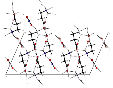
Figure 2
organic papers
o1006
B. Sridharet al. 2C3H7NO2H+NO3ÿ Acta Cryst.(2001). E57, o1004±o1006All the H atoms were located and re®ned isotropically. The CÐH and NÐH bond lengths are 0.94 (2)±0.97 (2) and 0.87 (2)±0.89 (2) AÊ, respectively.
Data collection: CAD-4 Software (Enraf±Nonius, 1989); cell re®nement: CAD-4 Software; data reduction: CAD-4 Software; program(s) used to solve structure: SHELXS97 (Sheldrick, 1997); program(s) used to re®ne structure:SHELXL97 (Sheldrick, 1997); molecular graphics:PLATON(Spek, 1999); software used to prepare material for publication:SHELXL97.
BS and RKR thank the Department of Science and Tech-nology (DST), India, for ®nancial support.
References
Enraf±Nonius (1989).CAD-4Software. Version 5.0. Enraf±Nonius, Delft, The Netherlands.
GoÈrbitz, C. H. & Etter, M. C. (1992).Acta Cryst.C48, 1317±1320.
Jeffrey, G. A. & Saenger, W. (1991). In Hydrogen Bonding in Biological
Structures. pp. 40±42. Berlin, Heidelberg, New York: Springer-Verlag.
Johnson, C. K. (1976).ORTEPII. Report ORNL-5138. Oak Ridge National Laboratory, Tennessee, USA.
Kay, M. I. (1977).Ferroelectrics,17, 415.
Lakshminarayanan, A. V., Sashisekaran, V. & Ramachandran, G. N. (1967). In
Conformation of Biopolymers, edited by G. N. Ramachandran. London:
Academic Press.
North, A. C. T., Phillips, D. C. & Mathews, F. S. (1968).Acta Cryst.A24, 351± 359.
Pandiarajan, S., Sridhar, B. & Rajaram, R. K. (2001). Acta Cryst. E57. Submitted.
Sheldrick, G. M. (1997). SHELXL97 and SHELXS97. University of GoÈttingen, Germany.
Spek, A. L. (1999). PLATON for Windows. Utrecht University, The Netherlands.
Srinivasan, N. & Rajaram, R. K. (1997).Acta Cryst.C53, 1711±1713. Srinivasan, N., Sridhar, B. & Rajaram, R. K. (2001a).Acta Cryst.E57, o875±
o877.
Srinivasan, N., Sridhar, B. & Rajaram, R. K. (2001b).Acta Cryst.E57, o888± o890.
Srinivasan, N., Sridhar, B. & Rajaram, R. K. (2001c).Acta Cryst.E57, o916± o918.
supporting information
sup-1
Acta Cryst. (2001). E57, o1004–o1006supporting information
Acta Cryst. (2001). E57, o1004–o1006 [doi:10.1107/S1600536801015793]
Bis(
β
-alanine) hydrogen nitrate
B. Sridhar, N. Srinivasan and R. K. Rajaram
S1. Comment
In amino acid–inorganic acid complexes, when the number of H atoms liberated from the inorganic acid is less than the
number of amino acids, the H atom is shared by two amino acids, resulting in short symmetric O—H···O hydrogen bonds,
as evidenced in triglycine sulfate (Kay, 1977), leading to phase transitions. In order to look for similar compounds, a
systematic study of the behaviour of hydrogen bonding in amino acid–inorganic acid complexes was undertaken. In this
context, the crystal structure of L-phenylalanine L-phenylalaninium perchlorate (Srinivasan & Rajaram, 1997), hydrogen
bis[L-lysinium (2+)] dichloride perchlorate (Srinivasan et al., 2001a), L-lysine L-lysinium dichloride nitrate (Srinivasan
et al., 2001b), L-phenylalanine-nitric acid (2/1) (Srinivasan et al., 2001c) and bis(L-proline) hydrogen perchlorate
(Pandiarajan et al., 2001) have been reported. A similar stucture, L-phenylalanine L-phenylalaninium formate, has been
reported by Gorbitz & Etter (1992). As part of this programme, the crystal structure of β-alanine with nitric acid was
undertaken to study the nature of the hydrogen bonding in the presence of an inorganic acid.
The asymmetric unit contains one β-alanine residue and a nitrate anion which lies on the twofold axis. The backbone
conformation angles ψ1 and ψ2 are -5.2 (2) and 174.9 (1)°, respectively, for the alanine residue. The straight-chain
conformation angle χ1 is in gauche I form [63.6 (2)°]. This tendency of twisting of the C—N bond is found in various
amino acids (Lakshminarayanan et al., 1967).
The nitrate anion plays a vital role in forming hydrogen bonds with the alanine residue and stabilizing the structure. The
H1B atom, which lies on a center of symmetry, links the two alanine residues through a strong symmetric O—H···O
hydrogen bond [O1B···O1B(1/2 - x, 1/2 - y, 1 - z) 2.467 (2) Å]. The large Uiso value of H1B suggests that this atom may
have positional or flip–flop disorder, leading to the switching of roles of the cation and zwitterion in a time-averaged
equilibrium (Jeffrey & Saenger, 1991). A similar feature of short hydrogen bonding has been observed in L-phenylalanine
L-phenylalaninium formate, L-phenylalanine L-phenylalaninium perchlorate, hydrogen bis[L-lysinium (2+)] dichloride
perchlorate, L-lysine L-lysinium dichloride nitrate, L-phenylalanine-nitric acid (2/1) and bis(L-proline) hydrogen
perchlorate. In these compounds, the hydrogen bond can be termed as a possible symmetric hydrogen bond. At low
temperature, the crystal of (I) may go into non-centrosymmetric space group Cc, triggering a structural phase transition
leading to interesting physical properties.
The O1 and O2 atoms of the nitrate anion, as acceptors, links the amino N atom in a three-centred hydrogen bond
involving the alanine residue. This O2 atom of the nitrate anion, sitting on the twofold axis, links two symmetry-related
β-alanine residues. A three-centred hydrogen bond is observed involving the alanine residue (amino N atom) and the
carboxyl O1A (intramolecular hydrogen bond) and O1B (Z2 head-to-tail sequence) atoms. A glide-related head-to-tail
supporting information
sup-2
Acta Cryst. (2001). E57, o1004–o1006S2. Experimental
Crystals of (I) were grown from an aqueous solution of a 2:1 stoichiometric ratio of β-alanine and nitric acid by slow
evaporation.
S3. Refinement
All the H atoms were located and refined isotropically. The C—H and N—H bond lengths are 0.94 (2)–0.97 (2) and
[image:5.610.123.484.179.313.2]0.87 (2)–0.89 (2) Å, respectively.
Figure 1
The molecular structure of (I) with the atom-numbering scheme and 50% probability displacement ellipsoids (Johnson,
1976).
Figure 2
[image:5.610.120.490.364.650.2]supporting information
sup-3
Acta Cryst. (2001). E57, o1004–o1006β-Alanine β-alaninium nitrate
Crystal data
2C3H7NO2·H+·NO3−
Mr = 241.21 Monoclinic, C2/c a = 19.791 (1) Å b = 5.3220 (3) Å c = 10.974 (1) Å β = 113.923 (6)° V = 1056.57 (13) Å3
Z = 4 F(000) = 512
Dx = 1.516 Mg m−3
Dm = 1.502 Mg m−3
Dm measured by flotation using a mixture of
carbon tetrachloride and xylene Mo Kα radiation, λ = 0.71073 Å Cell parameters from 25 reflections θ = 11.3–14.0°
µ = 0.14 mm−1
T = 293 K Needle, colorless 0.6 × 0.4 × 0.2 mm
Data collection
Enraf-Nonius sealed tube diffractometer
Radiation source: fine-focus sealed tube Graphite monochromator
ω–2θ scans
Absorption correction: ψ scan (North et al., 1968)
Tmin = 0.863, Tmax = 0.970
941 measured reflections
914 independent reflections 798 reflections with I > 2σ(I) Rint = 0.014
θmax = 25.0°, θmin = 2.3°
h = 0→23 k = 0→6 l = −13→11
3 standard reflections every 60 min intensity decay: none
Refinement
Refinement on F2
Least-squares matrix: full R[F2 > 2σ(F2)] = 0.033
wR(F2) = 0.092
S = 1.09 914 reflections 104 parameters 0 restraints
Primary atom site location: structure-invariant direct methods
Secondary atom site location: difference Fourier map
Hydrogen site location: inferred from neighbouring sites
All H-atom parameters refined w = 1/[σ2(F
o2) + (0.0459P)2 + 0.682P]
where P = (Fo2 + 2Fc2)/3
(Δ/σ)max < 0.001
Δρmax = 0.31 e Å−3
Δρmin = −0.18 e Å−3
Extinction correction: SHELXL97, Fc*=kFc[1+0.001xFc2λ3/sin(2θ)]-1/4
Extinction coefficient: 0.103 (7)
Special details
Geometry. All e.s.d.'s (except the e.s.d. in the dihedral angle between two l.s. planes) are estimated using the full covariance matrix. The cell e.s.d.'s are taken into account individually in the estimation of e.s.d.'s in distances, angles and torsion angles; correlations between e.s.d.'s in cell parameters are only used when they are defined by crystal symmetry. An approximate (isotropic) treatment of cell e.s.d.'s is used for estimating e.s.d.'s involving l.s. planes.
Refinement. Refinement of F2 against ALL reflections. The weighted R-factor wR and goodness of fit S are based on F2,
conventional R-factors R are based on F, with F set to zero for negative F2. The threshold expression of F2 > σ(F2) is used
only for calculating R-factors(gt) etc. and is not relevant to the choice of reflections for refinement. R-factors based on F2
are statistically about twice as large as those based on F, and R- factors based on ALL data will be even larger.
Fractional atomic coordinates and isotropic or equivalent isotropic displacement parameters (Å2)
x y z Uiso*/Ueq
supporting information
sup-4
Acta Cryst. (2001). E57, o1004–o1006O1B 0.21345 (6) 0.1038 (2) 0.41024 (9) 0.0381 (4)
H1B 0.2500 0.2500 0.5000 0.116 (14)*
C11 0.18276 (8) 0.2140 (3) 0.29461 (14) 0.0321 (4) C12 0.13324 (9) 0.0474 (3) 0.18276 (14) 0.0369 (4) H12A 0.0973 (11) −0.033 (4) 0.2091 (19) 0.050 (5)* H12B 0.1631 (11) −0.083 (4) 0.1728 (18) 0.045 (5)* C13 0.09337 (9) 0.1853 (3) 0.05321 (15) 0.0377 (4) H13A 0.0660 (10) 0.324 (4) 0.0621 (17) 0.042 (5)*
H13B 0.0597 (11) 0.077 (4) −0.015 (2) 0.050 (5)*
N11 0.14535 (8) 0.2912 (3) −0.00011 (14) 0.0389 (4)
H1A 0.1805 (12) 0.375 (4) 0.062 (2) 0.052 (5)*
H2B 0.1670 (11) 0.179 (4) −0.0288 (19) 0.055 (6)*
H3C 0.1196 (13) 0.386 (4) −0.071 (2) 0.061 (6)*
N1 0.5000 0.0750 (4) 0.7500 0.0379 (5)
O1 0.45215 (7) 0.1894 (3) 0.65579 (12) 0.0553 (4)
O2 0.5000 −0.1571 (4) 0.7500 0.0753 (7)
Atomic displacement parameters (Å2)
U11 U22 U33 U12 U13 U23
O1A 0.0592 (8) 0.0348 (7) 0.0367 (6) −0.0082 (5) 0.0143 (5) −0.0005 (5) O1B 0.0423 (6) 0.0377 (7) 0.0277 (6) −0.0011 (5) 0.0074 (4) 0.0007 (4) C11 0.0317 (7) 0.0343 (9) 0.0303 (8) 0.0006 (6) 0.0126 (6) −0.0012 (6) C12 0.0394 (8) 0.0352 (8) 0.0318 (8) −0.0027 (7) 0.0100 (7) −0.0014 (6) C13 0.0314 (8) 0.0471 (10) 0.0316 (8) −0.0014 (7) 0.0097 (6) −0.0003 (7) N11 0.0390 (8) 0.0441 (9) 0.0320 (8) −0.0008 (7) 0.0127 (6) −0.0002 (6) N1 0.0377 (10) 0.0440 (11) 0.0341 (9) 0.000 0.0168 (8) 0.000 O1 0.0497 (7) 0.0541 (8) 0.0507 (8) 0.0099 (6) 0.0086 (6) 0.0069 (6) O2 0.0956 (16) 0.0438 (11) 0.0539 (12) 0.000 −0.0034 (11) 0.000
Geometric parameters (Å, º)
O1A—C11 1.2204 (19) C13—H13A 0.94 (2)
O1B—C11 1.3030 (17) C13—H13B 0.96 (2)
O1B—H1B 1.2333 (10) N11—H1A 0.87 (2)
C11—C12 1.509 (2) N11—H2B 0.87 (2)
C12—C13 1.507 (2) N11—H3C 0.89 (2)
C12—H12A 0.97 (2) N1—O2 1.235 (3)
C12—H12B 0.94 (2) N1—O1i 1.2419 (15)
C13—N11 1.486 (2) N1—O1 1.2419 (15)
C11—O1B—H1B 112.62 (10) N11—C13—H13B 107.2 (12)
O1A—C11—O1B 123.06 (13) C12—C13—H13B 111.9 (12)
O1A—C11—C12 122.09 (13) H13A—C13—H13B 107.4 (15)
O1B—C11—C12 114.84 (13) C13—N11—H1A 110.2 (13)
C13—C12—C11 113.58 (13) C13—N11—H2B 113.9 (14)
C13—C12—H12A 109.3 (11) H1A—N11—H2B 106.2 (19)
supporting information
sup-5
Acta Cryst. (2001). E57, o1004–o1006C13—C12—H12B 111.1 (11) H1A—N11—H3C 113 (2)
C11—C12—H12B 107.1 (11) H2B—N11—H3C 105.5 (19)
H12A—C12—H12B 106.2 (16) O2—N1—O1i 119.35 (10)
N11—C13—C12 112.02 (13) O2—N1—O1 119.35 (10)
N11—C13—H13A 105.2 (11) O1i—N1—O1 121.3 (2)
C12—C13—H13A 112.7 (11)
O1A—C11—C12—C13 −5.2 (2) C11—C12—C13—N11 63.60 (19)
O1B—C11—C12—C13 174.93 (13)
Symmetry code: (i) −x+1, y, −z+3/2.
Hydrogen-bond geometry (Å, º)
D—H···A D—H H···A D···A D—H···A
O1B—H1B···O1Bii 1.23 (1) 1.23 (1) 2.467 (2) 180
N11—H1A···O1A 0.87 (2) 2.29 (2) 2.8971 (18) 126.5 (17) N11—H1A···O1Biii 0.87 (2) 2.34 (2) 3.0517 (18) 139.1 (17)
N11—H2B···O1Biv 0.87 (2) 2.01 (3) 2.877 (2) 175 (2)
N11—H3C···O1iii 0.89 (2) 2.10 (3) 2.908 (2) 150 (2)
N11—H3C···O2v 0.89 (2) 2.40 (2) 3.0779 (15) 133.2 (18)