Ganesh et al. World Journal of Pharmaceutical Research
EBOLA VIRUS-A PROTOTYPE PATHOGEN CAUSING
HAEMORRHAGIC FEVER, AN EPIDEMIC HEALTH THREAT IN
PUBLIC - WORLD’S NEXT AIDS- STATUS OF IT'S VACCINE AND
RESEARCH DEVELOPMENT
Mr. Ganesh Mhaske*, Ms. Shweta Dighe1, Mrs. Bindu Ram2, Mr. Ashish Chimbalkar3, and Mr. Dinesh Choudhary4
1,2,3,4
Siddhant College of Pharmacy, Sudumbare, Pune.
ABSTRACT
As the discovery of Ebola prototype pathogen as Ebola viral disease in 1976, the global spread of Ebola virus is reaching pandemic proportions, indicating a global developmental and threat in the community. The safety preventive measures, which can broadly and effectively control the spread of Ebola virus, can take it away from pandemic. In last few years, we observed that significant progress has been made in the areas like basic virology, pathogenesis of virus immunology and the development of various kinds of anti-retroviral drugs. However, the development of Ebola vaccine can be the measure to control its spread and treatment also. It is difficult to develop Ebola virus vaccine because of abrupt onset of Ebola hemorrhagic fever having incubation period of
2-21 days, ban on conduct of clinical trial on the existing animal model and some logistical problems; it may also face the problems like high variability in the genetic mutation or genome of virus, lack of immune correlates of protection etc. The Ebola vaccine development needs more volunteers or candidates to be tested in phase I clinical trial. Manufactures of vaccines needs to produce protective immune reaction toward the surface protein, multiple vaccine concepts, subunit vaccines, including DNA vaccines, live recombinants. A number of candidate of EVD vaccines have been tested in animals, promising results are obtained in non-human primates. The article reviews, the art of development of Ebola virus therapy, considering Ebola vaccine, antiretroviral drugs and creating awareness of prevalence of the disease.
Keywords:Ebola virus, Ebola hemorrhagic fever (EHF),Antiretroviral drugs,Ebola Vaccines.
World Journal of Pharmaceutical Research
SJIF Impact Factor 5.045
Volume 4, Issue 2, 861-873. Research Article ISSN 2277– 7105
Article Received on 21 Nov 2014,
Revised on 16 Dec 2014, Accepted on 10 Jan 2015
*Correspondence for
Author
Mr. Ganesh Mhaske
Siddhant College of
Pharmacy, Sudumbare,
Ganesh et al. World Journal of Pharmaceutical Research
INTRODUCTION
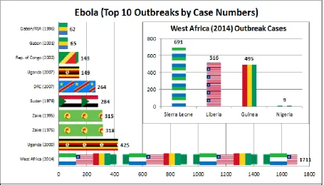
The Ebola species reported in 1976 in the Democratic Republic of the Congo near Ebola, a small river. Since then, outbreaks have appeared sporadically.[1] From 1976 (when it was first identified) through 2013, the WHO reported a total of 1,716 cases and since from the beginning the year, Ebola has killed nearly 3900 people in West Africa and can become world’s next AIDS.[2]
Ebola hemorrhagic fever remains a plague for the population of equatorial Africa, since 2000 with an increasing number of outbreaks. All human cases due to the emergence or re-emergence of Ebola species. Case fatality rate of African species in man are as high as 90%, as no prophylaxis or effective development of Ebola virus therapy is available. This infection is characterized by immune suppression and systemic inflammatory responses which causes impairment of vascular, coagulation and immune system that leads to multiorgan failure, resembling septic shock.[3]
Ebola hemorrhagic fever is caused by any of five genetically distinct members belonging to family Filoviridae: Zaire Ebola virus (EBOV); Sudan Ebola virus (SUDV); Tai forest Ebolavirus, (TAFV); Bundibugyo Ebolavirus (BDBV) and the fifth Reston Ebolavirus (RESTV). Reston Ebolavirus (RESTV) has only caused disease in nonhuman primates (NHP). Zaire, Sudan and Bundibugyo Ebolavirus are responsible for the most of the EHF outbreaks. EBOV constitutes serious threat in both human and NHP’s in sub-Saharian Africa.
Ebola hemorrhagic fever has been associated with large human outbreaks with case fatality rates as high as 90%. The case fatality rate of EBOV in NHP is unknown but some ecological data informs that it has contributed to declines of up to 98% of local great ape population in Gabon and the republic of Congo.[4]
Table 1: Species of genus Ebolavirus.
Species Virus Region Fatality rate Zaire Ebola virus
Sudan Ebola virus Bundibugyo Ebolavirus
Tai forest Ebolavirus Reston Ebolavirus
EBOV SUDV BDBV TAFV RESTV
Africa Africa Africa Africa Asia
60-90% 40-60%
25%, based on one outbreak
Unknown, only one known infection in Ivory Coast
Ganesh et al. World Journal of Pharmaceutical Research
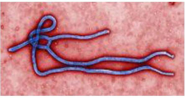
[image:3.595.149.447.145.304.2]Ebola virus constitutes the family Filoviridae in the order of mononegavirales. Ebola virus particles have a uniform diameter of 80 nm but can vary in length, with lengths up to 14000 nm.”Fig.1”
Figure 1: Ebola virus.
The genome consist of seven genes in order 3’ leader, nucleoprotein(NP), virion protein (VP)
35, VP40, glycoprotein, VP30, VP24, RNA- dependent RNA polymerase(L)-5’ trailer. In addition, EBOV expresses at least one nonstructural soluble GP (sGP) encoded by the GP
gene. The GP, with variable contributions from other viral proteins such as NP, appears to be the key immunogenic protein in vaccine protection.[5] With the exception of glycoprotein gene, all genes are monocistronic, encoding for one structural protein. An important distinction of Ebola virus from other mononegavirales is the production of a soluble glycoprotein, which is the primary product of the GP gene, and gets secreted to large quantities from infected cells.
Despite important achievements during the past two decades to unravel the molecular biology and pathogenesis of Ebola virus, we are still unclear about virulence factor and host responses, which seem to be partly detrimental to the host.[6]
Ebola, a small river in Democratic Republic of the Congo, the first case recognized on 26th august 1976. Mabalo Lokela, a 44 year old school teacher suffered from hemorrhagic fever,
The EVD emerged in the human population in the winter of 1976.[7]
Ganesh et al. World Journal of Pharmaceutical Research
unsafe injection and transfusion practices in postcolonial Africa. Today, EBOLA/EVD is the leading cause of death in sub-Saharan Africa and the fifth biggest killer in the world.[8]
The virus may be acquired upon contact with blood or bodily fluids of infected animals. Spreading through the air has not been documented in the natural environment. Fruit bats are considered to be a most likely reservoir of the EBOV, carrier and may spread the virus without being affected. Three types of fruit bats (Hysignathus monstrosus, Epomops franqueti and Myonycteris torquata) have been identified as being in contact with EBOV. Antibodies against Ebola Zaire and Reston viruses have been found in fruit bats in Bangladesh, thus identifying potential virus hosts and signs of the Filoviruses in Asia.[9] Once human infection occurs, male survivors may be able to transmit the disease via semen for nearly two months. To confirm the diagnosis, blood samples are tested for viral antibodies, viral RNA or virus itself.
Proposed plan of action
Any virus has two key features-infectivity which refers to the speed with which the virus can spread, and virulence which indicate how fatal the infection can. Ebola is dangerous because it is highly virulent (it has fatality rate up to 90% as per the WHO), but its infectivity is not very high. Ebola is not a respiratory disease or air born infection. Comparing the situation with other viral infection carriers did not show any symptoms (H1N1), on the other hand, any one infected by the Ebola virus becomes contagious only when they begin to show symptoms. This makes the process of detection much easier.[10]
Although progress has been made in understanding virus biology, no licensed vaccines or treatments currently exist, as there is a desperate need for a vaccine that not only prevents it’s transmission but also to control future incidences. Though inhalable Ebola vaccine passes
test on animals with long-lasting protection after single inhaled dose. A next stage of vaccine
research in human subjects entered in phase-I clinical trial.[11]
Hoped to have vaccine will be available by Nov, 2014. Because DNA vaccines, adenovirus-based vaccine, and VSIV-adenovirus-based vaccines have entered clinical trials. Healthcare professionals would have to be alert and recognize the symptoms early.[12]
Ganesh et al. World Journal of Pharmaceutical Research
vesicular stomatitis virus, Venezuela equine encephalitis virus replicons, recombinant parainfluenza virus type 3, replication-defective recombinant adenovirus (rAd), and DNA combined with rAd prime-boost strategie.While many of these approaches have been evaluated in a nonhuman primate model, only DNA/rAd-, rAd-, or vesicular stomatitis virus-based vaccines have shown efficacy in primates.[13]
[image:5.595.73.529.246.506.2]This article emphasizes on the current status of Ebola outbreaks, its vaccines and research development. ”Fig.2”
Figure 2: Ebola outbreaks.
[image:5.595.70.534.594.765.2]Plan of action
Table 2:-Laboratory tests used in diagnosis include
Timeline of Infection Diagnostic tests available Within a few days after symptoms begin
Later in disease course or after recovery
Retrospectively in deceased patients
1. Antigen-capture enzyme-linked immunosorbent assay (ELISA) testing 2. IgM ELISA
3. Polymerase chain reaction (PCR) 4. Virus isolation
5. IgM and IgG antibodies
6. Immunohistochemistry testing 7. PCR
Ganesh et al. World Journal of Pharmaceutical Research
[image:6.595.69.529.147.691.2]The rVSV platform has shown complete efficacy as a preventive single-shot vaccine in 3 relevant animal models, including some non-human primate models.
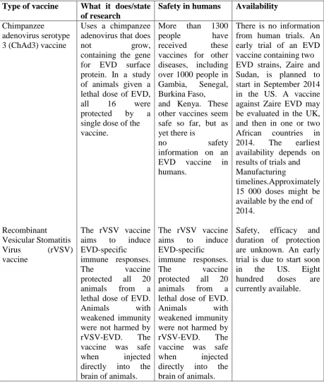
Table 2:-Overview of scientific information on unproven vaccines under development.
Type of vaccine What it does/state of research
Safety in humans Availability
Chimpanzee
adenovirus serotype 3 (ChAd3) vaccine
Recombinant
Vesicular Stomatitis Virus (rVSV) vaccine
Uses a chimpanzee adenovirus that does
not grow,
containing the gene for EVD surface protein. In a study of animals given a lethal dose of EVD, all 16 were protected by a single dose of the vaccine.
The rVSV vaccine aims to induce EVD-specific
immune responses. The vaccine protected all 20 animals from a lethal dose of EVD. Animals with weakened immunity were not harmed by rVSV-EVD. The vaccine was safe when injected directly into the brain of animals.
More than 1300 people have received these vaccines for other diseases, including over 1000 people in Gambia, Senegal, Burkina Faso, and Kenya. These other vaccines seem safe so far, but as yet there is
no safety
information on an EVD vaccine in humans.
The rVSV vaccine aims to induce EVD-specific immune responses. The vaccine protected all 20 animals from a lethal dose of EVD. Animals with weakened immunity were not harmed by rVSV-EVD. The vaccine was safe when injected directly into the brain of animals.
There is no information from human trials. An early trial of an EVD vaccine containing two EVD strains, Zaire and Sudan, is planned to start in September 2014 in the US. A vaccine against Zaire EVD may be evaluated in the UK, and then in one or two African countries in 2014. The earliest availability depends on results of trials and Manufacturing
timelines.Approximately 15 000 doses might be available by the end of 2014.
Safety, efficacy and duration of protection are unknown. An early trial is due to start soon in the US. Eight hundred doses are currently available.
Ganesh et al. World Journal of Pharmaceutical Research
The platform has shown partial to complete efficacy in post exposure treatment against homologous filovirus challenge. Knowledge gained from these data should advance the development of rVSV-based vaccines for human use.
Ganesh et al. World Journal of Pharmaceutical Research
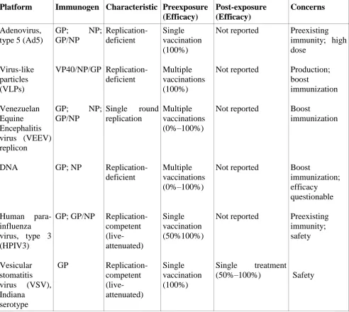
Table 3: Most promising vaccine platforms with efficacy in non-human primates.
Platform Immunogen Characteristic Preexposure (Efficacy)
Post-exposure (Efficacy)
Concerns
Adenovirus, type 5 (Ad5)
Virus-like particles (VLPs) Venezuelan Equine Encephalitis virus (VEEV) replicon
DNA
Human para-influenza virus, type 3 (HPIV3)
Vesicular stomatitis virus (VSV), Indiana
serotype
GP; NP; GP/NP
VP40/NP/GP
GP; NP; GP/NP GP; NP GP; GP/NP GP Replication-deficient Replication-deficient Single round replication Replication-deficient Replication-competent (live-attenuated) Replication-competent (live-attenuated) Single vaccination (100%) Multiple vaccinations (100%) Multiple vaccinations (0%–100%) Multiple vaccinations (0%–100%) Single vaccination (50%100%) Single vaccination (100%) Not reported Not reported Not reported Not reported Not reported
Single treatment (50%–100%)
Preexisting immunity; high dose Production; boost immunization Boost immunization Boost immunization; efficacy questionable Preexisting immunity; safety Safety
Note: GP- glycoprotein, NP- nucleoprotein, VP 40- virion protein 40 k Da.
Study population demographics
Ganesh et al. World Journal of Pharmaceutical Research
Figure 3: Kinetics and frequency of antibody responses.
(A) Percentages of responders following the third vaccination by the IP-Western assay for all subjects are shown. The y axis represents the percentage of responders with a positive assay, and the x axis represents the vaccine dose group. White bars, GP (Z); black bars, GP (S/G); gray bars, NP (Z). (B) Kinetics of the antibody response for all subjects is shown over the 52 weeks of the study. The geometric mean titer of the log10 reciprocal dilution and standard
deviation of the antibody response to GP (S/G) are plotted against the number of weeks after initial vaccination for each of the three dose levels. Vaccinations were given at 0, 4, and 8 weeks. The threshold for positivity in this assay was a reciprocal dilution of 30 and is shown as a dashed line. Of note, only six of eight subjects in the 8-mg dose group received all three vaccinations in the series, yet all vaccines are included in the immunogenicity analysis.
Vaccine safety
Due to a theoretical concern over GP-mediated cytopathicity, coagulation parameters of study subjects were closely monitored. At enrollment and throughout the study, D-dimer, prothrombin time, partial thromboplastin time, fibrinogen, complete blood count, and red blood cell smears were evaluated. There were no reportable coagulation laboratory abnormalities.[14]
Two subjects were withdrawn from the vaccination schedule due to serious adverse events that were assessed as “possibly” related to vaccination: a grade 4 creatine phosphokinase
Ganesh et al. World Journal of Pharmaceutical Research
eruption 3 weeks after the second vaccination, both in 8-mg recipients. Of note, the grade 4 creatine phosphokinase elevation was associated with vigorous exercise. These events resolved without sequelae, and these subjects continued to participate in the study and attended all study visits. Although only six of eight subjects in the 8-mg dose group received all three injections, the immunogenicity and safety laboratory values for all subjects are included in the analyses. One subject in the 2-mg dose group chose to withdraw after the second vaccination; another subject (in the placebo group) withdrew after the third injection. Neither of these subjects returned for further visits and, therefore, they were not included in
the immunogenicity analysis due to a lack of samples at time points following their withdrawal. As a result, 20 of 21 vaccines had immune responses assessed. All subjects are represented in the safety data through the time points available.
The diary cards showed that 90.5% (19/21) of subjects who received vaccine (at any dose level) experienced at least one local injection site symptom (mild to moderate pain/tenderness, mild induration, or mild skin discoloration) following a vaccination. The
systemic symptoms recorded on diary cards included malaise, myalgia, headache, nausea, and fever, as well as local injection site symptoms. The study vaccinations were well-tolerated and safe in healthy subjects, ages 18 to 44 years.
DISCUSSION – PUBLIC HEALTH IMPACT
The subsequent signs and symptoms includes systemic (prostration) , gastrointestinal (anorexia, nausea, vomiting, abdominal pain, diarrhea), respiratory (chest pain, shortness of
breath, cough, nasal discharge), vascular (congectival injection, postural hypotension, oedema), and neurological (headache, confusion and coma), manifestations.
Patients develop clinical signs during infections and die typically between day 6 and 16 with hypovolemic shock and multiorgan failure. Hemorrhages can be serious but are only present
in fewer than half of patient.
Ganesh et al. World Journal of Pharmaceutical Research
People who are living in or travelling to affected areas of Africa may be at risk of infection; however, this risk is extremely low unless there has been direct exposure to the bodily fluids of an infected person (including unprotected sexual contact with confirmed cases up to three months after they have recovered), or infected animal (alive or dead).
Caring for relatives with EVD is a known risk factor for infection, and healthcare workers, particularly those in resource poor settings with inadequate infection control are also at risk. During outbreaks of Ebola HF, the major prevalence of the virus includes healthcare workers, their family and friends. Medical professionals are also at high risk of infection.
CONCLUSION
The broad immunogenicity of this Ebola virus DNA vaccine suggests that immunization by plasmid DNA delivery is a viable platform and merits further development. Nonhuman primate studies have shown that a rAd5 vaccine effectively prevents disease, and DNA vaccination prior to boosting with rAd5 also confers protection and markedly increases the magnitude of the immune response. Therefore, future formulations of this DNA product will include multiple GP constructs encoding GP in either its wild-type form or a modified form to optimize vaccine potency. Because Ebola virus from Ivory Coast has been observed in only one limited outbreak and is closely related to Ebola virus Zaire, it is not included in vaccine formulations.
Since the prophylactic efficacy of an Ebola virus vaccine cannot feasibly or ethically be demonstrated in a human trial, the combination of safety and immunogenicity data from phase I, II, and III human trials and efficacy data from nonhuman primate studies will ultimately need to be utilized to obtain licensure of an Ebola virus vaccine under the Animal Rule.
The successful evaluation of a DNA vaccine to multiple Ebola virus subtypes reported here provides the opportunity for further clinical evaluation of candidate Ebola virus DNA vaccines alone or in combination with Ebola virus rAd vaccines as a heterologous prime-boost strategy.
Ganesh et al. World Journal of Pharmaceutical Research
public health challenges at the dawn of this year, so that Ebola could not become world’s next AIDS.
AKNOWLEDGEMENT
An enormous body of the work has been contributed to the knowledge of status of Ebola vaccine and Research Development. With that said, we acknowledge the research that has been carried out is mentioned in this review. This work has been carried out at Siddhant College of Pharmacy, Sudumbare, Pune.
REFERENCES
1. World Health Organization, Background document- Potential Ebola therapies and vaccines, September 2014; 1-16.
2. Ebola virus disease. wikipedia.org/wiki/Ebola_virus_disease.
3. Feldmann H, Geisbert TW. Ebola hemorrhagic fever. The Lancet, Elsevier Ltd., 2011; 377(9768): 849-862.
4. Margaret C. Ebola virus disease in West Africa- No Early end to the outbreak. World Health Organization, 2014; DOI: 10.1056/NEJMp 1409859.
5. Geisbert TW, Feldmann H. Recombinant vesicular stomatitis virus-based vaccines against Ebola and Marburg virus infections. The journal of infectious diseases, 2011; 204(3): 1075-1081.
6. Feldmann H, Geisbert TW, Jahrling PB, Klenk HD, Netesov SV, Peters CJ, Sanchez A,Swanepoel R, Volchkov VE. Family Filoviridae. In: Fauquet CM, Mayo MA, Maniloff
J,Desselberger U, Ball LA. Virus Taxonomy – Eighth Report of the International Committee on Taxonomy of Viruses. San Diego, US: Elsevier/Academic Press, 2005; 645–53. ISBN 0-12-370200-3.
7. Tyagi S, Kumar S. Clinical aspects of Ebola Hemorrhagic fever. International journal of pharma and Bioscience, 2010; 1(3): 1-9.
8. Girard MP, Osmanov SK, Kieny MP. A review of vaccine research and development: The human immunodeficiency virus (HIV). Vaccine, Elsevier, Science Direct, 2006; 24: 4062-4081.
9. Report on the EBOLA Virus disease epidemic in Liberia,
http://www.mohsw.gov.lr/…/SITRep%20222%20Dec%2023%202014%20.
Ganesh et al. World Journal of Pharmaceutical Research
11.World Health Organization, Ebola reston in pigs and humans, Philippines. Weekly Epidemiological records, 2009; 84(7): 49-50.
12.Olival KJ, Islam A, Yu M, Anthony SJ, Epstein JH, Khan SA, Khan SU, Crameri G, Wang LF, Lipkin WI, Luby SP, Daszak P. Ebola virus antibodies in fruit bats, Bangladesh. Emerging Infect. Dis. 2013; 19(2): 270–273. DOI:10.3201/eid1902.120524 13.Geisbert TW, Lewis MG. Vesicular stomatitis virus-based Ebola vaccine is well tolerated
and protects immunocompromised nonhuman primates. PLOS Pathog, 2008; DOI: 10.1371/journal.ppat.1000225.