REGULATION OF THE EXPRESSION OF BDNF AND
ITS RECEPTORS IN THE DEVELOPING NERVOUS
SYSTEM
Michelle Yvonne Robinson
A Thesis Submitted for the Degree of PhD
at the
University of St Andrews
1996
Full metadata for this item is available in
St Andrews Research Repository
at:
http://research-repository.st-andrews.ac.uk/
Please use this identifier to cite or link to this item:
http://hdl.handle.net/10023/14772
Regulation
of
the Expression
of
BDNF and its Receptors
In
the Developing Nervous System
Michelle Yvonne Robinson
Submitted
for
the Degree of Ph.D,
April, 1996
School of Biological and Medical Sciences
ProQuest Number: 10166380
All rights reserved
INFORMATION TO ALL USERS
The quality of this reproduction is dependent upon the quality of the copy submitted.
In the unlikely event that the author did not send a com plete manuscript and there are missing pages, these will be noted. Also, if material had to be removed,
a note will indicate the deletion.
uest
ProQuest 10166380
Published by ProQuest LLO (2017). Copyright of the Dissertation is held by the Author.
All rights reserved.
This work is protected against unauthorized copying under Title 17, United States C ode Microform Edition © ProQuest LLO.
ProQuest LLO.
789 East Eisenhower Parkway P.Q. Box 1346
Contents
Acknowledgements
7
Declarations
8
Copyright
9
List of Figures
10
Abbreviations
13
Abstract
16
CHAPTER 1
17
General Introduction
Early Neural Development 17 The Neurotrophic Theory 18
The Structurally-Related Family of Neurotrophic Factors 20 Nerve Growth Factor (NGF) 21
Brain-Derived Neurotrophic Factor (BDNF) 23 Neurotrophin-3 (NT-3) 23
Neurotrophin-4/5 (NT-4/5) 24 Neurotrophin-6 (NT-6) 24
Neurotrophic Factor Expression 25 Neurotrophic Factor Receptors 26 The Low-Affinity p75 Receptor 26
trk Tyrosine Kinase Receptors 28
Developmental Expression of trk Receptors 30
Signal Transduction by the trk Receptor 34
Neurotrophic Factors and Nervous System Development 36 Proliferation and Differentiation of Neuronal Precursors 37
Eaiiy Neuronal Maturation 38
The Onset of Neurotrophic Factor Responsiveness 39
Neurotrophic Factor Requirements During Eaily Stages of Target Innervation 41
Neurotrophic Factor Requirements During Neuronal Death 43 Neurotrophic Factors Not Related to NGF 49
Ciliaiy Neurotiophic Factor (CNTF) 50 Fibroblast Growth Factor (FGF) 51
Glial Cell Line-Derived Neurotrophic Factor (GDNF) 52 Aims of Project 52
CHAPTER 2
54
Materials and Methods
Molecular Biology Techniques
54
Subcloning of cDNA Fragments 54 Plasmid Vectors 54
Digestion of DNA by Restriction Endonucleases 55 Purification of Digested Insert and Vector Fragments 56 Ligation of DNA Fragment and Vector 57
Bacterial Host Strain 58
Preparation of Competent Host Cells and Transformation 59 Small-Scale Isolation of Plasmid DNA by Alkaline Lysis 61
Isolation of Plasmid DNA with Polyethylene Glycol Purification 63 Agarose Gel Electrophoresis 65
Sequencing of Double-Stranded DNA 66
Reverse Transcription-Poîymerase Chain Reaction Assay 71 Precautions Against Contamination of PCRs 73
Precautions in Handling RNA 74 PCR Primer Choice 76
Practical Aspects of the Assay 77
Prepaiation of RT-PCR Contiol Templates 79 Extraction of Total RNA 83
Preparation of Labelled Primers 84 Reverse Transcription 85
PCR 86
Electrophoresis of PCR Products 89
Dissection and Ceil Culture Techniques
90
Dissection Techniques 90
Sympathetic Ganglia and Dorsal Root Ganglia (DRG) 91 Cranial Sensoiy Ganglia 92
Ciliary Ganglion 94 Tissue Dissociation 94
Separation of Neurons from Non-Neuronal Ceils 95 Cell Culture 96
CHAPTER 3 99
In Vivo p75 Expression in Sensory, Sympathetic, and Parasympathetic Neurons and In Viti'o Regulation of p75 Expression in
Sympathetic and Dorsal Root Ganglion Neurons Introduction 99
M ethods 102
In Vivo p75 Expression in Sensory, Sympathetic, and Parasympathetic Neurons 102
Ganglion Neurons 102 R esults 103
Specificity and Reliability of Quantitation of p75 mRNA by RT-PCR 103
In Vivo p75 Expression in Sensory, Sympathetic, and Parasympathetic Neurons 105
In Vitro Regulation of p75 Expression in Sympathetic and DRG Neurons 107
D iscussion 111 C onclusion 118
CHAPTER 4
119
Synthesis of BDNF by NGF-Dependent Embiyonic Sensory Neurons In tro d u ctio n 119
M ethods 121
Isolation and Purification of Neurons 121 Measurement of BDNF in Isolated Neurons 121 Neuronal Cultures 122
R esults 123
BDNF mRNA Expression by Different Kinds of Sensoiy Neurons 123 BDNF Synthesis by NGF-Dependent Sensory Neurons 126
D iscussion 129 C onclusion 131
CHAPTER 5
133
Timing and Regulation of trkB and BDNF mRNA Expression in Placode-Derived Sensory Neurons and Their Targets In tro d u ctio n 133
Embiyonic Tissues 136 Neuronal Cultures 136
Measurement of Specific mRNA Transcripts 137 R esults 137
Expression of trkB mRNA in Developing Nodose and Vestibular Ganglia 137
Expression of p75 mRNA in Developing Nodose and Vestibulai* Ganglia 137
Expression of BDNF mRNA in the Peripheral and Central Taigets of Vestibulai* and Nodose Ganglia 140
Regulation of trkB mRNA in Cultured Nodose Neurons 143 D iscussion 143
Acknowledgements
Declarations
I, Michelle Yvonne Robinson, hereby notify that this thesis, which is approximately 50,000 words in length, has been written by me, that it is the record of work carried out by me and that it has not been submitted in any previous application fo a higher degree.
Date lo/if-j Signature of candidate
I was admitted as a research student at St. George’s Hospital Medical School in June, 1993, and as a candidate for the degree of Ph.D. at the University of St. Andrews in November, 1993; the higher study for which this is a record was carried out in St. George’s Hospital Medical School and the University of St. Andrews between 1991 and 1996,
Date I o / / c) 4 Signature of candidate
I hereby notify that the candidate has fulfilled the conditions of the Resolution and Regulations appropriate for the degree of Ph.D. in the University of St. Andrews and that the candidate is qualified to submit this thesis in application for that degree.
In submitting this thesis to the University of St. Andrews I understand that I am giving peiTnission for it to be made available for use in accordance with the regulations of the University Libraiy for the time being in force, subject to any copyright vested in the work not being affected thereby. I also understand that the title and abstract will be published, and that a copy of the work may be made and supplied to any hona fide
library or research worker.
Figure 1, page 93
(A) Camera lucida drawing of the ventral aspect of the lumbosacral region of an ElO chick embryo after evisceration showing the location of the dorsal root ganglia (DRG). (B) Schematic illustration of an ElO chick embryo showing the locations of tlie cranial sensory ganglia.
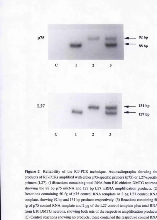
Figure 2, page 104
Reliability of the RT-PCR technique. Autoradiographs showing the products of RT- PCRs amplified with either p75- or L27-specific primers.
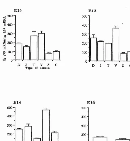
Figure 3, page 106
Bar charts showing p75 mRNA expression relative to L27 mRNA in purified preparations of chicken sensory, sympathetic, and parasympathetic neurons at ElO, E12, E14, and E16.
Figure 4, page
Phase contrast photomicrograph of E7 chicken sympathetic neurons suiwiving without neurotiophic factors on a polyomithine-laminin substratum.
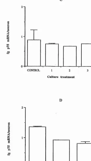
Figure 5, pages 109-110
Regulation of in vitro p75 mRNA levels in E7 chicken sympathetic neurons. Graphs show (A) effect of NGF, CNTF, and SCF; (B) effect of RA; (C) effect of TGF-pl, -p2, and -p3; and (D) effect of depolarization by KCl and of depolarization in the presence of CNTF.
Figure 6, page 112
combinations.
Figure 7, page 124
Autoradiographs showing the products of RT-PCRs amplified with BDNF-specific primers.
Figure 8, page 125
Bar charts showing BDNF mRNA levels relative to L27 mRNA in purified DMTG, jugular, TMN, and VLTG neurons from ElO, E12, and E14 chicken embiyos.
Figure 9, page 127
Bar chart showing percent survival of ElO DMTG, jugulai*, TMN, and VLTG neurons after 48 hours in control cultures and cultures supplemented with 5 ng/ml NGF and 5 ng/ml BDNF.
Figure 10, page 128
Photomicrographs of co-cultures of TMN and DMTG neurons. (A) Phase contrast view showing all neurons and their processes. (B) The same field as in (A) viewed by rhodamine epifluorescence showing Dil-labelled TMN neuron.
Figure 11, page 130
Bar chai't showing percent survival of ElO TMN neurons cultured alone or co-cultured with a ten-fold greater number of ElO DMTG neurons.
Figure 12, page 138
Figure 13, page 139
Graph of the level of full-length trkB mRNA relative to L27 mRNA (fg mRNA per pg L27 mRNA) in the vestibular and nodose ganglia of chicken embryos at stages
17 to 24.
Figure 14, page 141
Graph of the level of p75 mRNA relative to L27 mRNA (fg p75 mRNA per pg L27 mRNA) in the vestibulai* and nodose ganglia of chicken embryos at stages 17 to 24 (stage 20 was the earliest stage at which nodose ganglia were studied).
Figure 15, page 142
Graph of the level of BDNF mRNA relative to L27 mRNA (fg BDNF mRNA per ng L27 mRNA) in the heart, hindbrain, and otic vesicle of chicken embiyos at stages 16 to 24.
Figure 16, page 144
Abbreviations
a/b FGF Acidic/basic fibroblast growth factor BDNF Brain-derived neurotrophic factor ChAT Choline acetyl transferase
CIAP Calf intestinal alkaline phosphatase
CMF-PBS Calcium- and magnesium-free phosphate-buffered isotonic saline CNTF Ciliary neurotrophic factor
ddNTP Dideoxynucleotide triphosphate DEPC Diethylpyrocarbonate
Dil r , r-Dioctadecyl-3, 3,3', 3-tetramethylindocarbocyanine perchlorate DG Diacylglycerol
DMSO Dimethyl sulphoxide
DMTG Dorsomedial trigeminal ganglion DNase 1 Deoxyribonuclease 1
dNTP Deoxynucleotide triphosphate DRG Dorsal root ganglion
DTT Dithiothreitol E Embryonic day
EDTA Ethylenediaminetetraacetic acid
Erk 1,2 Extracellular signal-regulated kinase 1,2 FGF(-S) Fibroblast growth factor(-5)
G ABA y-aminobutyric acid
GAP Ras GTP-ase activating protein
GDNF Glial cell line-derived neurotrophic factor HIFCS Heat-inactivated foetal calf serum HIHS Heat-inactivated horse serum IMS Industrial methylated spirits IL-6 Interleukin-6
BPTG Isopropyl-p-D-thiogalactopyranoside L broth/agar Luiia broth/agar
LIF Leukaemia inhibitoiy factor
MAPK Mitogen-activated protein Idnase
MAPKK Mitogen-activated protein kinase kinase MOPS 4-Moipholinepropanesuifonic acid
NGF Nerve growth factor NPY Neuropeptide Y
NT-3, -4/5, -6 Neurotrophic factor-3, -4/5, -6
OD Optical density PEG Polyethylene glycol
PI-3 kinase Phosphatidylinositol-3 kinase PLC-yl Phospholipase C-yl
P-ORN Poly-DL-ornithine RA Retinoicacid
RNase Ribonuclease
RT-PCR Reverse transcription-polymerase chain reaction SCF Stem cell factor
SDS Sodium dodecyl sulphate SH2 Src homology 2
SNT 5MC-associated neurotrophic-factor-induced tyrosinephosphorylated target SOS Son of sevenless
SCG Superior cervical sympathetic ganglion TAE buffer Tris-acetate-EDTA buffer TBE buffer Tris-boric acid-EDTA buffer
TGF-p (1, 2, 3) Transforming growth factor-p(l, 2, 3) TEMED N, N, N', N'-Tetramethylethylenediamine TH Tyrosine hydroxylase
VLTG Ventrolateral trigeminal
VRC Vanadyl-ribonucleoside complex
BDNF binds to two ti’ansmembrane receptors: trkB, which is a tyrosine kinase essential for signalling, and p75, which is a common nenrotiophin receptor whose role is contoversial. To determine the relationship between BDNF synthesis, BDNF receptor expression, and neuronal responsiveness, the expression of BDNF, trkB, and p75 mRNAs were studied for different populations of sensory neurons whose axons reach their targets and become dependent on BDNF for suiwival at different times. BDNF mRNA was expressed in the peripheral and central tai'gets of these neurons prior to the arrival of sensory axons. The onset of BDNF responsiveness was preceded by the expression of first p75 mRNA then trkB mRNA, and neurons that start responding to BDNF early were the first to express trkB mRNA. BDNF upregulated
trkB mRNA expression just shortly before the onset of BDNF dependence.
BDNF is expressed not only in sensory neuron targets but in some of these neurons themselves. To determine whether BDNF is synthesized in NGF-dependent or BDNF-dependent neurons, BDNF mRNA expression was studied in purified populations of cranial sensory neurons that depend on either NGF or BDNF for survival. During the period of neuronal death, BDNF mRNA expression was highest in NGF-dependent cutaneous sensory neurons, lower in BDNF-dependent cutaneous sensory neurons, and undetectable in BDNF-dependent proprioceptive neurons. In co culture, NGF-dependent neurons promoted the survival of BDNF-dependent neurons by the production and release of BDNF, implying a paracrine role for BDNF duimg the period of naturally occurring neuronal death.
CHAPTER 1
General Introduction
Early Neural Development
The following is a brief account of the events occurring in early neural development. These are described more fully in a review by Schoenwolf and Smith (1990). The onset of neural development is marked by the formation of a dorsally- positioned groove along the ectodermal surface of the gastrula. The neural groove then widens, accompanied by the thickening of the ectoderm lateral to it which forms the neural plate on the dorsal side of the embryo. The margin of the neural plate bears ridges called neural folds which start fusing near the anterior end to foim the neural tube. Most of the cells from which the brain and spinal cord are formed come from the neural tube. The beginning of brain development is maiked by a slight anterior swelling of the neural tube.
During neural tube closure, a group of cells, the neural crest, detach from the margin of the neural ectoderm and foim the peripheral nervous system. They come to lie in a band along the length of the neural plate and finally position themselves dorsally and laterally to the closing neural tube. The cells then undergo a period of migration into the periphery followed by differentiation into spinal and autonomic ganglia, glial cells of the autonomic and sensory neivous system, and other non-neuronal tissues such as melanocytes, chromaffin cells of the adrenal medulla, cartilage, blood-foiming cells, connective tissue covering the brain and spinal cord, and pai'ts of the facial bones.
The Neurotrophic Theory
During development, neurons extend axons towards their target fields to establish contacts with tai'get cells and make appropriate synaptic connections. Since more neurons aie generated than required, a mechanism must exist for controlling the number that finally malce successful synaptic connections in the target field. This process is marked by a distinct period of neuronal cell death, which commences shortly after the first neurons innervate their targets (Cowan et al, 1984). It has been assumed that the target field itself controls the size of neuronal populations that innervate it by synthesizing a limiting supply of a neurotrophic factor on which the neurons are dependent for survival. This has been embodied in the neurotrophic theory (Davies, 1988a; Barde, 1989; Oppenheim, 1991; Thoenen, 1991).
Most of the studies supporting the theory have been on neiwe growth factor (NGF), the first neurotrophic molecule to be identified and completely chai'acterized. It was shown that NGF is produced by cells in the target field of neurons that require it and that its availability regulates neuronal survival. NGF is bound by the neuron and the NGF-receptor complex internalized and transported to the cell soma (Levi- Montalcini, 1987). It has also been demonstrated that NGF is produced in the target field in proportion to the density of innervating neurons (Heumann et al, 1994; Shelton and Reichai'dt, 1984), especially duiing development (Harper and Davies, 1990).
An alternative proposal to limited production of neurotrophic factor is one of limited access to it. Enough neurotrophic factor would be produced to support all the developing neurons and whether or not a neuron survives would be governed by its ability to form sufficient neuronal branches and synaptic sites through which to obtain the factor (Oppenheim, 1989). Studies in support of this theory make use of hyper- innervated embryonic chicken muscle produced by phannacological paralysis. It has been demonstrated that extract from this muscle promotes motoneuron survival with a similar efficiency to that from noimal embryos both in vitro (Tanaka, 1987) and in vivo
However, further studies on the nervous system and other NGF-related neurotrophic molecules have shown that the control of target inneiwation is not so simplistic. The members of the NGF family of neurotrophic factors (or neurotrophins) are brain-derived neurotrophic factor (BDNF), neurotrophin-3, -4/5, and -6 (NT-3, -4/5, and -6). Both NGF, its related neurotrophic molecules, and their receptors will be discussed in more detail in later sections of this Chapter.
Recent studies have exposed the limitations of the neurotrophic theory. Apart from obtaining neurotrophic support from innervated target cells (the classical retrograde mechanism) (Levi-Montalcini, 1987; Thoenen et ai, 1987), neurons might also obtain it from other neurons which are afferent (an anterograde mechanism). Oppenheim (1991) has described how a number of neuronal types degenerate when afferent input is lesioned, providing strong evidence for an anterograde mechanism. Neurotransmitter molecules, which are transported anterogradely, may also influence neuronal survival. Lipton and Kater (1989) have documented the effects on neuronal survival and differentiation of a variety of neurotransmitters.
A further source of neurotrophic support may come from cells ensheathing neiwes. Heumann eta l (1987), have demonstrated that a high rate of NGF synthesis is maintained by peripheral nerve-ensheathing cells duiing development which decreases in adulthood. It also increases briefly when the nerve is lesioned. This suggests that the nerve cells receive a local supply of NGF prior to target innervation during development or before re-contacting target organs during regeneration.
Some types of neuron have been found to synthesize both neurotrophic factors and their respective receptors and so respond to their own neurotrophic factors in an autocrine mechanism. Dorsal root ganglion (DRG) neurons, sympathetic neurons, and hippocampal pyramidal neurons synthesize both BDNF and its receptor, trkB (Ernfors
other neurons within a population, possibly via a paracrine mechanism of neuronal support. Autocrine or paracrine support mechanisms would obviously provide a function distinct from that of neuron-target interaction. They may play a role in maintaining neurons prior to tai'get field innervation (Korsching, 1993).
The Structurally-Related Family of Neurotrophic Factors
The work of Hamburger and his colleagues provided evidence that neurons require more than nutrients in order to survive. Theii* experiments involved the removal of a wing or limb bud in the chick embryo prior to innervation, with resultant death of the innervating sensory, autonomic, and motor neurons. These results suggested that the peripheral tissues might produce certain substances essential for the maintenance of neurons (review by Levi-Montalcini, 1987). Experiments directed at understanding the nature of this peripheral influence led to the discoveiy of the first neurotrophic factor, NGF, in the early 1950's, Levi-Montalcini and Hamburger (1951,1953) obseiwed that transplantation of a mouse sarcoma into chick embryos after limb bud removal resulted in the enlargement of sympathetic and dorsal root ganglia and hyper-innervation of the tumour. Studies of the substance released by the sarcoma were initially earned out using neuiite outgrowth assay techniques for chick sympathetic and dorsal root ganglia (Levi-Montalcini etal., 1954) and ultimately led to the purification of NGF.
Nerve Growth Factor (NGF)
As the first neurotrophic factor to be isolated, NGF remains the best characterized with respect to structure and function. It promotes the survival of sympathetic neurons (Chun and Patterson, 1977; Greene, 1977), certain types of sensoiy neurons (Davies and Lindsay, 1985), and basal forebrain cholinergic neurons (Hatanakaefa/., 1988).
The NGF protein was purified from the submandibular gland of the male mouse (Angeletti and Bradshaw, 1971; Angeletti et al, 1973), where it is found in great quantities. Why NGF is located here in such quantity (approximately 0.1% of total protein; Cohen et al, 1960) is uncertain. Aloe eta l (1986) suggest that it might be important in the aggressive behaviour of male mice as the salivary glands release large quantities of NGF into the bloodstream during fighting. This, however, is not found in other aggressive animals.
Significant levels of NGF are also found in other exocrine tissues and their secretions. These include snake venom (Bailey et al 1975), guinea pig prostate (Chapman et al, 1981), bull semen and seminal vesicles (Hoffman and Unsicker, 1982), and the submandibular gland of the African rat Mastomys natalensis (Aloe et a l, 1981). No explanation has been found for the presence of NGF in these regions; there appeal's to be no influence on the developing or mature nervous system (Thoenen and Barde, 1980; Haiper and Thoenen, 1981). Most studies have been carried out using mouse NGF because the submandibular gland of male mice is such a rich source of the factor.
Protein Structure
each containing three intrachain disulphide bonds (Bradshaw et al, 1993). Loss of these bonds results in loss of biological activity (Greene and Shooter, 1980; Thoenen and Barde, 1980).
X-ray diffraction studies have recently been used to determine the three- dimensional structure of the mature molecule (McDonald et al, 1991). Dimer foimation occurs through hydrophobic contacts between the flat surfaces of each monomer. The hydrophobic core of each monomer bears the majority of amino acids common to aU the neurotrophic factors, including the disulphide bonds. Several amino acids that are conserved in the neurotiophic factors contiibute to NGF structure by forming hydiogen bonds (review by Ebendal, 1992). A group of positively-chaiged amino acids has been shown to be important in the interaction of NGF with the low-affinity receptor (Ibdnez
et a l, 1992). Of the regions that differ between the neurotrophic factors, one forms three closely-associated p-hairpin loops in the NGF protein. It is possible that these regions are responsible for the different spécificités each factor has for its high-affinity receptor (review by Ebendal, 1992).
Gene Structure
The mouse NGF gene is 45 kb in length and consists of five exons separated by four introns. The mature protein is translated from the transcript of a single 3' exon. The remaining 5' exons are smaller and undergo alternative splicing to produce at least four transcripts that encode precursor proteins that differ in their amino termini (Selby
while the other transcript is predominant in all other tissues.
Brain-Derived Neurotrophic Factor (BDNF)
BDNF was the next neurotrophic factor to be characterized. It promotes the survival of sensory neurons that are unresponsive to NGF (Davies et al, 1986a; Lindsay e ta l, 1985), the dopaminergic neurons of the substantia nigra (Hyman et al,
1991), and developing motoneurons under certain expeiimental conditions (Oppenheim
e ta l, 1992).
Molecular Structure
BDNF was first isolated from pig brain (Baide et al, 1982). It was found to be a 12.3-kDa basic protein. Molecular cloning (Leibrock et al, 1989) has shown that the BDNF protein has approximately 50% amino acid identity with NGF.
BDNF gene sequence information has been determined for a number of other species, including human and rat (Maisonpiene etal, 1991) and chicken (Maisonpierre
et a l, 1992). The rat BDNF gene has been shown to span over 40 kb of genomic DNA and consists of four short 5' exons and one 3' exon encoding the mature BDNF protein (Timmusk et a l, 1993). Eight different BDNF mRNAs with four 5' ends and two alternative polyadenylation sites are transcribed from this gene. BDNF mRNAs containing the first three 5’ exons are expressed predominantly in the brain, whereas transcripts containing the fourth exon aie found predominantly in the lung and heart
N eurotrophin-3 (NT-3)
NT-3 was the third member of the NGF gene family to be identified and, in contrast to NGF and BDNF, this did not require prior purification of the NT-3 protein. Primaiily, the research groups involved made use of the polymerase chain reaction (PCR) technique and the nucleotide sequence homology between NGF and BDNF (Hohn et al, 1990; Maisonpierre era/.,1990; Rosenthal et al, 1990).
enteroceptive sensory neurons (Hohn et ai, 1990), and embiyonic motoneurons in vitro (Henderson etaLy 1993).
Neurotrophiiî-4/S (NT-4/5)
Hallbdok et al. (1991) took advantage of the regions of conservation between NGF, BDNF, and NT-3 in a PCR-based search for further members of the NGF gene family. As a msult, NT-4 was identified and sequenced for the clawed toad Xenopus.
Mammalian versions of NT-4 have since been isolated from the rat and human genomes
(Ip et a l y 1992). Ip et al (1992) have also isolated a pseudo-NT-4 gene containing
several frame shifts, an internal stop codon, only four of the six essential cysteine residues, and no cleavage site to process the mature neuroti'ophic factor. The biological activity of recombinant Xenopus NT-4 is similar to that of BDNF in that it promotes the
in vitro survival of sensory neurons (Hallbook et a l y 1991). NT-4 has also been shown to support the survival of embryonic rat motoneurons in vitt'o (Henderson et al y
1993).
Berkemeier et a l (1991) also used a PCR strategy to isolate DNA fragments encoding a protein they refeired to as neurotiophin-5 (NT-5). This protein has the same sequence as that described for human NT-4 by Ip et al (1992). Berkemeier et al
(1991) claim that NT-5 is a distinct neurotrophic factor from NT-4 as it is expressed at low levels in a few peripheral organs and can stimulate sympathetic neurons when in the form of a recombinant protein; properties not exhibited by Xenopus NT-4. However, because the sequences of NT-5 and human NT-4 are the same, mammalian NT-4 is referred to as NT-4/5.
N euFotrophm -6 (NT-6)
NT-6 was cloned from a genomic libraiy of the teleost fish Xiphorus maculatis
survival of a similar range of neurons to NGF.
Neurotrophic Factor Expression
in the Central Nervous System
NGF, BDNF, and NT-3 appear to be synthesized primarily in specific neurons in the brain and, therefore, target-derived influences are achieved through interactions between neurons (Ayer-LeLievre et al.y 1988; Gall and Isackson, 1989; Ernfors et al.y
1990a,b; Philips et al. y 1990; Isackson et al.y 1991). Several studies have demonstrated the changes in NGF mRNA and protein levels in valions regions of the rat brain during embryonic and early postnatal development (Laige et a l y 1986; Lu et a l y 1989; Mobley
et a l y 1989). NGF mRNA and protein levels aie initially low and uniform throughout
the brain and do not reach a maximum until postnatal day 21 in the neocortex and hippocampus. During development, BDNF is expressed initially at low levels but increases to become the most widespread neurotrophic factor in different areas of the brain. It reaches its peak level of expression two weeks after birth. NT-3 mRNA is weakly expressed in the hippocampus and cerebellum and reaches its maximum level of expression shortly after birth. NGF, BDNF, NT-3, and NT-4 are expressed in the adult brain (Persson and Ernfors, 1992). NT-6 expression has been detected in a small region of teleost cerebellum beneath the midbrain tectum during organogenesis, in 8- day old fish, and in the adult (Gotz et al.y 1994).
in Peripheral Tissues
proprioceptive neurons projecting to skeletal muscle and the somatosensory fibres of the nodose ganglion (Thoenen, 1991). NT-6 expression has been detected in adult teleost gill, liver, and eye with weak expression in skin, spleen, heait, and skeletal muscle (Gotz et aly 1994).
Neurotrophic Factor Receptors
The use of highly purified 125% NGF in binding and cross-linking studies has facilitated the characterization of NGF receptors in the late 1970's (Sutter e t a l y 1979;
Schechter and Both well, 1981). It is now well established that members of the NGF family of neurotrophic factors recognize two types of receptor, distinguishable by their pharmacological properties (review by Meakin and Shooter, 1992). One type of receptor binds all the factors with low affinity and has a dissociation constant of lO'^M. It is a ceU surface protein with a moleculai* weight of 75 kDa in the human, although the rat protein is slightly larger, and is refened to as p75 for all species. The p75- neurotrophic factor moiety is not internalized. The precise role of p75 in mediating the biological effects of neurotrophic factors is not certain but it may form part of a complex or interact in some other way with the second class of receptors. These are products of the trk oncogenes, a family of related tyrosine kinase-bearing receptors. They show specific high affinity binding for the neurotrophic factors (the dissociation constants being in the low picomolar range for NGF and NT-3 but undeteimined for BDNF) and aie capable of internalizing bound factor. Neurotrophic factor binding induces autophosphorylation of an intracellular tt'k tyrosine Idnase residue.
The Low-Affinity p7S Receptor
workers (1989). p75 mRNA size in chicken is 4.5 kb; in human, 3.8 kb; and in rat, 3.6 kb. The molecules differ at their 3' noncoding regions which accounts for the size differences.
The human p75 gene encodes a glycoprotein consisting of a single transmembrane polypeptide of 427 amino acid residues, 399 of which make up the mature protein. Rat p75 cDNA codes for a protein of 425 amino acids, including a 29 amino acid signal peptide. The extracellular domain comprises 222 amino acids, the transmembrane domain 22 amino acids, and the intracellular domain 152 amino acids. The amino acid sequences for human and rat p75 are almost identical but the chicken sequence has more variation. In all three species, the extracellular domain bears four conserved cysteine repeats. It has been demonstrated that these are responsible for ligand recognition since a secreted 168 amino acid receptor without all residues carboxy terminal to this domain still binds NGF (Welcher etal, 1991; Yan eta l, 1991). There is also considerable homology between the three species within the transmembrane domain and in the C-terminus of the intracellular domain. The role of the intracellular domain remains to be fully elucidated. However, it has been shown that deletions in this region interfere with the ability of p75 to form functional high affinity NGF receptors (Hempstead et aly 1989).
A soluble, truncated form of p75 has been isolated from conditioned media of p75-producing cells and in various biological fluids (Distefano and Johnson, 1988). This comprises the extracellular domain only and is thought to be produced by post- translational processing as opposed to differential splicing. The biological significance of the truncated receptor remains unclear, but it binds NGF, BDNF, and NT-3 with equal affinity to the full-length transmembrane receptor. Truncated p75 may limit the activity of neurotrophic factors by competing for factors with the full-length receptor or act as a neurotrophic factor transporter (review by Barker and Murphy, 1992).
trk Tyrosine Kinase Receptors
malignantly active in a colon carcinoma patient (review by Barbacid et a l y 1991).
Martin-Zanca et al (1989) showed that this proto-oncogene encodes a 140 kD tyrosine kinase (designated p i40^^^^) which exhibits the structural characteristics of a growth factor receptor (review by Chao, 1992). In situ hybridization studies using mouse embryos showed that the trkA gene is highly expressed in NGF-dependent neurons, for example, sensory neurons of spinal ganglia and a proportion of neurons from neural crest-derived cranial sensory ganglia (Martin-Zanca et al, 1990). Further studies established that pMO^'"^^ is a component of the high affinity NGF receptor. NGF stimulates tyrosine autophosphorylation of p i40^^^^ in PC 12 cells, in spinal ganglia sensory neurons, and in NIH 3T3 cells transfected with trkA cDNA (Kaplan et al,
1991a,b).
The observation that genes encoding trk tyrosine kinase receptors for BDNF and NT-3 should be structurally similar to that for pl40^^^^ led to the isolation of cDNA clones for trkB and trkC by low stringency hybridization with trkA probes. The intracellular tyrosine kinase domains of trkA, trkB, and trkC share 85% sequence homology. trkB and trkC encode the tyrosine kinase receptors pl45^^^^ and pl45^^^ respectively, both of molecular weight 145 kD. pl45^''^® has been shown to be the signalling receptor for BDNF (Klein et al, 1991a; Glass et al, 1991; Soppet et al,
1991; Squinto etal, 1991) and NT-4 (Berkemeier a/., 1991; Ip eta l, 1992; Klein et aly 1992). pl45^^*^ has been shown to be the receptor for NT-3 (Lamballe et al,
1991). There appears to be some cross-talk between NT-3 and pl40^^^^ and pl45^^^®. NT-3 induces in vitro tyrosine phosphorylation of these receptors with similar efficiency to their cognate ligands NGF and BDNF (or NT-4) respectively (Klein et al,
1991b; Soppet et al, 1991; Squinto et a l, 1991). Glass et a l (1991) have demonstrated that NT-3 induces tyrosine phoshorylation of these receptors much less efficiently in NIH 3T3 cells. Ip et a l (1993b) have shown that NT-3 exerts its neurotrophic effects on PC 12 cells and cultured neuronal cells bearing p l45^^*C receptors.
rat (Meakin et al, 1992) and the other in the human (Martin-Zanca et al, 1989). The isoforms differ in that the rat molecule codes for an additional 6 amino acids in the extracellular domain. They are produced by alternative splicing and both appear to generate functional receptors. However, the one coding for the additional 6 amino acids is expressed predominantly in neuronal tissues while the other is expressed mainly in non-neuronal tissues (Barker et al, 1993). The pl40^^*^ receptor comprises a glycosylated peptide chain with a single transmembrane domain (review by Meakin and Shooter, 1992). It is larger than p75, containing approximately 790 amino acid residues,
Middlemas and co-workers (1991) have shown that the rat trkB gene expresses multiple transcripts. Two of these are predicted to encode the full-length receptor and are 9.0 and 4.8 kb in size. There are a number of C-terminally truncated transcripts of 7.5, 7.0, 2.4, 0.9, and 0.7 kb in length. These possess the extracellular domain but lack the tyrosine kinase domain. A 1.6 kb mRNA was also isolated that comprises the tyrosine kinase domain only. These forms are possibly generated through alternative splicing. Northern blot analysis has shown that at least 5 rr^B transcripts are present in the chick (Déchant et al, 1993a). These are estimated to be 9.0, 8.1, 6.3, 5.0, and 1.6 kb in length. The 9.0 kb transcript is the only one possessing a tyrosine kinase domain and has only been detected in neuronal structures. The shorter transcripts lacking the tyrosine kinase domain are expressed predominantly in non-neuronal tissues.
only the ti'uncated ones are detected in astrocytes, peripheral nerves, and non-neuronal tissues. Those containing the amino acid inserts are still capable of autophosphorylation in response to NT-3 but cannot mediate proliferation in fibroblasts or neuronal differentiation in PC 12 cells.
Developmental Expression of trk Receptors
trkA
In mouse, trkA is initially expressed during early neurogenesis at embryonic day (E) 9.5 (Martin-Zanca et al., 1990; Wyatt and Davies, 1993). By E13.5, its expression is confined to certain structures of the nervous system. These include the DRG and cranial sensory ganglia (for example, the trigeminal, superior, and jugular ganglia). In older embryos (E17.5), trkA expression is also detectable in sympathetic ganglia (Maitin-Zanca et al., 1990) and in defined structures of the central nervous system, for example, the cholinergic neurons of the basal forebrain and the striatum (Steininger 1993).
trkB
Mouse trkB expression is first detected in both the central and peripheral nervous system at E8.5. It is expressed in many structures, for example, the forebrain, caudal midbrain, hindbrain, spinal cord, trigeminal ganglion, and differentiating neural crest cells which form the dorsal root ganglia (Klein et al., 1990b). trkB expression is found to be widespread in the nervous system thioughout embryonic development.
In adult mouse, the highest expression levels are found in the brain and spinal cord (Klein et al., 1990b). Expression is also detected in most structures of the cerebrum including the cortical layers, thalamus, and hippocampus. Non-neuronal tissues express trkB, for example, tongue, whisker pad mesenchyme, and outer dermal papilla (Klein eta l, 1990a).
In situ hybridization studies have shown that the truncated transcripts are located predominantly in the choroid plexus and ventricles. In general, the full-length receptor is more abundant in non-neuronal cells, for example, astrocytes, oligodendrocytes, and Schwann cells (Frisen et at., 1993).
In chicken, trkB transcripts encoding the full-length receptor have been detected during early embryonic development in most sensory ganglia. No expression is detected at any stage in BDNF non-responsive sympathetic and ciliary neurons (Déchant et al, 1993a). Expression is first detected in the trigeminal ganglion at E3.5. At E5, expression is restricted to the ventral region of the ganglion; expression in the dorsal region is poor at early stages of development but increases at later stages. In the nodose ganglion, transcripts are first detected at E4. Expression in the vestibular ganglion is high throughout development. In the DRG, expression is first detected at the mid-thoracic level at E3.5, predominantly in the ventral and lateral regions of the ganglion. Expression is also detected at lower levels in the ventral horn of the spinal cord, particularly in areas with motoneurons. Transcripts encoding truncated receptors have been found in non-neuronal structures, for example, mesenchymal cells surounding the spinal cord.
trkC
vestibular-acoustic, nodose, and geniculate cranial sensory ganglia. M C transcripts have also been located in structures outside the nervous system, for example, facial structures including vibrissae of the snout, dental papillae, and posterior tongue, and body cavity structures such as the submandibular gland and the walls of arteries and urinogenital tracts. In adult mouse, trkC transcripts can be detected in many stmctures of the central nervous system (Lamballe et al, 1994).
The Roie of p75 in Neurotrophic Factor Binding and Responsiveness The role of p75 in mediating the effects of neurotrophic factors remains unclear and the available evidence is conflicting. Weskamp and Reichardt (1991) have shown that a mutated NGF with an impaired p75 binding site is still capable of inducing neurite outgrowth and cell survival in PCI 2 cells and primaiy cultures of neurons. Also, blocking the ligand binding domain of p75 using antibodies has shown that p75 is not necessary for NGF action. However, other studies in which p75 has a deletion mutation have shown that high-affinity binding is abolished unless intact p75 is co expressed with pl40^'^^ (Hempstead et al, 1990; Battleman et al, 1993).
There is an indication that the number of high-affinity bnding sites formed is influenced not simply by the co-expression of p75 and pl40^'^^ but by whether these molecules are co-expressed in the relevant ratio. p75 expression in NGF-responsive neurons is markedly higher than that of trkA. For example, in PC 12 cells and neonatal sympathetic ganglia, p75 mRNA levels aie five- to ten-fold in excess of trkA mRNA levels (review by Chao, 1994). Also, Hempstead and co-workers (1992) have shown that overexpression of trkA in PC 12 cells increases the number of high- and low- affinity binding sites, indicating that neurotrophic factor binding is influenced by the levels of both p75 and trk components.
receptors is necessary for NGF binding to activate them. Given that a certain ratio of p75 to trk may be required to determine neurotrophic factor specificity, the numbers of p75 and trk molecules involved in the functional receptor are likely to be complex. A model in which p75 first binds neurotrophic factors and then presents and transfers them to adjacent trk receptors has not been substantiated.
As p75 binds all neurotrophic factors, it has been suggested that it might be involved in the process by which the trk receptors show specificity for certain factors (Rodriguez-Tébar et al, 1992). For example, p75-deficient fibroblasts transfected with
trkB will respond to BDNF, NT-3, and NT-4/5, whereas p75-expressing PC 12 cells transfected with trkB wiU only respond to BDNF (Ip et al, 1993b). These results indicate that the trk receptors may respond to different neurotrophic factors more selectively when p75 is also expressed. The co-expression of p75 with p i40^^^^ can also result in increased sensitivity to neurotrophic factors (Scheibe and Wagner, 1992; Davies e t a l1993b).
Using mice with a null mutation for p75, Lee and co-workers (1992) gained further insight into the role of p75 during embryonic development. These animals showed extensive loss of cutaneous sensory neurons along with a lack of sympathetic innervation in certain targets, especially the pineal gland and a subset of footpad sweat glands (Lee et al, 1994a). The effect of the p75 null mutation on sensory neuron survival has been studied in primary cultures of trigeminal neurons from mice homozygous for the mutation (Davies et al, 1993b). These neurons were less sensitive to NGF compared with wild-type neurons, although the survival responses to BDNF or NT-3 were not affected. This suggests that both p75 and trkA play an important role in regulating the sensitivity of these neurons to NGF during development. Neuronal survival may be controlled through receptor expression, with the p75 to trk ratio providing a means of modulating receptor specificity to neurotrophic factors.
Signal Transduction by the trk Receptor
receptor to the nucleus. To date, much of the work with neurotrophic factors has concentrated on NGF signalling, using mostly PCI2 cells. The binding of NGF to the
trkA. receptor initiates the tyrosine kinase activity of the receptor, leading to tyrosine phosphorylation of the receptor and intracellular proteins. This provides a means of regulating cellular protein activity and initiates gene transcription, which in turn conttols the growth, migration, morphology, and survival of neurons.
The binding of NGF to trkA is believed to induce the formation of trkA dimers or oligomers with subsequent transphosphorylation on receptor tyrosine residues (Jing
et al, 1992). As discussed previously, it appears that p75 may play a role in regulating neuronal specificity and sensitivity to neurotrophic factors. Receptor transphosphorylation plays a dual role. It firstly activates the receptor and, secondly, provides recognition sites for the binding of cellular signalling proteins. NGF induces phosphorylation on human trkA on at least five tyrosine residues. Two of these phosphotyrosines bind at least two intracellular proteins involved in the NGF signalling process. These include the SHC protein group and phospholipase C-yl (PLC-yl) (Stephens et al, 1994). These proteins contain in their non-catalytic regions an approximately 100 amino acid domain referred to as src homology 2 or SH2 (hence the abbreviation SHC for the former) which enables them to complex with specific sequences of receptors with phosphorylated tyrosine residues (Songyang etai, 1993). The SHC gene encodes three proteins of similar size and sequence and these are the major substrates for NGF-induced tyrosine kinase activity in PC 12 cells. Other proteins complexing with the trkA receptor include phosphatidylinositol-3 kinase (PI-3 kinase) and extracellular signal-regulated kinase 1 (Erk 1) (Loeb etal, 1992).
may also serve to activate the protein by inducing phosphorylation on tyrosine residues, as has been found for PI-3 kinase, PLC-yl, and the SHC proteins (Shoelsen et al,
1991).
The Roles o f PLC-yl, PI-3 Kinase, SHC Proteins, and Erk 1
The activities of PLC-yl, PI-3 kinase and the SHC proteins are stimulated in response to NGF binding to the trkA receptor. PLC-yl catalyzes the hydrolysis of phosphatidylinositol 4,5-bisphosphate to diacylglycerol (DG) and inositol trisphosphate (IP3), both of which are important second messengers (Rhee and Choi, 1992). DG activates protein kinase C and IP3 causes a transient increase in intracellular calcium levels. Other functions associated with PLC-yl include changes in ion fluxes and intracellular pH, cytoskeletal rearrangements, and stimulation of gene transcription (review by Kaplan and Stephens, 1994).
In PC 12 cells and fibroblasts, PI-3 kinase catalyzes the formation of phosphoinositides (Whitman et a l, 1987). The precise role of these second messengers remains uncertain. In mammalian cells, they may mediate the transport of activated tyrosine kinase receptors to specific intracellular sites (Kapellar et al, 1993).
SHC proteins induce neurite outgrowth when overexpressed in PC 12 cells (Rozakis-Adcock et al, 1992), They also play a part in activating the Ras protein (a later target of trkA activity to be discussed in the following section) (Suen et al, 1993).
Erk 1 is a serine/threonine kinase and part of the Ras signal transduction pathway. The trkA receptor associates with activated forms of Erk 1 which are not phosphorylated on tyrosine residues and so is not one of its direct substrates.
The Ras Signal Transduction Pathway
Signalling events occurring downstream of PLC-yl, the SHC proteins, and PI- 3 kinase involve a small p21Ras G protein, activators of Ras, and serine/threonine kinases activated either directly or indirectly by Ras activity (Li et al, 1992). Stimulated
PI-3 kinase, and the SHC proteins. The SHC proteins then bind to Grb2 (another protein bearing src homology regions) and SOS (son of sevenless). The latter is a protein serving as an exchange factor for the activation of p21Ras. It enhances the exchange of GDP against GTP on the p21Ras protein which oscillates between inactive GDP-bound and active (signalling) GTP-bound forms (McCormick, 1994). The activation of p21Ras by exchange factors or by inhibition of RasGTP-ase activating proteins (GAP) is also involved in raising levels of GTP-bound p21Ras during NGF activity in PC12 ceUs (Li et al., 1992).
In PC 12 cells, the signal transduction cascade from the cytoplasm (with activated p21Ras) to the nucleus is a sequence of phosphorylation events which leads to fibre outgrowth. This involves the serine/threonine kinase proto-oncogene Raf(c-Raf, b-Raf) which activates mitogen-activated protein kinase kinase (MAPKK) by serine phosphorylation. MAPKK then activates mitogen-activated protein kinase (MAPK) by phosphorylation on threonine and tyrosine residues. In PC 12 cells, MAPK comprises the extracellular signal-related kinases Erk 1 and Erk 2 (review by Heumann, 1994).
There is evidence for a signal transduction pathway that functions independently of Ras, involving SNT (fwc-associated neurotrophic factor-induced tyrosine phosphorylated target). This is a nuclear protein that appears to be tyrosine phosphorylated in NGF-treated PC12 cells and in neurotrophic factor-treated neuronal cultures (Rabin et al, 1993). Studies using PC 12 cells expressing trkA mutants which prevent Ras signalling but not SNT tyrosine phosphorylation, suggest that SNT forms part of a signalling cascade that by-passes Ras (Stephens et al, 1994).
Neurotrophic Factors and Nervous System Development
poorly. More recently, gene targeting has confinned the role of neurotrophic factors in nervous system development through the production of mice that have a null mutation in genes encoding either the factors or their receptors (the so-called 'knockout' mice).
It is now clear that neurotrophic factors have a complex role in shaping the developing neiwous system. They not only promote the survival of developing neurons but regulate the proliferation and differentiation of neuron progenitor cells and determine other neuronal characteristics throughout life. A neuronal population may respond to more than one neurotrophic factor or may depend on different factors at different stages of development.
Proliferation and Differentiation of Neuronal Progenitors
NGF has been shown to influence in vitro proliferation of progenitor cells from embryonic rat striatum (Cattaneo and McKay, 1990). However, studies of striatal neurons from NGF 'knockout' mice (Crowley et al, 1994) and trkA 'knockout' mice (Smeyne et al, 1994) have indicated that NGF may not be involved in regulating the proliferation of neuronal precursors since the resultant neuronal population in the null mutant animals is not diminished in compared to those in wild type animals. Further studies of different neuronal populations from these mice should confirm any possible proliferative roles for NGF.
found a maiked decrease in the number of neurons in these ganglia. This suggests that NT-3 may play a role in maintaining these neurons prior to their receiving target- derived influences, or it may be that NT-3 regulates neuronal proliferation during gangliogenesis.
Kalcheim and Gendreau (1988) have studied possible roles for NGF and BDNF in promoting differentiation of neuronal precursors. Using the expression of substance P as a maiker for sensory neurons, they found that the number of these neurons increased in response to BDNF and not NGF in cultures of post-migratory tmnk neural crest cells. This indicates that BDNF may play a pai't in the differentiation and/or survival of neural crest-derived sensoiy neurons. Using expression of the SSEA 1 epitope as a marker for sensoiy neurons, studies by Sieber-Blum (1991) in the quail have suggested that BDNF directs pluripotent neural crest cells to differentiate along the primary sensoiy neuron lineage. However, it required three weeks of exposure to BDNF for the proportion of cells expressing SSEA 1 to increase significantly, while, in vivo, neuronal differentiation takes place a few days after neural crest migration. It is possible, therefore, that these results may not be developmentally relevant. In mice with a null mutation in the BDNF gene (Ernfors et al, 1994a; Jones et al, 1994) or the trkB
gene (Klein eta l, 1993), there are marked reductions in the sensory neurons, though these may be attributed to losses due to poor survival. To determine whether differentiation plays a part in reducing neuronal numbers in these mice, further detailed studies must be carried out to find out when the reduction takes place.
Early Neuronal Maturation
innervated their targets and are not in contact with target-derived neurotrophic factors, being independent of neurotrophic factors for survival. These early DRG neurons do, however, express BDNF mRNA which indicates that they may promote maturation by producing their own BDNF (Wright et a l, 1992). BDNF may be acting via an autocrine mechanism since early DRG neurons still mature when cultured in isolation and maturation is inhibited by antisense BDNF oligonucleotides (Wright et al, 1992).
The Onset of Neurotrophic Factor Responsiveness
Prior to innervating their targets, neurons may survive and extend axons without neurotrophic factor support. Evidence for this has come from studies using sensory ganglion explants (Davies eta l, 1981; Davies and Lumsden, 1984). Neurites emerge from these explants in neurotrophic factor-free medium at the stage when the ganglia extend axons towards their targets in vivo. In dissociated cultures of early sympathetic (Emsberger et al, 1989a) and sensory ganglia (Ernsberger and Rohrer, 1988), in which few non-neuronal cells are present, neurons survive independently of neurotrophic factors, ruling out the possibility that early neurons in ganglion explants might obtain neurotrophic support directly from non-neuronal cells. Wright et al
(1992) have shown that early sensory neurons survive without neurotrophic factors in single cell cultures in defined medium and, therefore, do not obtain neurotrophic support from other cells or from factors in poorly defined serum supplements.
There is also a possibility that these early neurons are dependent on neurotrophic factors for survival but receive them via an autocrine mechanism. A number of studies have shown that some developing neurons do express BDNF mRNA and/or NT-3 mRNA (Ernfors et al, 1992; Schecterson and Bothwell, 1992). To date, however, the only study showing firm evidence of an autocrine mechanism in developing neurons is of one promoting neuronal maturation in early DRG neurons and not survival (Wright etal, 1992). Although antisense BDNF oligonucleotides delay a maturational change in these neurons, they do not have any effect on survival.
neuronal axons reach their targets. Evidence for this comes from studies by Davies and Lumsden (1984) in which trigeminal ganglion explants start showing extensive neurite outgrowth in response to NGF at about the stage when trigeminal neurons begin innervating their peripheral targets in vivo. More recent studies by Buchman and Davies (1993) have shown that early trigeminal neurons in low-density cultures survive independently of neurotrophic factors at the stage when their axons are growing towards their targets in vivo and become neurotrophic factor dependent at the stage when their peripheral axons reach their targets in vivo. NGF dependency is preceded by a short period of dependency on BDNF, NT-3, or NT-4/5 (Buchman and Davies, 1993; Davies etal, 1993a).
That neurons only become neurotrophic factor dependent when their axons reach their targets has been further substantiated by studying other types of cranial sensory neurons. Extensive in vitro work has been carried out using embryonic chicken vestibibular, geniculate, petrosal, and nodose ganglia. These ganglia are derived from thickened regions of head ectoderm (the neurogenic placodes) and are bom during the same period in development. Their axons, however, have different distances to grow before reaching their central and peripheral targets and this is reflected in differences in their ability to survive in culture without BDNF before becoming BDNF dependent. Vestibular neurons have the closest targets and only survive for a short time without BDNF. Nodose neurons have the most distant targets and can survive the longest time in the absence of BDNF. Geniculate and petrosal neurons have intermediate target distances and survive without BDNF for intermediate times (Vogel and Davies, 1991).
Evidence for the Intrinsic Timing o f Responsiveness in Neurons
timing the onset of neurotrophic factor dependency. This would presumably ensure that neurons do not survive if their axons fail to reach their targets. The onset of BDNF responsiveness in early nodose neurons can be brought forward in time by transiently exposing them to BDNF within 24 hours of when they would be expected to start responding to BDNF in vitro (Vogel and Davies, 1991). It seems likely that neurons innervate their targets within a 'window' of neurotrophic factor independence and become responsive partly under the influence of target-derived factor- the duration of this 'window' being controlled by an intrinsic timing programme.
Neurotrophic Factor Requirements During Early Stages of Target Innervation
There is in vitro evidence that cells of the sympathetic lineage also change neurotrophic factor survival requirements early in development. Many proliferating sympathetic neuroblasts survive in the absence of neurotrophic factors (DiCocco- Bloom and Black, 1988). However, NT-3 and not BDNF or NGF has been shown to increase the survival of these cells (DiCocco-Bloom et a l, 1993). Later in development, sympathetic neurons require NGF for survival, while only much higher concentrations of NT-3 are effective (Déchant et al, 1993b). It has not been directly demonstiated whether sympathetic neuroblasts/neurons undergo a change in survival requirement from NT-3 early in development to NGF in later development. However, the fact that neonatal mice with a null mutation in the NT-3 gene possess only half the number of neurons in their sympathetic ganglia compared with wild type mice provides further evidence for this (Ernfors et al, 1994b). NGF-responsive jugular* neurons are also transiently supported by BDNF and NT-3 early in their development (Buj-Bello et a l, 1994). In vitro, embryonic DRG neurons are initially neurotrophic factor independent but subsequently respond to either NGF or BDNF before a proportion of the neurons lose their BDNF-responsiveness (Emsberger and Rohrer, 1988). It is not Icnown whether early DRG neurons ar e transiently BDNF responsive before becoming NGF responsive, but in vivo studies have shown that BDNF and not NGF supports early DRG neurons (Kalcheim et al, 1987).
However, BDNF-responsive embryonic chicken ventrolateral trigeminal neurons do not respond to NGF or NT-3 in early development. Similarly, BDNF- and NT-3-responsive nodose neurons do not show an early response to NGF (Buj-Bello et a l, 1994). However, it is possible that these neurons are supported by other survival factors early in development.
The reason why some neurons respond to different neurotrophic factors at different stages of development is not known for certain. For mouse trigeminal neurons
are regulated by naturally occuning cell death. The initial period of responsiveness to BDNF, NT-3, and NT-4/5 may be to delay neuronal death until most of the axons have reached their targets so that a maximum number of neurons can compete for NGF simultaneously, giving a greater selection,
N eurotrophic Factor Requirements During Neuronal Death
The availability of neurotrophic factors to developing neurons during naturally occuning cell death determines the final size of a neuronal population. Neurotrophic factor requirements of neurons during this period have been studied in culture and in vivo by the use of blocking antibodies to factors and gene tar geting to 'knockout' either the factors or their receptors. These give further insight into the role of neurotrophic factors in controlling neuronal survival.
NGF Responsive Neurons
For sympathetic ganglia, the period of neuronal death occurs a few days after birth as their time course of development is delayed compared to that for sensory neurons, which undergo cell death before birth. The first demonstration that sympathetic neurons require NGF for sirrvival came from the work of Levi-Montalcini (1987) in which anti-NGF antiserum was injected into neonatal rodents, destroying the paravertebral sympathetic chain. This observation has been confirmed by studies using mice with null mutations in the NGF (Crowley et al, 1994) and trkA genes (Smeyne et al, 1994). In these mice, the sympathetic chain has been almost completely eliminated by 10 to 14 days after birth.
to painful stimuli and showed selective loss of small-diameter DRG neurons. The study by Davies and Lindsay (1985) has shown that only small-diameter cutaneous cranial sensory neurons respond to NGF in vitro.
The observation that NGF is necessary for the survival of nociceptive and thermal receptive neurons is borne out by the phenotype of mice with null mutations in the NGF and trkA genes (Crowley et al, 1994; Smeyne et al, 1994). After birth, these mice are insensitive to painful and thermal stimuli and show selective loss in small- diameter DRG neurons. Conversely, transgenic mice overexpressing NGF exhibit extreme hyperalgesia to noxious mechanical stimulation (Davis etal, 1993). Mice with a null mutation in the p75 gene have an impaired ability to detect thermal stimuli, indicating a loss in nociceptive and thermal receptive neurons (Lee et al, 1992). This strengthens the idea that p75 acts to modulate the functions of trkA, possibly increasing neuronal responsiveness to NGF during critical periods in development.
BDNF Responsive Neurons
BDNF supports the survival of proprioceptive neurons from the trigeminal mesencephalic nucleus (TMN) and proprioceptive neurons in DRG, which relay information about joint movement and position (Davies et a l 1986a,b; review by Davies, 1992). Neurons from the vestibular, geniculate, petrosal ganglia, from the ventrolateral region of the trigeminal ganglion in the chicken, and a sub-population of nodose ganglion neurons are also BDNF-responsive (review by Davies, 1992). BDNF has been shown to promote the survival of motoneurons innervating skeletal muscle. BDNF applied to chicken embryos in ova at the time of naturally occurring death rescues approximately one third of the motoneurons that normally die during this phase (Oppenheim, 1992). Tliis is not observed with NGF. Axotomized motoneurons in neonatal rats can be partially rescued from death by the local application of BDNF, but not NGF, to the cut nerve (KoHatsos et ai, 1993). In vitro studies have indicated that several populations of neurons in the central nervous system are responsive to BDNF, including dopaminergic neurons in the midbrain and granule cells in the cerebellum (review by Korsching, 1993). The high levels of BDNF expression in hippocampal and cortical neurons suggest that BDNF might play a role in their development (Ernfors
etal., 1990a; Phillips etal., 1990).
Studies using mice with null mutations in the BDNF (Jones et al, 1994) and
proprioceptive TMN neurons in BDNF 'knockout' mice and also some loss of DRG neurons with both 'knockouts’, in agreement with the observation that BDNF supports a proprioceptive sub-population of these (review by Davies, 1992). This deficit in proprioceptive neurons might contribute to some of the movement abnormalities observed in BDNF 'knockout' mice. There is a significant reduction in the number of nodose ganglion neurons. The other BDNF-responsive cranial sensory neurons have not been studied in these mice.
Surprisingly, motoneurons in BDNF 'knockout' mice aie not affected (Jones et al, 1994), although previously mentioned studies have shown that this factor promotes their survival (Oppenheim, 1992). trkB 'knockout' mice, however, exhibit a significant loss in motoneurons (Klein et al., 1993). This is consistent with the study of Henderson el al. (1993) which shows thatNT-4/5 promotes motoneuron survival. This suggests that NT-4/5 may be more important for motoneuron survival in vivo than BDNF.
The neuronal populations in the central neiwous system thought to be BDNF responsive (midbrain dopaminergic neurons, cerebellar granule cells, and hippocampal and cortical neurons) are present in normal numbers in both BDNF and trkB
development, or perhaps the role of BDNF is not one of regulating expression but of directing development along certain cell lineages (Snider, 1994). The levels of the calcium binding proteins pai valbumin and calbindin are also reduced in the cortical and hippocampal regions of BDNF 'knockout' mice and so BDNF may regulate these (Snider, 1994). Calcium binding proteins buffer increased intracellular calcium levels caused by neuronal activity (Baimbridge eta l, 1992).
NT-3 Responsive Neurons
In vitro studies have shown that NT-3, in addition to BDNF, promotes the survival of proprioceptive neurons in the TMN and a proprioceptive neuronal sub population in DRG (Hohn et al, 1990; Hory-Lee etal, 1993). Studies using mice with null mutations in the NT-3 (Ernfors eta l, 1994b) and trkC (Klein et al, 1994) genes have confirmed these in vitro observations. For both null mutations, the animals show a phenotype of abnormal postures and movements, indicative of deficits in proprioception. Dil labelling techiques have shown that the trkC 'knockout' mice have no DRG axon collaterals (group la afferents) projecting to the spinal cord (Klein et al,
1994), while the NT-3 'knockout' animals have only a few (Ernfors et a l, 1994b). DRG in NT-3 'knockout' mice have reduced staining for carbonic anhydrase and parvalbumin, which are markers for proprioceptive DRG neurons (Ernfors et al,
1994b). Also, muscle spindles (sensoiy end organs involved in proprioception) fail to develop in these animals (Ernfors et al, 1994b). Animals heterozygous for the NT-3 mutation show a 50% reduction in the number of muscle spindles which further illustrates that neurotrophic factors are present in limiting quantities during development. NT-3 'knockout' mice also have a significant reduction in proprioceptive TMN neurons (Ernfors eta l, 1994b).
type animals (Ernfors et al, 1994b). It has not been verified whether this reduction is due to the increased death of post-mitotic sympathetic neurons. Kalcheim et al. (1992) have shown that NT-3 is a mitogen for neural crest cells in vitro and so the loss in sympathetic neurons in NT-3 'knockout* mice could be due to the reduced development of sympathetic neuronal precursors. Also, a proportion of proliferating sympathetic neuronal precursors are supported by low concentrations of NT-3 in vitro (Birren et al.,
1993; Déchant et a t, 1993b; DiCicco-Bloom et al., 1993) and so the loss of sympathetic neurons could be due to the poor survival of these precursors.
Henderson et al. (1993) have shown that NT-3 promotes the survival of embryonic rat motoneurons in vitro. NT-3 'knockout' mice have apparently normal numbers of facial nucleus motoneurons (Ernfors et al, 1994b), while the spinal nerve roots of trkC 'knockout' mice show a significant loss of motoneurons (Klein et al,
1994), as determined by myelinated axon counts. This may be indicative of the different neurotrophic factor requirements of cranial and spinal motoneurons. However, there is a possibility that any reduction in motoneuron number might be due to the lack of group la afferents in NT-3 and trkC 'knockout' mice as these fibres aie thought to play a part in promoting motoneuron suiwivial (Okado and Oppenheim,
1984).
NT-4/5 Responsive Neurons