Frank G. Sheilock1 John V. Crues2
Received May 27, 1987; accepted after revision August 6,1987.
This work was supported in part by a special research grant from the Medical Systems Group, General Electric Co., Milwaukee, WI, and by PHS grant 1 R01 CA4414-01 awarded by the National Cancer Institute.
1 Division of Cardiology, Department of Medi-cine, Cedars-Sinai Medical Center, and University of California at Los Angeles, School of Medicine. Address reprint requests to F. G. Shellock, Cedars-Sinai Medical Center, 8700 Beverly Blvd., Room 5314, Los Angeles, CA 90048.
2 Department of Diagnostic Radiology, Cedars-Sinai Medical Center and University of California at Los Angeles, School of Medicine, Los Angeles, CA 90048.
AJNR 9:287-291, March/April 1988 0195-6108/88/0902-287
© American Society of Neuroradiology
Temperature Changes
Caused
by
MR Imaging of the
Brain
with a
Head Coil
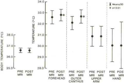
Tissue heating caused by exposure to RF radiation is a primary safety concern in MR imaging. Therefore, to determine temperature changes caused by high field strength MR imaging of the brain with a head coil, we measured body and skin temperatures in 35 patients immediately before and after clinical MR imaging. MR imaging was performed with a 1.5 T MR system using a 28-cm, open-bore RF transmit/receive head coil specifically designed for examinations of the brain. The average body temperature was 36.6 ± 0.2°C before MR imaging and 36.6 ± 0.2°C immediately afterward (mean ± SO, P
=
not significant). The average forehead skin temperature increased from 32.6 ± 0.6 to 32.8 ± O.soC (p < .01), and the average outer canthus skin temperature increased from 32.1 ± 0.6 to 32.7 ± 0.6°C (p < .01) after MR imaging. The highest skin temperature recorded was 34.2°C, and the largest temperature change was +2.1°C. There were no statistically significant changes in the average skin temperatures of the upper arm and hand.We conclude that patients undergoing MR imaging of the brain with a head coil at the RF radiation exposure we studied experience no significant changes in average body temperature and statistically significant increases in local (Le., areas within the head coil) skin temperatures. The observed elevations in skin temperatures were physiolog-ically inconsequential.
MR imaging requires exposing subjects to static, gradient, and RF electromag-netic fields. Tissue heating, which is believed to be caused predominantly by absorption of RF energy, is a primary safety concern in MR imaging [1-3]. Excessive increases in body and/or skin temperatures can occur if the total RF power absorbed by biological tissues is sufficient to overwhelm head-dissipating mechanisms [4,5].
Little is known about the thermo physiologic consequences of MR imaging in the clinical setting. Since one of the most common MR imaging examinations involves imaging the brain with a head coil, we sought to characterize the temperature responses of patients undergoing this type of diagnostic procedure.
Materials and Methods
Thirty-five patients referred for MR imaging of the brain for suspected disease were studied under a protocol approved by the Institutional Review Board. There were 19 males and 16 females, ages 13 to 87 years old (average age, 48 years). All patients were thoroughly screened (i.e., no pacemakers, aneurysm clips, etc.) to determine if they could safely undergo MR imaging.
A 1.5-T superconducting magnet-operating at 64 MHz for proton imaging was used in this investigation along with a 28-cm open-bore, linear drive, RF transmit/receive coil specif-ically designed for head imaging.
The MR imaging protocol used conventional RF pulse sequences, as follows: spin echo, sagittal plane, TR = 600, TE = 25, acquisition matrix = 128 x 256, slice thickness = 10 mm,
average number of slices = 20, average imaging time = 2:40 min; spin echo, axial plane, TR = 2000, TE = 30 and 60, acquisition matrix = 256 x 256, slice thickness = 5 mm, average number of slices = 40, average imaging time = 8:40 min.
288
SHELLOCK AND CRUES
AJNR:9, March/April 1988The Signa MR system is equipped to monitor and display the estimated whole body average and local specific absorption rates (SAR) for each patient based on the subject's weight and the scan parameter information [15]. The estimated whole body average SAR for the above imaging protocol was 0.06 Wjkg and the local SAR was 2.54 Wjkg. The Food and Drug Administration recommends that exposure to RF power during MR imaging should not exceed a whole body average SAR of 0.4 Wjkg or 2.0 Wjkg in any 1 g of tissue (i.e., local SAR). Note that the scan parameters used in this study were well below the whole body average SAR but slightly above the local SAR recommended by the FDA.
Physiological Measurements
Either most physiological monitoring devices are adversely affected by the electromagnetic fields used during MR imaging or the presence of the monitors can distort the image quality by producing unwanted artifacts [6, 7]. Therefore, all the instruments used in this investigation were thoroughly evaluated in pilot studies before clinical use to ensure that there were no unfavorable interactions between the monitors and the MR imaging system. Calibration procedures were performed on a frequent and regular basis.
Body Temperature. Since we were particularly concerned about temperature changes that occurred within the immediate area of the head coil, body temperature was measured in the sublingual pocket. The sublingual pocket is the portion of the oral cavity that has the warmest and most stable temperature, located near the vasculature at the joint of the tongue and the floor of the mouth [8]. Temperatures were obtained with an electronic thermometert that has an accuracy
and resolution of 0.1 °C.
Skin Temperature. Skin temperatures were obtained with a non -contact, fast-response (i.e., less than 0.1 sec), infrared thermometer' [9]. The resolution and accuracy of this instrument is 0.1°C. Skin temperatures were measured in less than 30 sec from the following sites: forehead, outer canthus, upper arm, and hand. These skin temperature sites were selected in an attempt to obtain representa-tive information from surface areas located in the immediate vicinity of the RF power deposition within the head coil (i.e., outer canthus and forehead) as well as from remote, peripheral surface areas (i.e.,
upper arm and hand).
Heart Rate and Blood Pressure. Since the circulatory system is involved in the regulation of thermal responses [10], heart rate and blood pressure (systolic and diastolic) were determined noninvasively with an Omega 1400 blood pressure monitor. § This monitor provides
semicontinuous recordings of heart rate and blood pressure by using the oscillometric technique. The monitor was modified for use during MR imaging by the addition of an 18-ft pneumatically filled hose, which positioned the electronic components at a magnetic fringe field of approximately 200 G [7]. Heart rate and blood pressure measure-ments were not attenuated by the extra length of hose. The metal couplings of the blood pressure cuff were replaced by plastic fittings.
Experimental Protocol
Patients wore lightweight cotton hospital gowns and all had similar amounts of skin surface area uncovered during the procedure. The patients were exposed to a room temperature of 21.0 ± 1.0°C, a relative humidity of 45% ± 5%, and an air flow of less than 0.1 Mj
tMark X electronic thermometry system, Electromedics, Englewood, CO. • Medi-Therm, Everest Interscience, Tustin, CA.
fin Vivo Research Laboratories, Broken Arrow, OK.
sec for 15 min to allow for temperature equilibration. Body and skin temperatures, heart rate, and blood pressure (determined while the patient was in a supine position) were obtained immediately before MR imaging of the brain and within 60 sec after completion of the procedure. The thermometry probe used for measurement of the sublingual pocket temperature was inserted at the above-mentioned times and was not left in place during the scan because of the known adverse interactions between the MR imaging electromagnetic fields and electronic equipment [7].
Statistical Analysis
Variables obtained before MR imaging were compared with those obtained afterward by means of a standard paired Student t test [11]. Data are reported as mean ± 1 SD.
Results
The average body temperature measured in the sublingual pocket was unchanged after MR imaging of the brain with the head coil (Fig. 1). There was a statistically significant increase (p
<
.01) in the average skin temperatures obtained from the forehead and outer canthus while the average skintemperatures of the upper arm and hand were essentially
unchanged (Fig. 1). The highest temperature measured on
any skin surface was 34.2°C (measured at the outer canthus
site) and the largest change in skin temperature we observed was +2.1 °C (measured at the outer canthus site).
None of the patients had evidence of any temperature-related cutaneous flushing or erythema after MR imaging of
the head with the head coil. Fourteen (40%) of the 35 patients had observable signs of sweat on their forehead. None of the patients reported feeling uncomfortable as a result of the temperature changes.
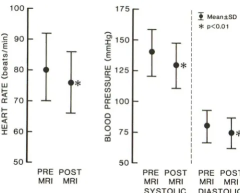
There was a statistically significant decrease (p
<
.01) in the average heart rate, with an average change of 4 beats/ min (Fig. 2). There was also a statistically significant decrease(p
<
.01) in the average systolic and diastolic blood pressures,with average changes of 11 and 5 mm Hg, respectively (Fig. 2).
34.0
I Meant.SO * p<O.Ol
33.0
I
1
*
1*
G
I
e..w 32.0
CI:
I I
~~
G 38.0 CI: w 31.0
~ a.
w ::;
CI: W
~
>-~ 37.0 30.0
CI:
I I
w
a.
::;
36.0 29.0
w
>-0
0 35.0 28.0
m
PRE POST PRE POST PRE POST PRE POST PRE POST
MRI MRI MRI MRI MRI MRI MRI MRI MRI MRI
FOREHEAD OUTER UPPER HAND
CANTHUS ARM
[image:2.612.313.554.540.702.2]100 175
I
Mean.±SDC 90
'E
--
C/)(0
OJ 80
.0 ~ W
*
ti:
a: 70
I
*
p<O.Ol~
:¥
150I·
E E
~ 125
::l
(f) (f)
w
a: 100
I-a: «
w
60 I
a.. Cl
I
I·
0 0 75
-.J
In
50 50
PRE POST PRE POST PRE POST MRI MRI MRI MRI MRI MRI
SYSTOLIC DIASTOLIC
Fig. 2.-Average heart rate, systolic, and diastolic blood pressures before and after MR of the brain at 1.5 T using a head coil (n = 35).
Discussion
Body Temperature. Patients undergoing MR imaging of the
head at 1.5 T with a head coil had no significant change in average body temperature. During conditions of heat loading,
an increase in body temperature will occur only if the capacity for heat loss is exceeded by the amount of heat gained [5,
12-14]. Since the primary source (i.e., RF radiation) of heating during this MR imaging procedure was relatively small and localized over a limited area of the body, it is not surprising
that we did not observe substantial changes in internal body
temperature.
Skin Temperature. Skin temperatures increased signifi-cantly in the area surrounded by the head coil (i.e., forehead
and outer canthus) but were unchanged at the peripheral sites (hand and upper arm) after MR imaging of the brain. RF radiation induced heating at 64 MHz appeared to be confined
to the outermost tissues, similar to what we reported in our previous investigation [15]. Our observance of forehead
sweating indicates that normal thermoregulatory mechanisms were activated to counterbalance and prevent any excessive
skin temperature elevations.
Although we did not perform experiments on control
sub-jects (i.e., without RF), it is doubtful that the increases in skin
temperatures and localized sweating that we observed were
caused by factors others than the RF radiation because our
experiments were conducted under strict environmentally controlled and consistent conditions and no other potential heating factors were operative. For instance, one might
sus-pect that radiative mechanisms may have been responsible
for the observed increases in skin temperatures. However, the head coil used in this study did not have any components
that were close enough to the skin surfaces we examined
(i.e., outer canthus and forehead) to effect heat gain.
Of interest is the fact that the predominant heating effects
occurred within the transmit/receive head coil. Although this
does not appear to present a potential problem with tissue
areas that have an adequate capacity to dissipate heat,
certain thermal-sensitive tissues (such as the eye or the testis) may not tolerate localized heating as well. Therefore, when
determining safe exposure levels to RF radiation during MR
imaging, practitioners should consider that localized tissue
heating may occur with transmit/receive coils.
The increases in skin temperatures we observed, although
statistically significant, were not considered physiologically stressful or hazardous. Skin temperature in humans is nor
-mally 5-1 DoC lower than deep body temperature and varies
according to environmental conditions [5, 12-14]. The highest
skin temperature we observed under the constraints of this
study was 34.2°C. This temperature level is minor as
com-pared with the upper limit at which painful sensations or
damage occur, which is approximately 43-45°C [16].
Because the primary biological effect of RF radiation is
tissue heating, various regulatory agencies have provided advice for safe exposure levels. The National Radiological Protection Board (NRPB) in the United Kingdom has specified
acceptable limits of exposure to the nonionizing radiation
used during clinical MR imaging, which indicate exposure to
RF radiation "should not result in a rise in body temperature
of more than 1°C as shown by skin and rectal temperature"
[17]. According to the results of our study, high-field MR imaging of the brain with a head coil would definitely be
unacceptable in certain patients because the resulting
eleva-tions in skin temperature would exceed this recommended
exposure criterion. However, we feel that the safety guidelines
provided by the NRPB are too conservative and do not take
into account important aspects of temperature regulation.
Although we agree with the NRPB's choice of using tem -perature parameters to indicate the acceptable levels of
ex-posure to RF radiation (because patients will have different
temperature responses to a heat load depending on their
heat-dissipating capabilities, which in turn are dependent on
the ambient conditions, the presence of clinical conditions
associated with heat intolerance, the amount of body fat, age,
etc.), we disagree with the NRPB's recommended limit of an
increase of 1 °C for both rectal and skin temperatures because
skin temperature is considerably more labile than internal
body temperature. From a thermal tolerance standpoint, an
increase of 1°C in skin temperature is relatively insignificant
and does not compare with a 1°C increase in body tempera
-ture.
Recently, Kido et al. [18] examined temperature responses to high-field MR imaging of the brain in normal volunteers and reported that the "mean temperature rise (measured in the axilla) was always less than 0.2°C." However, it is difficult to compare the results of our study with those of Kido et al. [18] for the following reasons: (1) it is unknown whether a head
coil was used during the imaging procedure, (2) temperature was measured in the axilla, which is not considered a repre-sentative site of either body or skin temperature, and (3) a conventional thermistor probe was used, which is an unsat -isfactory technique for measuring temperature during expo -sure to RF radiation because the presence of wire leads are known to distort the field and can also cause concentrations of RF power [19, 20].
[image:3.614.57.297.77.270.2]290 SHELLOCK AND CRUES AJNR:9, March/April 1988
with a head coil were significantly higher than those recorded at the completion of the scan. Kido et al. [18] reported a
significant decrease in heart rate but no change in mean blood
pressure during MR imaging of the brain. In our previous
study [15], we did not observe any statistically significant changes in heart rate or blood pressure during MR imaging with a body coil at RF radiation exposures (i.e., whole body
average SAR) between 0.42-1.20 W jkg, during which both
body and skin temperatures increased significantly.
At first glance, it appears as though cardiovascular re-sponses to MR imaging are somewhat confusing. However, it is known that heart rate and blood pressure elevations can occur in patients awaiting diagnostic tests as a result of
tension and apprehension caused by the imminence of the
clinical procedure [21, 22]. The relative changes in heart rate and blood pressure we observed in this study may be partially explained by a similar mechanism. The fact that there were no apparent changes in heart rate and blood pressure in our previous study may be due to slight, off-setting increases in these parameters as a cardiovascular response to the in-creases in body and skin temperatures.
Since cardiovascular changes may also result from
expo-sure to static magnetic fields [23-25], we cannot entirely rule
out the possibility that heart rate and blood pressure were altered by the high-field MR imaging system used in this
study. Further examination of static magnetic field effects on
the cardiovascular system is warranted.
Whatever the cause, the MR imaging clinician should be
aware of this observed phenomenon. The relative increases
in heart rate and blood pressure at the beginning of MR imaging have important implications for cardiac-gated studies (i.e., repetition time is dependent on heart rate) as well as for
any interventional studies involving MR imaging in which
cardiovascular parameters are evaluated.
Potential Limitations. A potential. limitation of this study is related to our assumption that there was enough "thermal
inertia" in human tissues to allow meaningful temperature
data to be obtained immediately before and after MR imaging.
Since this does appear to be a reasonable presumption [26],
there should only have been a minimal amount of cooling of
the skin between the time the scanning procedure was
com-pleted and the short period (i.e., 60 sec) that transpired before
skin temperatures were measured. Therefore, it is unlikely
that significant temperature changes would have occurred.
Optimally, continuous, real-time temperature recordings
should be determined during investigations of tissue heating
associated with MR imaging. However, as previously
indi-cated, it is not possible to use conventional thermistors or
thermocouples in the electromagnetic environment used for
MR imaging because (1) the thermal conductivity of the
electrical leads can cause a perturbation of the temperature
measurements (particularly the surface temperature
measure-ments), (2) the metallic leads attract RF noise and can be
heated artifactually by induced eddy currents, (3) the RF field
is distorted by the presence of metallic leads, and (4) it is
possible for the static magnetic field to perturb the
tempera-ture recording as well as to disrupt the operation of the
measurement device [19, 20]. Recently, a fluoroptic
thermom-etry instrument has been developed that is compatible with
MR imaging systems [20] and we intend to use this instrument
in future studies.
Another limitation is the fact that we did not evaluate
temperature responses to an exceptionally high exposure to
RF radiation. One of the primary intentions of this study was
to determine the thermophysiologic responses to a typical MR procedure in a clinical situation; therefore, we did not
choose to vary the pulsing parameters in order to deposit
additional RF power. It is possible that other scanning pro-cedures may be substantially more taxing on a patient if a
higher exposure to RF radiation is absorbed for a longer
period of time.
In conclusion, we have observed that, within the constraints of this study, there is no change in average body temperature and statistically significant increases in local skin
tempera-tures within the head coil during high field strength MR
imaging of the brain. In relative terms, the skin temperature changes we observed were not considered harmful to the
patients. However, additional studies examining temperature
responses to MR imaging are needed to determine the overall safety of MR imaging with respect to thermally induced changes, especially since it is possible to perform imaging procedures at RF power levels higher than the one evaluated in this study.
ACKNOWLEDGMENTS
We gratefully acknowledge the help provided by all the magnetic resonance imaging technologists at Cedars-Sinai Medical Center and by the General Electric service engineers. We are also indebted to Daniel Joe Schaefer and Christopher J. Gordon for their critique of this manuscript.
REFERENCES
1. Budinger TF. Nuclear magnetic resonance (NMR) in vivo studies: known thresholds for health effects. J Comput Assist Tomogr 1981;5:800-811
2. Saunders RD, Smith H. Safety aspects of NMR imaging. Br Med Bull
1984;40:148-154
3. Shellock FG. Biological effects and safety aspects of magnetic resonance
imaging. Diagn Imag 1987;9:96-101
4. Spiegel RJ, Deffbaugh OM, Mann JE. A thermal model of the human body exposed to an electromagnetic field. Bioelectromagnetics 1980;1 : 253-270
5. Gordon CJ. Thermal physiology. In: Biological effects of radiofrequency
radiation. Washington, DC: EPA-600/8-83-026A, 1984:4-1-4-28
6. Reis R. Potential interference with medical electronic devices. Bull NY Acad Med 1979;55:1216-1221
7. Shellock FG. Monitoring during MRI: an evaluation of the effect of
high-field MRI on various patient monitors. Med Electronics 1986;100:93-97
8. Gerbrandy J, Snell ES, Cranston WI. Oral, rectal, and oesphageal temper-ature in relation to central tempertemper-ature control in man. Clin Sci
1954;13:615-619
9. Shellock FG, Rubin SA, Everest CEo Surface temperature measurement by IR. Med Electronics 1984;86:81-83
10. Rowell LB. Cardiovascular aspects of human thermoregulation. Circ Res
1983;52: 367 -379
11. Winer BJ. Statistical principles in experimental design. New York: McGraw
Hill, 1971
12. Bligh J. Temperature regulation in mammals and other vertebrates. New
York: Elsevier, 1973:80-84
13. Houdas Y, Ring EFJ. Temperature distribution. In: Human body tempera
14. National Council on Radiation Protection and Measurements. Thermoreg-ulatory responses in human beings. In: NCRP Report No. 86, Biological effects and exposure criteria for radiofrequency electromagnetic fields.
Bethesda: National Council on Radiation Protection and Measurements, 1986:221-250
15. Shellock PG, Crues JV. Temperature, heart rate, and blood pressure changes associated with clinical MR imaging at 1.5 T. Radiology 1987;
163: 259-263
16. Hardy JD, Wolff HG, Goodell H. Pain sensations and reactions. New York: Hafner, 1967
17. National Radiological Protection Board ad hoc Advisory Group on Nuclear
Magnetic Clinical Imaging. Revised guidelines on acceptable limits of exposure during nuclear magnetic resonance clinical imaging. Br J Radiol
1983;56:974-977
18. Kido OK, Morris TW, Erickson JL, Plewes DB, Simon JH. Physiological changes during high field strength MR imaging. AJNR 1987;8:263-266
19. National Council on Radiation Protection and Measurements. Radiofr
e-quency electromagnetic fields. In: NCRP Report No. 67, Quantities and units, biophysical interaction, and measurements. Bethesda: National
Council on Radiation Protection and Measurements, 1981: 88-91
20. Wichersheim KA, Sun MH. Fluoroptic thermometry. Med Electronics 1987; 101 :84-91
21. Hickam JB, Gargill WH, Golden A. Cardiovascular reactions to emotional
stimuli. Effect on the cardiac output, arteriovenous oxygen difference, arterial pressure, and peripheral resistance. J Clin Invest 1948;27:290 -298
22. Konzett H. Cardiovascular parameters and methods of measuring emo-tions. In: Levi, L. ed. Emotions- their parameters and measurement. New York: Raven, 1975:369-378
23. Vardanyan VA. Effect of a magnetic field on blood flow. Biofizika
1973;18:491-496
24. Beischer DE, Knepton JC. Influence of strong magnetic fields on the electrocardiogram of squirrel monkeys. Aerospace Med 1964;35:939-944
25. Jehenson P, Duboc 0, Levergne T, Guize L, Guerlin F, Syrota A. Effect of
a 2-T magnetic field on human cardiac rhythm. Radiology 1986;161 :280
-281 (abstr)