By studying the electrical activity of muscles during a given behaviour, one can reveal both how this activity produces integrated movements and how the outputs of the central nervous system are organized. We have taken this approach in analyzing the escape behaviour of the cockroach Periplaneta americana.
This insect detects an approaching predator by sensing the air displacement produced by the forward movement of the predator’s body (Camhi and Tom, 1978; Camhi et al. 1978). Wind-sensitive hair receptors located on the cerci, two posterior abdominal appendages, detect both the predator’s approach and its direction (Nicklaus, 1965; Dagan and Camhi, 1979; Westin, 1979; Hamon et al. 1992). In response, the cockroach first turns away from the attack and then runs (Camhi and Tom, 1978; Camhi et al. 1978).
The nerve circuit for escape comprises at least five tiers of cells essentially organized in a linear array (Camhi, 1993; Ritzmann, 1993): sensory neurones to giant interneurones (GIs) to thoracic interneurones (TIs) to local interneurones to motor neurones. The TIs also bypass the local interneurones to interact synaptically with motor neurones. Most of the sensory-to-GI connections, and all known GI-to-TI connections, are excitatory. From the TIs onwards, there is both excitation and inhibition.
A key question that we address here is how the cockroach
uses the wind information encoded by these neurones to execute a left versus a right turn. Of particular interest is the observation that the cockroach discriminates behaviourally relatively well between wind from the front left versus the front right. Specifically, puffs of wind from only 15 ˚ left of ‘head-on’ evoke right turns, and those from 15 ˚ right of head-on evoke left turns, on approximately 90 % of trials (Camhi and Tom, 1978).
In general, where fine sensory discrimination occurs, one might expect to discover neurones that are sharply tuned to the discriminated parameters. An enigma in the cockroach system is that, to date, neurones sharply tuned for wind direction have not been found. Of approximately 220 sensory cells on each adult cercus, each responds broadly to wind over roughly 180 ˚, centred around a given best excitatory direction (BED) (Westin, 1979; Dagan and Camhi, 1979). Among the GIs, four of the seven cells on each side of the nerve cord are similar to the sensory cells in the breadth of their directional response, and the three remaining GIs are much more broadly tuned. Significantly for the present study, none of the GIs responds exclusively to wind from just one side of the body (Kolton and Camhi, 1995). Moreover, of the neurones beyond the GIs in the escape circuit, none of those so far recorded from, which are thought to be involved in setting the initial turn direction, show a response selective for wind from one side. This
JEB9795
The cockroach responds to wind from the front left by making an escape turn to the right, and vice versa. So far, no interneurones in the escape system are known that respond only to wind from the left or only to wind from the right. In this study, we used electromyographic recordings to determine whether motor neurones respond in this direction-selective manner during escape behaviour.
In the mesothoracic coxal–femoral joint, whose movement direction is diagnostic for escape direction, the fast motor neurones of one muscle respond selectively to one wind direction, and those of the antagonistic muscle
respond selectively to wind from the other direction, resulting in an appropriate turning response. This rules out an alternative hypothesis, a co-activation mechanism of specifying turn direction. These results suggest that it would be fruitful to search among the interneurones of the escape system for additional cells and circuit properties that could give rise to this sharp directional discrimination.
Key words: directional behaviour, giant interneurones, escape behaviour, cockroach, Periplaneta americana.
Summary
PRODUCING DIRECTED BEHAVIOUR: MUSCLE ACTIVITY PATTERNS OF THE
COCKROACH ESCAPE RESPONSE
RAFAEL LEVI ANDJEFFREY M. CAMHI*
Department of Cell and Animal Biology, Life Sciences Institute, Hebrew University, Jerusalem 91904, Israel
Accepted 20 October 1995
*Author for correspondence.
includes TIs, local interneurones and motor neurones (Westin et al. 1988).
It is not intuitively obvious how neurones so broadly tuned, each responding to both left and right wind, manage to produce sharp behavioural discrimination of wind direction from 15 ˚ left versus 15 ˚ right of head-on. One likely possibility is that there are neurones, not yet identified, that do respond selectively to wind from just one of these two directions. In this report, we begin the search for such neurones by examining the motor outputs, using electromyograms. Our goal was to determine whether there are motor neurones that respond, for example, when wind from 15 ˚ left evokes a right turn, but not when wind from 15 ˚ right evokes a left turn. Such a finding would then motivate a search for pre-motor interneurones that may also discriminate sharply between these two directions.
As an alternative to such sharp neural discrimination, the cockroach could use a strictly mechanical means of producing directed leg movements, even if the motor neurones are not selective for wind from the left or right. For instance, when a wind puff arrives from, say, 15 ˚ left of head-on, its activation of left GIs would give rise to a motor output for a right turn. However, as the right GIs are also activated, and produce almost as many spikes as their left homologues (Camhi and Levy, 1989; Kolton and Camhi, 1995), a motor output for a left turn, slightly weaker than that for the right turn, might also be produced. Both motor outputs would then simultaneously activate antagonistic muscles in the legs. According to this hypothesis, the stronger muscle contractions for a right turn would mechanically override those for a left turn, and so the cockroach would turn right.
Perhaps the best-known example of such co-contraction of antagonist muscles is that underlying the jump of the locust, in which the leg extensor muscles develop the power needed for the jump by first contracting isometrically against co-activated, antagonistic flexors. When the flexor activity ceases, the stored extensor force is suddenly released (Heitler, 1974). This co-contraction in the locust hindleg lasts for about 500 ms, a situation that clearly could not exist in the cockroach’s rapid escape behaviour. However, there is no a priori reason to rule out a role for short-term co-contraction in the cockroach, in which the more strongly contracted muscle simply overrides its antagonist. The main reason for considering this possibility in the cockroach is that, as stated above, wind from either side of the body appears to excite bilateral homologous neurones at all levels of the escape circuit.
A critical test for distinguishing between these two possible mechanisms – neural versus mechanical discrimination of left versus right – is to determine whether there is co-activation of antagonistic leg muscles (i.e. those for a right and those for a left turn) at the onset of an escape turn. We have chosen to study the coxal–femoral (CF) joint of the mesothoracic legs, because its direction of movement is diagnostic for a left versus a right turn. In response to wind puffs from the front left, the initial movement of the CF joint of the left leg is an opening (95 % of the trials; Nye and
Ritzmann, 1992). For wind from the front right, the initial movement of this joint is usually a closing (76 % of the trials; Nye and Ritzmann, 1992).
We show here that there is no co-activation of the fast opener and closer muscles of this joint. Rather, when the wind onset is from 15 ˚ left and the cockroach makes a right turn, one set of muscles is activated, and for the opposite condition, the antagonistic set is activated. This indicates the presence of some mechanism for directional sharpening in the escape system at a neural level up to, and perhaps including, the motor neurones.
Materials and methods
We used adult cockroaches, Periplaneta americana L., that we reared in the laboratory. We used only males, as is standard in our intracellular studies where males are chosen because of the relative absence of internal fat, simplifying the dissection. We rear the cockroaches in large, screen-covered barrels, at a temperature of 18–22 ˚C, with a 12 h:12 H L:D photoperiod. We provide them with rat chow and water ad libitum.
We fixed the cockroaches, using a drop of wax under the abdomen, to a lubricated, transparent plastic plate, permitting free movement of the legs. We have previously shown that the escape movements of the legs relative to the body in this situation are virtually identical to those of a free-ranging cockroach (Camhi and Levy, 1988). We monitored the wind-evoked escape movements reflected from a mirror angled at 45 ˚ below the plastic plate, using a high-speed video recorder at 250 frames s21(NAC, Tokyo). Digitizing two points that we
labelled with white dots on the coxa, and two more points on the femur, allowed us to calculate the angle of the CF joint, using the hardware/software package Movias (NAC).
We activated escape behaviour using a controlled wind puff that rose to a peak of 2.3 m s21within 50 ms and arrived at the
cockroach from an angle of 45 ˚ above horizontal. The details of the wind-producing system have been described elsewhere (Camhi and Levy, 1988).
We recorded muscle activity using a pair of 50mm copper electrode wires, insulated to their tips. Signals were amplified by Grass P511 amplifiers, using a bandpass of 30 Hz to 30 kHz, and were stored on an instrumentation tape recorder (Hewlett Packard 3968A). Analysis was carried out off-line using the hardware/software package Computerscope (RC Electronics) on a PC platform.
We analyzed data only from cockroaches that walked normally on the spot, showed generally normal behaviour, and gave sharp escape responses to wind puffs. We also eliminated data from recordings where cross-talk between the recording channels prevented clear discrimination of the EMG signals. We only analyzed data from trials in which both mesothoracic CF joints made initial movements in the direction expected relative to the wind direction on the basis of the behavioural tests described in the previous paragraph. These criteria were fulfilled by trials in approximately one-third of the cockroaches we prepared for recording. We recorded the EMGs from one leg only, and produced opposite CF joint movements in this leg by presenting wind stimuli from both 15 ˚ left and 15 ˚ right of head-on.
Results
It was important first to verify that, as has been reported by others (Nye and Ritzmann, 1992), we could obtain differential movement responses of the mesothoracic legs in response to wind from our two test directions, 15 ˚ left and 15 ˚ right of head-on. Thus, in a group of six cockroaches, we delivered these two wind stimuli in a randomized sequence and video-taped the resulting behaviour. In response to the 15 ˚ left wind, the initial movement of the CF joint of the left leg was an opening in 85 % of the trials (N=18 trials); and in response to the 15 ˚ right wind, the initial movement was a closing in 86 % of the trials (N=23 trials). These values compare favourably with the values of 95 % and 76 %, respectively, obtained previously (Nye and Ritzmann, 1992).
In video recordings of the joint movements of mesothoracic legs that also carried EMG electrodes, we measured the angular sizes of the initial closings and openings of the CF joint. The initial opening movement was 61±16 ˚ (mean ± S.D., N=19 in six animals), whereas the initial closing movement was
smaller, 22±118.0 ˚ (N=23 in seven animals). This difference is explained below.
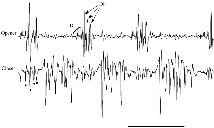
We were able to discriminate clearly in our EMG recordings between the electrical responses of the opener and closer muscle, as could be seen during bouts of running, which we recorded for each cockroach. The two muscles were reciprocally activated during running (Fig. 1). These records resemble in several ways published records from the metathoracic legs (Delcomyn, 1973; Pearson, 1972), as follows: (1) the opener activity begins with a burst of small spikes (unit Ds of the metathorax) which are followed by much larger spikes (Df of the metathorax); (2) the small unit, as for the metathoracic Ds, is also active spontaneously during standing (not shown); (3) the closer activity appears to consist of several fast units, based on the range of spike amplitudes and spike overlaps (see, for instance, the first closer burst). In the trials we accepted for analysis, cross-talk between the recording channels was minimal and did not interfere with the interpretation of spike patterns. We could also discern that each burst in the wind-evoked escape responses in the opener muscle began with small Ds spikes, after which Df began its short spike burst. The closer muscle’s response was more complex and appeared to consist of activity in several large units.
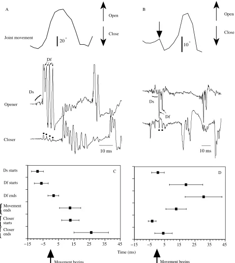
In all 19 trials (in six animals) in which the CF joint initially opened, the first identifiable EMG response was in the opener units Ds and Df, without any accompanying closer spikes. Rather, the closer response was delayed until at least the end of opener activity (Fig. 2A,C). In 17 of these 19 trials, the first opener unit to respond was Ds. In the two remaining trials we could not identify the small Ds spikes, perhaps because they occurred later and were obscured by the much larger Df spikes. The general pattern was several Ds spikes at the outset, followed by 1–6 Df spikes (Fig. 2A).
In all 23 trials (in seven animals) with initial joint closing, a very different pattern of activity was seen (Fig. 2B,D). The first units to be activated were closers. These produced a brief
Opener
Closer
100 ms Ds
Df
[image:3.609.196.567.511.734.2]{
Closer starts Closer ends Movement ends Ds starts
Df starts
Df ends
Movement begins Movement begins
Joint movement
Opener
Closer
20
10 °
°
10 ms 10 ms
Open
Close
A B
Open
Close
Ds
Ds
Df Df
B B
B B B B
−15 −5 5 15 25 35 45
B B
B
B B
B
−15 −5 5 15 25 35 45
D C
Time (ms)
{
{
{
{
{
Fig. 2. (A,B) Wind-evoked EMGs (lower panels) and movement responses (upper panels) of the CF joint in the left mesothoracic leg. (A) Joint opening in response to wind from the left front. Stimulus onset occurred approximately 20 ms before the onset of the traces. The four dots above the closer trace indicate four small peaks of electrical cross-talk from the four large Df spikes in the opener trace. (B). Joint closing, in a different cockroach, in response to wind from the front right. Stimulus onset occurred roughly 20 ms before the onset of the traces. The arrow in the top trace points to the onset of evoked joint closing. The initial joint closing movement is usually small and, as shown here, is followed by a large joint opening movement. Two dots under the closer trace indicate cross-talk from the two large Df spikes. (C) Summary of all 19 joint opening responses. The mean time of each event shown on the left is indicated ± 1 S.D. All events are shown relative to the time when the joint movement
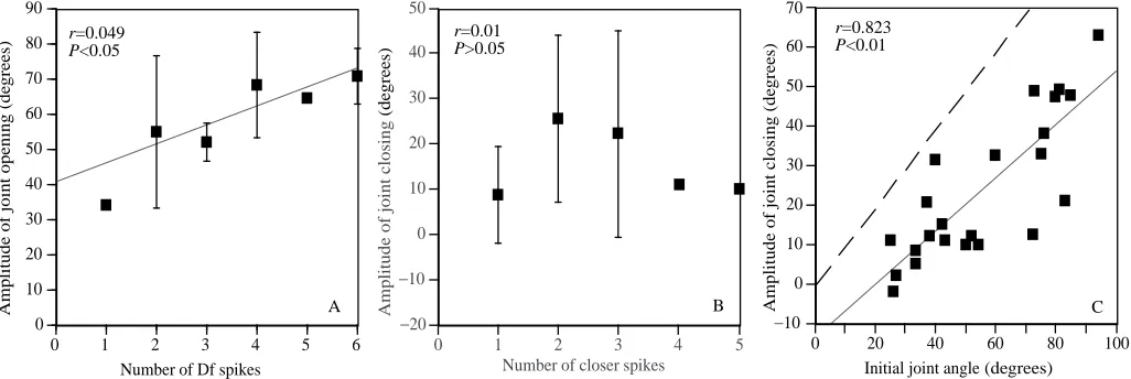
[image:4.609.61.539.73.608.2]burst lasting, on average, only 7 ms. Among the opener units, in 15 of the 23 trials, we were able to identify the small Ds spikes above the background noise caused by cross-talk from the closer muscles. In 10 of these 15 trials, Ds began its activity only after the end of the closer burst (although the mean time of Ds onset for all 15 trials was actually 2 ms before the end of the closer burst). The large Df unit did not begin to fire until a mean of 16 ms after the end of the closer burst (Fig. 2B,D). The initial burst of activity in Ds and Df, during both opening and closing of the CF joint (Fig. 2A,B), resembles that during running (Fig. 1, top trace), in that the burst begins with several Ds spikes and this is followed by Df spikes. Thus, it may well be that the same central pattern generator that produces running (Pearson and Fourtner, 1975) also gives rise to the first leg movements during wind-evoked escape behaviour. It is known that the inter-leg coordination during the onset of escape behaviour is different from that during running (five or all six legs push on the ground simultaneously in escape, whereas there is alternating tripod stepping in running; Camhi and Levy, 1988). However, the output to each individual leg could be derived from that leg’s own central oscillator, and the phasing of the different oscillators could change as the initial step of escape grades into tripod running. The amplitude of the initial opening angle in the CF joint is correlated with the number of Df spikes (P<0.05, F-test), confirming the importance of Df in producing this movement (Fig. 3A). The regression line does not extrapolate to a y-intercept of zero, suggesting that a single Df spike may be much more effective than following spikes in producing an angular change. In addition, this non-zero y-intercept could reflect in part the role of Ds in producing joint opening, although one would expect this slow unit to produce much less motor effect than the fast Df.
The comparable graph for the initial closing angle of the CF joint does not show a significant correlation with the number of spikes in the closer muscle (Fig. 3B). Thus, some factor other than the number of spikes in this muscle appears to determine the extent of this movement. To determine what this factor might be, we plotted, for all trials with initial joint closings, the angle of closing as a function of the initial joint angle (Fig. 3C). This gave a highly significant correlation (P<0.01). The dashed line in Fig. 3C is the predicted line for complete joint closing from any initial angle. The points fall along a line that is almost parallel to this line, showing that the joint closed, whatever its starting angle on a given trial, to a fairly narrow range of final angles. On the basis of measurements from the regression line, the final angle ranged from approximately 10 ˚ for those trials where the leg was already nearly closed, to approximately 45 ˚ for those trials where the leg started at approximately 90 ˚ open.
On the majority of trials, the leg started at an angle of less than 60 ˚ open, a situation associated with small closing responses. This accounts for the observation that, on trials where the joint closed, the angular change of the joint was generally smaller than on trials where the joint opened, as stated above and as seen in Fig. 2A,B.
Discussion
This study shows that one group of motor neurones is turned on by a wind stimulus from 15 ˚ left and a different group is turned on by a wind stimulus from 15 ˚ right. Consequently, we can rule out any major role for a co-activation and mechanical overriding mechanism of directional discrimination, at least for the mesothoracic CF joint.
[image:5.609.53.566.486.658.2]As mentioned in the Introduction, none of the interneurones of
Fig. 3. Control of the amplitude of initial CF joint movement. (A) Amplitude of initial opening, as a function of the number of Df spikes in the burst. Means ±S.D. are shown. N=19 trials in five cockroaches. (B) Amplitude of initial closing as a function of the number of large closer spikes in the burst. N=23 trials in seven cockroaches. Correlation not significant. (C) Amplitude of initial closing as a function of the joint’s initial angle. The dashed line indicates the predicted regression line for complete closing from any angle. The actual linear regression, which is almost parallel to the dashed line, indicates that the joint closes, from whatever initial angle, to a small range of joint angles. In fact, from initial joint angles of 25–95 ˚ (a range of 70 ˚), the final angles (initial angle minus amount of closing, based on the regression line) are 10–45 ˚, a range of only 40 ˚.
B B B B B −20 −10 0 10 20 30 40 50
0 1 2 3 4 5
Amplitude of joint closing
(de
grees)
Number of closer spikes
B B B B B B B B B B B BB B B B B B B B B B B −10 0 10 20 30 40 50 60 70
0 20 40 60 80 100
Amplitude of joint closing (de
grees)
Initial joint angle (degrees) B
B B
B B B
0 10 20 30 40 50 60 70 80 90
0 1 2 3 4 5 6
Amplitude of joint opening (de
grees)
Number of Df spikes
r=0.823 P<0.01 r=0.01 P>0.05 r=0.049 P<0.05
the escape system that have yet been studied shows sharp directional discrimination that could easily account for this directional tuning at the motor neuronal level. This raises two possibilities. Either there are interneurones yet to be found that are highly directional and that have an important influence on the motor neurones we recorded from here, or the motor neurones’ directional tuning is an emergent property of its synaptic inputs from interneurones with broad directional tuning and possibly from synaptic interactions among the motor neurones themselves. It remains to be determined whether one or both of these possibilities accounts for the directional motor output.
Other motor systems are known in which interneurones that are directionally broadly tuned give rise to directionally refined movements. A key example is hand-reaching movements in the monkey, which are controlled by neurones of the motor cortex whose directional tuning is roughly cosine-like, as is that of several cockroach GIs and TIs (Kolton and Camhi, 1995; Westin et al. 1988). The monkey’s motor cortex appears to control the direction of movement through the group action of many broadly tuned neurones, each promoting its own ‘preferred’ direction of hand movement with a strength proportional to the cell’s spike frequency. Quantitatively, this system integrates the contributions of the several participating neurones through a population vector code (Georgopoulos, 1994).
Likewise, in the cockroach, a population code among the GIs, similar to that in the monkey cortex, appears to control the direction of the escape turn (Levi and Camhi, 1994; R. Levi and J. M. Camhi, in preparation). The work we report in the present paper indicates that the output of this code is a directionally highly tuned response in the leg motor neurones. In the monkey, there is little information as to how the spinal synaptic outputs of neurones from the motor cortex organize the directional motor output. In the cockroach, many of the interneurones postsynaptic to the GIs have been identified (Ritzmann, 1993), as have many of the motor neurones themselves, and it may be now possible to determine the synaptic organization underlying the directional motor tuning. This work was supported by grant J94-13 from the Whitehall Foundation.
References
CAMHI, J. M. (1993). Neural mechanisms of behavior. Current Opin.
Neurobiol. 3, 1011–1019.
CAMHI, J. M. AND LEVY, A. (1988). Organization of a complex movement: Fixed and variable components of the cockroach escape behavior. J. comp. Physiol. A 163, 317–328.
CAMHI, J. M. ANDLEVY, A. (1989). The code for stimulus direction in a cell assembly in the cockroach. J. comp. Physiol. A 165, 83–97. CAMHI, J. M. AND TOM, W. (1978). The escape behavior of the cockroach Periplaneta americana. I. Turning response to wind puffs. J. comp. Physiol. A 128, 193–201.
CAMHI, J. M., TOM, W. ANDVOLMAN, S. (1978). The escape behavior of the cockroach Periplaneta americana. II. Detection of natural predators by air displacement. J. comp. Physiol. A 128, 203–212. CARBONELL, C. S. (1947). The thoracic muscles of the cockroach
Periplaneta americana (L). Smithson. Misc. Collns 107, 1–23. DAGAN, D. AND CAMHI, J. M. (1979). Responses to wind recorded
from the cercal nerve of the cockroach Periplaneta americana. II. Directional selectivity of the sensory neurons innervating single columns of filiform hairs. J. comp. Physiol. A 133, 103–110. DELCOMYN, F. (1973). Motor activity during walking in the cockroach
Periplaneta americana. II. Tethered walking. J. exp. Biol. 59, 643–654.
GEORGOPOULOS, A. P. (1994). New concepts in generation of
movement. Neuron 13, 257–268.
HAMON, A., GUILLET, J. C. AND CALLEC, J. J. (1992). Patterns of monosynaptic input to the giant interneurons 1–3 in the cercal system of the adult cockroach. J. comp. Physiol. A 174, 91–102. HEITLER, W. J. (1974). The locust jump: Specialization of the
metathoracic femoral–tibial joint. J. comp. Physiol. 89, 93–104. KOLTON, L. AND CAMHI, J. M. (1995). Cartesian representation of
stimulus direction: Parallel processing by two sets of giant interneurons in the cockroach. J. comp. Physiol. 176, 691–702. LEVI, R. ANDCAMHI, J. M. (1994). Testing for a population vector
code for wind direction in the cockroach giant interneurons. Soc. Neurosci. Abstr. 20, 418.9.
NICKLAUS, R. (1965). Die Erregung einzelner Fadenhaare von Periplaneta americana in Abhangigkeit von der Grosse und Richtung der Auslenkung. Z. vergl. Physiol. 50, 331–362. NYE, S. W. ANDRITZMANN, R. E. (1992). Motion analysis of leg joints
associated with escape turns of the cockroach, Periplaneta americana. J. comp. Physiol. A 171, 183–194.
PEARSON, K. G. (1972). Central programming and reflex control of
walking in the cockroach. J. exp. Biol. 56, 173–193.
PEARSON, K. G. AND FOURTNER, C. R. (1975). Nonspiking
interneurons in walking system of the cockroach. J. Neurophysiol.
38, 33–52.
RITZMANN, R. E. (1993). The neural organization of cockroach escape and its role in context-dependent orientation. In Biological Neural Networks in Invertebrate Neuroethology and Robotics (ed. R. D. Beer, R. E. Ritzmann and T. McKenna), pp. 113–138. San Diego, CA: Academic Press.
WESTIN, J. (1979). Responses to wind recorded from the cercal nerve of the cockroach Periplaneta americana. I. Responses properties of single sensory neurons. J. comp. Physiol. A 133, 97–102. WESTIN, J., RITZMANN, R. E. ANDGODDARD, D. G. (1988).