Imaging White Matter Tracts and Nuclei of the Hypothalamus: An
MR-Anatomic Comparative Study
Marc J. Miller, Leighton P. Mark, F. Zerrin Yetkin, Khang-Cheng Ho, Victor M. Haughton, Lloyd Estkowski, and Eric Wong
PURPOSE: To evaluate the MR appearance of the hypothalamus and its associated white matter
tracts. METHODS: Coronal and sagittal spin-echo images were obtained in cadaver brains. Gross and histologic sections were made of the cadaver brains. The size, shape, signal intensity, course, and pattern of structures in the hypothalamic region were identified in MR images by comparison with the anatomic sections. RESULTS: The mamillary bodies, paraventricular zone of hypothalamic nuclei, postcommissural fornix, mamillothalamic fasciculus, and anterior commissure were iden-tified on the MR images. CONCLUSION: This study suggests that, with MR imaging of sufficiently
high resolution, some of the tracts and nuclei in the hypothalamus may be identified.
Index terms: Brain, anatomy; Brain, magnetic resonance; Hypothalamus; White matter
AJNR Am J Neuroradiol 15:117-121, Jan 1994
The borders of the normal hypothalamus have
been dem
o
nstrated with magnetic resonance
(MR) (1). The MR appearance of the
individual
hypothalamic nuclei and white matter tracts
as-sociated with the hypothalamus have not been
described. We studied the MR appearance of
white matter tracts and nuclei in the
hypothala-mus by comparing high
-
resolution MR imaging
with anatomic sections
.
Materials and Methods
Three routine autopsy cases without neurologic disease and without significant postmortem neuropathologic change were selected for this study. Brains were harvested via craniotomy and fixed in 10% buffered formalin solution for 3 weeks. In two specimens, cylinders of brain tissue 10 em in height and 2 em in diameter (with the long axis in an anteroposterior direction) containing the hypothalamus were harvested with a specially designed plug cutter at-tached to an electric drill. The third cylinder was harvested in a similar orientation by means of gross dissection. These cylinders were introduced into 30-cc polypropylene con-tainers with 10% buffered formalin solution to fill the remaining volume and sealed.
The cylinders were imaged in a 1.5-T Signa imager (General Electric Medical Systems, Milwaukee, Wis) with a
Received November 11, 1992; accepted pending revision January 12, 1993; revision received February 2.
From the Departments of Radiology (L.P.M., F.Z.Y., V.M.H.), Pathol-ogy (K.-C.H.), and Biophysics (L.E., E.W.), The Medical College of Wis-consin (M.J.M.), Milwaukee, Wis.
Address reprint requests to: Victor M. Haughton, MD, Department of Radiology, Medical College of Wisconsin, Froedtert Memorial Lutheran Hospital, 9200 West Wisconsin Ave, Milwaukee, WI 53226.
AJNR 15:117-121, Jan 1994 0195-6108/94/1501-0117
© American Society of Neuroradiology
117
2.5-cm-diameter solenoid coil. Spin-echo images were ob-tained in coronal planes with the following imaging param-eters: 1000/54/2 or 2000/80/2 (repetition time/echo time/ excitations), 256 X 256 matrix, 1-mm section thickness, 2- or 3-cm field of view, and 0 skip. Sagittal planes were selected with reference to the coronal images, and sagittal images were obtained with the same parameters.
Subsequently, the three specimens were embedded in paraffin and sectioned in sagittal planes with a microtome in 6-JLm thicknesses at selected variable intervals. Coronal sections were obtained at the level of the mamillary bodies in three other routine autopsy cases. The sections were stained with hematoxylin and eosin and luxol fast blue and photographed with a 35-mm camera and macro lens.
White matter tracts in the hypothalamus were identified in anatomic sections as fiber bundles staining darkly with luxol fast blue stain and compared with structures visible on the exactly correlated MR images. The tract originating in the mamillary bodies and terminating in the thalamus, with a posterior and supralateral course, was defined as the mamillothalamic fasciculus (2). The fiber bundle terminat-ing in the mamillary bodies, with a superior course around the anterior perimeter of the third ventricle, was defined as the fornix (2). The tract lateral to the thalamus was defined as the internal capsule or capsulopeduncular transition ( 1 ). In MR images, the signal intensity, course, and pattern of structures corresponding to the mamillothalamic fasciculus and fornix were identified. Nuclei in the hypothalamus were identified by the diminished intensity of staining in relation to white matter tracts.
Results
118 MILLER AJNR: 15, January 1994
F
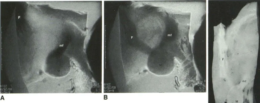
Fig. 1 . A and B, Parasagittal MR images and corresponding anatomic section (C) through the
hypothalamus. The images demonstrate the mamillary body (M) projecting into the interpeduncular
cistern {ic), the mamillothalamic fasciculus (mf), and the postcommissural fornix (f).
c
hypothalamic nuclei demonstrated a thin region of much higher intensity signal. The mamillary bodies produced a signal intermediate in intensity between the white matter tracts and hypothal-amic nuclei.
In sagittal MR images the hypothalamus, mam-illary bodies, fornix, mamillothalamic fasciculus, and anterior commissure are recognized. The section just off the midline (Fig 1 A) depicts the mamillothalamic fasciculus as a homogeneous, low-signal-intensity white matter tract with a pos-terosuperior course around the perimeter of the third ventricle to the thalamus. The postcommis-sural fornix appears as an inhomogeneous white matter tract composed of discrete bands of low-intensity signal interspersed with high-low-intensity signal structure. The mamillary bodies appear in this image as sharply defined spheroid structures of intermediate signal intensity protruding into the interpeduncular cistern. The hypothalamus is evident anterior to the mamillary bodies as a region of homogeneous, higher signal intensity.
The next lateral contiguous section (Fig 1 B) best depicts the course of both the mamillothal-amic fasciculus and the postcommissural fornix. A comparison of the two sharply defined white matter tracts demonstrates that the postcommis-sural fornix possesses greater signal intensity and heterogeneity than the mamillothalamic fascicu-lus. The mamillary bodies appear with interme
-Fig. 2. Parasagittal MR image with a 3-cm field of view in a different specimen illustrating the relationship of the anterior
commissure (ac) and optic chiasm (oc) to the postcommissural
fornix (f) and mamillary body (M).
diate signal intensity. The tracts and nuclei are identified by referring to the corresponding ana-tomic section (Fig 1 C).
The anterior commissure appears in the 3-cm field of view sagittal images as an ovoid, low-signal-intensity structure anteroinferior to the body of the fornix (Fig 2). In the 2-cm field of view images the region of the anterior commis-sure was not included. The postcommissural for-nix is demonstrated as a curving, low-intensity tract posterior to the anterior commissure.
[image:2.615.58.556.75.272.2] [image:2.615.345.530.349.508.2]AJNR: 15, January 1994
8
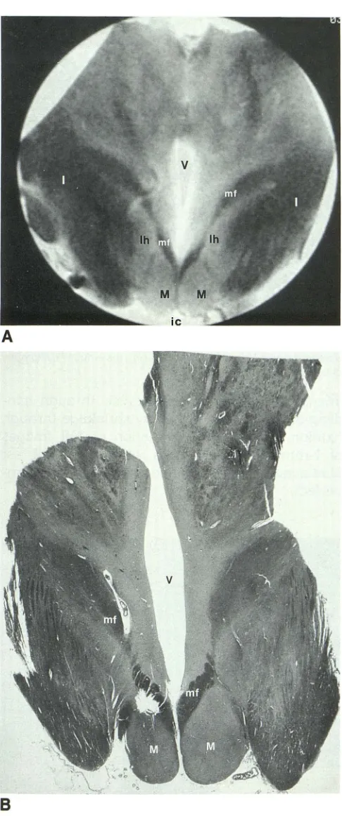
Fig. 3. A, Coronal MR image and B, corresponding anatomic section through the hypothalamus. The images illustrate the mamillothalamic fasciculus (mf) at the mamillary bodies (M), the internal capsule (/), the lateral hypothalamic nuclei (/h), the third ventricle ( V), and the interpeduncular cistern (ic). The irregular void in the anatomic section represents a sectioning artifact.
MR OF HYPOTHALAMUS 119
mamillothalamic fasciculus, fornix, and internal
capsule with respect to the third ventricle,
mam-illary bodies, and one another (Figs 3A and 36).
The third ventricle, sharply differentiated from the hypothalamus on each side, appears as a homogeneous midline area of moderately high signal intensity. The mamillary bodies, which
protrude into the interpeduncular cistern, are
ev-ident as homogeneous, spheroid structures of intermediate signal intensity. The internal cap-sule, seen lateral to the mamillary bodies, is demonstrated as a low-intensity signal structure interspersed with high-intensity bands. The mam-illothalamic fasciculus, originating at the
supra-medial aspect of the mamillary bodies, shows
low-intensity signal with marked contrast to sur-rounding tissue. The lateral hypothalamic nuclei appear as an area of intermediate signal intensity. The next contiguous anterior section (Fig 4A) illustrates the postcommissural fornix,
paraven-tricular zone of hypothalamic nuclei, and tuber
cinereum. The termination of the postcommis-sural fornix at the mamillary bodies is evident as
a low-intensity-signal region. The paraventricular
zone of hypothalamic nuclei, flanking the third ventricle, is included within a thin region of high-intensity signal. The tuber cinereum, seen pro-jecting into the interpeduncular cistern, appears
with high-intensity signal in this section.
In the next anterior contiguous section (Fig 4B), in the plane of the foramen of Monro, the postcommissural fornix is located along the lat-eral margins of the third ventricle and appears as a band with low signal intensity and sharp bor-ders. The internal capsule has a lower intensity
signal than either the thalamus, which borders it
medially, or the lenticular nucleus, which borders its lateral margin. Lateral to the hypothalamus, the optic tracts and anterior cerebral arteries are prominent low-signal-intensity landmarks. An an-atomic section corresponds to the images de-picted in Figures 4A and 4B (Fig 4C).
The most anterior of the contiguous sections
(Fig 5) demonstrates the internal capsule,
thala-mus, lenticular nucleus, anterior commissure, and
precommissural fornix. The internal capsule
de-fines the borders of the lenticular nucleus and
thalamus, which are superior and lateral to the
hypothalamus and of intermediate-intensity
sig-nal. The anterior commissure, a landmark near
[image:3.612.57.300.87.666.2]120 MILLER
as a well-delineated low-intensity structure. The precommissural fornix appears with low-intensity signal superior to the anterior commissure.
Discussion
Loes and colleagues ( 1) described the bound-aries and anatomy of the hypothalamus with standard spin-echo pulse sequences but did not describe the appearance of individual hypothal-amic nuclei or white matter tracts on MR. Others described a focus of high signal intensity in the hypothalamus with infundibular disease (3). This study compares anatomic specimens and high-resolution MR images of the hypothalamic region. Our observations may not be directly extrap-olated to clinical imaging, because experimental imaging parameters different from prevalent clin-ical imaging techniques were used to optimize resolution. The descriptions presented herein are based on the MR appearance of cadaveric fixed tissue. Consequently, our observations may not correlate precisely with the MR appearance of structures in the hypothalamic region in vivo. All possible anatomic structures and variations may not have been identified within the sam-ples studied because of technical inadequacies and normal anatomic variation. Anatomic dis-tortion may have been introduced into our
spec-AJNR: 15, January 1994
Fig. 5. Coronal MR image anterior to those in Figure 4 illus-trating the precommissural fornix (F), anterior commissure (ac), internal capsule (I), lenticular nucleus (In), thalamus ( 7), and optic chiasm (oc).
imens by the method of harvest, through han-dling of the specimens, or by shrinkage through fixation. Finally, the correlation of MR images of 1-mm section thickness with
6-,um
luxol fast blue anatomic sections necessarily would be im-perfect.Fig. 4. A, Coronal MR images immediately anterior to those in Figure 3 and B,
corresponding anatomic section, illustrating the relationship of the postcommissural fornix
(F), the internal capsule(/), and optic tracts (ot) to the third ventricle ( V). The paraventricular
8
zone of hypothalamic nuclei (arrows) and the tuber cinereum (tc) are evident in A. The
thalamus (T), hypothalamus (H), lenticular nucleus (In), anterior cerebral artery (aca), and
foramen of Monro (f) are evident in B. The irregular voids in the anatomic section represent
[image:4.613.316.557.76.308.2] [image:4.613.59.555.467.686.2]AJNR: 15, January 1994
Although the effects of formalin on contrast in cerebral tissues has not, to our knowledge, been systematically studied, the relative contrast be-tween gray and white matter in our experimental images resembles that in clinical images. This suggests that the anatomic detail in our study could be obtained clinically with the use of ap-propriate fields of view and surface coils to achieve suitable signal-to-noise ratios. Conse-quently, high-resolution MR imaging could be implemented to evaluate intrinsic hypothalamic
MR OF HYPOTHALAMUS 121
anatomy and pathology, and possibly to investi-gate sexual dimorphism in the hypothalamus (4).
References
1. Loes DJ, Barloon T J, Yuh WTC. et al. MR anatomy and pathology of the hypothalamus. AJR Am J Roentgenol 1991; 156:5 79-585 2. Miller RA, Burach E. Atlas of the central nervous system in man. 3rd
ed. Baltimore: Williams and Wilkins
3. El Gammal T. Brooks BS, Hoffman WH. MR imaging of the ectopic
bright signal of posterior pituitary regeneration. AJ!'IR Am J
f'leuro-radiol I 989; 10:323-328