1534
Use of the Alberta Stroke Program Early CT Score
(ASPECTS) for Assessing CT Scans in Patients with
Acute Stroke
J. H. Warwick Pexman, Philip A. Barber, Michael D. Hill, Robert J. Sevick, Andrew M. Demchuk, Mark E. Hudon, William Y. Hu, and Alastair M. Buchan
BACKGROUND AND PURPOSE: Clinicians are insecure reading CT scans by using the one-third rule for acute middle cerebral artery stroke (1/3 MCA rule) before treating patients with recombinant tissue plasminogen activator. The 1/3 MCA rule is a poorly defined volumetric estimate of the size of cerebral infarction of the MCA. A 10-point quantitative topographic CT scan score, the Alberta Stroke Program Early CT Score (ASPECTS), is described and illus-trated. A sharp increase in dependence and death occurs with an ASPECTS of 7 or less. We describe how to use ASPECTS and why it works with CT scans obtained on all commonly used axial baselines. We also describe interobserver reliability among clinicians from different specialties and with different experience in reading CT scans in the context of acute stroke.
METHODS: The six physicians who developed ASPECTS answered a questionnaire on pre-cisely how they interpret and use ASPECTS. The ASPECTS areas as interpreted by these physicians were compared with one another and with standards in the literature. k statistics were used to assess the interobserver reliability of ASPECTS versus the 1/3 MCA rule.
RESULTS: The exact methods of interpretation varied among the six individual observers, with either a 3:3 or 4:2 split on the specific questions. The overall interobserver agreement was good compared with that of the 1/3 MCA rule. Normal anatomic vascular and interobserver variations explain why ASPECTS can be applied with different CT axial baselines.
CONCLUSION: ASPECTS is a systematic, robust, and practical method that can be applied to different axial baselines. Clinician agreement is superior to that of the 1/3 MCA rule.
Intravenous recombinant tissue plasminogen acti-vator (tPA) improves outcome after acute ischemic stroke (1). The European Cooperative Acute Stroke Study (ECASS) trials pioneered the importance of assessing early CT ischemic changes to predict the benefit from intravenous thrombolysis (2, 3). In both trials, the randomization decision depended on whether more or less than one third of the territory of the middle cerebral artery (MCA) was involved.
Received November 2, 2000; accepted after revision March 27, 2001.
From the Departments of Clinical Neurosciences (J.H.W.P., P.A.B., M.D.H., A.M.D., A.M.B.) and Radiology (R.J.S., M.E.H., W.Y.H.), Foothills Hospital, Calgary, Alberta, Canada.
Supported in part by grants from the Heart & Stroke Foun-dation of Canada (M.D.H., A.M.B.), the Canadian Institutes of Health Research (M.D.H., A.M.D., A.M.B.), and the Alberta Heritage Foundation for Medical Research (M.D.H., A.M.D., A.M.B.).
Address reprint requests to Professor J. H. Warwick Pex-man, Department of Clinical Neurosciences, Room MRG005, Foothills Hospital, 1403 29th St NW, Calgary, Alberta, Canada T2N 2T9.
qAmerican Society of Neuroradiology
Unfortunately, subsequent research has shown that even experienced stroke physicians and radiologists have difficulty recognizing and quantifying these changes (4–8).
is-FIG1. ASPECTS study form and MCA variants.
A and B, Right hemisphere, observer variations: lower and up-per ASPECTS slices show as shaded areas the minimal and maximal variations in size of the cortical areas of the MCA (M1– M6) chosen by six expert observers. Left hemisphere, ASPECTS study form:A5anterior circulation;P5posterior circulation;C 5caudate head;L5lentiform nucleus;IC5internal capsule;I 5insular ribbon;MCA5middle cerebral artery;M15anterior MCA cortex;M25MCA cortex lateral to insular ribbon;M35 posterior MCA cortex;M4, M5, and M6 are anterior, lateral, and posterior MCA territories, respectively, approximately 2 cm su-perior to M1, M2, and M3, respectively, rostral to basal ganglia. C and D, Cortical MCA area variations with change of baseline. In the right hemisphere, the baseline is parallel to the inferior OML; in the left hemisphere, the baseline is the superior OML.
E and F, Normal vascular variations in MCA size on the two ASPECTS slices. The right hemisphere shows the larger normal variations described by van der Zwan (18) (light shading). The left hemisphere of each shows the smaller, textbook (17), vari-ations (dark shading).
chemia in the posterior circulation and 24 were treated outside the protocol). The baseline ASPECTS value correlated inversely with the se-verity of the stroke on the National Institute of Health Stroke Scale (NIHSS) (Spearman’s r 5
2.56, P , .001). The baseline ASPECTS value predicted functional outcome and symptomatic in-tracerebral hemorrhage (P , .001 and P 5 .012, respectively). The sensitivity of ASPECTS for functional outcome was .78 and specificity was .96; the respective values for symptomatic intracerebral hemorrhage were .90 and .61. A sharp increase in dependent or fatal outcomes occurred with ASPECTS of 7 of less. The inter- and intraobserver reliability of ASPECTS was good to excellent (k
5.71–.89) and consistently superior to the 1/3 rule between observer pairs from the same specialty.
In this article, we explain how ASPECTS is used in practice, illustrate the method with examples, de-fine landmarks and definitions, show why it can be used on CT scans obtained on all commonly used axial baselines, and emphasize that clinicians from different specialties and with varying levels of ex-perience in assessing acute ischemic CT changes can agree on an acceptable level.
Methods
Patients were recruited from both Calgary and Houston, TX. For the purposes of the current analysis, technical information was not available from CT scans obtained in Houston (n 5 70). CT scans obtained in Calgary (n5 86) were performed on fourth-generation helical scanners. For better definition, in-dividual scans were acquired using contiguous axial 6-mm sec-tions from the foramen magnum to the suprasellar region and 10-mm contiguous sections through the remainder of the brain. One sixth of the CT scans were obtained using the orbitomea-tal line (OML), one third using the superior OML, and half using the inferior OML. Scanner settings were kV5120, mAs 5400, and mA5200. The section time was 2 seconds, the matrix size was 512, and a small focal spot with an algorithm was used to reduce bone artifacts and give finer granularity. The mA was reduced near the vertex to 150, 130, and 120 mA for the upper three slices, respectively. Photography was done at a window level of 30 H with a window width of 75 H. All the CT scans were interpreted from film. The areas studied were in the part of the brain cut with 10-mm sections. Images were read in a darkened room, and were studied from near and far and even obliquely.
The interpreters used the ASPECTS method to read the CT scans. The ASPECTS was determined from two standardized axial CT cuts (Fig 1), one at the level of the thalamus and basal ganglion and one adjacent to the most superior margin of the ganglionic structures, such that they were not seen. On these two sections, which were, by definition, not continuous, the MCA territory was allotted 10 points. A single point was subtracted for an area of early ischemic change, such as focal swelling or parenchymal hypoattenuation, for each of the de-fined regions. A normal CT scan received an ASPECTS of 10 points. A score of zero indicated diffuse ischemic involvement throughout the MCA territory (Figs 2–4). Parenchymal hy-poattenuation was defined as a region of abnormally decreased attenuation of brain structures relative to attenuation of other parts of the same structures or of the contralateral hemisphere. Focal brain swelling or mass effect was defined as any focal narrowing of the CSF space due to compression by adjacent
structures, such as effacement of cortical sulci or ventricular compression (10).
neuroradi-FIG2. CT scans in a 65-year-old woman with left-sided hemiplegia, hemianopia, and neglect less than 3 hours after symp-tom onset.
A and B, Baseline CT scans show hy-poattenuation with swelling and efface-ment in regions M1, M2, insula (I), M4, and M5 (ASPECTS 55). Intravenous throm-bolysis was administered.
C and D, Follow-up CT scans show a large area of hypoattenuation involving much of the MCA territory. The patient was dependent at 3 months.
ologists had at least 2 years’ experience in assessing acute ischemic stroke with CT scans. Although radiology trainees participated in the validation of ASPECTS (9), they did not participate in the questionnaire.
The questionnaire, devised by a neuroradiologist who had no prior experience with ASPECTS, was developed to assist physicians who were inexperienced in the precise use of this technique. Because interobserver agreement in the original ASPECTS was good to excellent, it was hypothesized that all respondents would give similar answers.
Recent textbooks state that ischemic stroke can be seen as early as 6 hours after symptom onset (11, 12), quoting the abstracts from the classic articles of Tomura et al in 1988 (13) and Truwit et al in 1990 (14). However, the former group made a diagnosis in less than 3 hours in 13 of 15 patients with obscuration of the lentiform nucleus (from a total of 25 pa-tients), while the latter made it in 10 of 13 patients with loss of the insular ribbon (from a total of 27 patients). These de-scriptions formed the basis for diagnosis in two of the areas of ASPECTS. However, certain considerations remain; such as, did one lose a point if only a small part of the insular ribbon was involved? The questionnaire also sought to answer how many of the signs of ischemia (mass effect, abnormally low attenuation in white matter, loss of demarcation of the gray/ white junction) need to be present to lose a point in any area.
Furthermore, where were the boundaries of each M area, and did the interpreters really only look at two CT sections?
The two extreme axial baselines, the superior OML and the inferior OML, were drawn on an outline traced from a lateral pilot film. The two ASPECTS cuts were then drawn in cor-respondence with each baseline, and these cut lines were marked in thirds. It was then possible to measure the distances between the two anterior third and the two posterior third junc-tions, respectively, by using the scale on the pilot scan (Fig 5).
FIG 3. CT scans in a 68-year-old man with global aphasia and an NIHSS score of 7.
A and B, Baseline scans show a region of hypoattenuation involving the anterior insula (I) and hypoattenuation and swelling in the M2 region (ASPECTS58).
C and D, Follow-up scans confirm the area of infarction. The patient made a full neurologic recovery.
Results
How Different Physicians Interpreted ASPECTS (Table 1)
In the lentiform, caudate, and insular regions, all observers agreed upon several points. Hypoatten-uation of the insular ribbon was defined as loss of differential density between subcortical white and gray matter of the insular cortex (loss of gray/white matter differentiation). It was emphasized that ei-ther the anterior or posterior halves may be lost independently. Obscuration of the lentiform nucle-us was defined as a decrease in density of all or part of this nucleus and loss of gray/white matter differentiation in the area. Again, only part of the caudate head had be to obscured or show hypo-attenuation to lose a point. The caudate head often has a dual blood supply, from the MCA and ante-rior cerebral artery (Fig 1E). Comparison was al-ways made with the opposite hemisphere. Loss of a well-defined lateral margin of the lentiform nu-cleus in both hemispheres raised the differential di-agnosis of a CT scan of poor quality, new bilateral infarcts, or one new infarct along with a
contralat-eral old infarct (most likely). All viewers subjec-tively interpreted a lesion with very marked hy-poattenuation as an old lesion. Workstations were not used.
tem-FIG4. CT scans in a 79-year-old woman with left-sided weakness and NIHSS score of 15, 2 hours after symptom onset.
A and B, Baseline CT scans show hy-poattenuation of the right lentiform nucleus (L) and caudate nucleus (C) on two axial cuts (ASPECTS58).
C and D, Follow-up scans at 24 hours confirm the area of infarction. The patient made a full neurologic recovery after thrombolysis.
poral lobe as the anterior boundary of M2, but the oblique posterior boundary varied.
There was at least a centimeter difference in the size of each M area between the minimum and maximum chosen by the observers (Fig 1A and B, right hemisphere). For comparison, the right hemi-sphere of Figure 1C and D shows the usual MCA territory when the inferior OML is used (15) while the left hemisphere shows it using the superior OML baseline (16). Figure 1E and F shows cross-sectional representations of normal variations in MCA size derived from textbooks (17) in the left hemisphere and from van der Zwan (18) in the right hemisphere.
The internal capsule was scored variably. Two neurologists and one neuroradiologist assessed only the posterior limb of the internal capsule; the others assessed both limbs and deducted a point if any portion of it was affected. In their overall assess-ment, the observers disagreed on several points. Four of the six chose to score the CT scans without clinical information and then reread them knowing
which hemisphere was affected. This was a very deliberate attempt to avoid overreading. If one could not decide which hemisphere was involved when one read the scan without any clinical infor-mation, the ASPECTS was judged to be greater than 8. All neuroradiologists looked at all CT scans, whereas two of the three neurologists re-viewed only the two critical ASPECTS cuts. The neuroradiologists used this assessment to judge whether there was volume averaging or an infarct present, whereas the neurologists looked at either the opposite side or at adjacent sections to make this decision. Two neuroradiologists were slightly more conservative and one stroke neurologist was more aggressive at interpreting the CT scans than the other three.
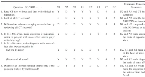
an-TABLE 1: Methods used to avoid overreading CT scans of infarcts less than 3 hours old
Question DO YOU. . . N1 N2 N3 R1 R2 R3 Y* D*
Comments Concerning Dissenters
1. Read CT first without, and then with clinical in-formation?
Y D Y Y Y D 4 2 N2 and R3 read CT knowing
symptoms
2. Look at all CT sections? D D Y Y Y Y 4 2 N1 and N2 used the two
ASPECTS sections in Fig 1 3. Differentiate volume averaging versus infarct by
reviewing all CT sections?
D D D Y Y Y 3 3 N1 and N2 compared opposite side; N3 looked at adjacent sections
4. In M1–M6 areas, make diagnosis if hypoatten-uation is present with mass effect and/or gray/ white blurring?
Y D D Y Y Y 4 2 N2 and N3 made diagnosis on the basis of hypoattenuation alone
5. In M1–M6 areas, make diagnosis with mass ef-fect plus hypoattenuation in
(A) one M area? Y D Y D D Y 3 3 N2, R1, and R2 made diagnosis
on the basis of mass effect alone
(B) several M areas? Y Y D Y D Y 4 2 N2 and R2 made diagnosis on
the basis of mass effect alone 6. Diagnose an internal capsular infarct only if the
posterior limb is hypoattenuated?
D Y Y Y D D 3 3 N1, R2, and R3 would have
made the diagnosis if only the anterior limb had been af-fected
Total in agreement
4 2 4 6 4 5
Note.—N indicates stroke neurologist, R5neuroradiologist, Y5agree with question, D5disagree with question, M5cortical middle cerebral artery.
*Average split among observers53 : 3 and 4 : 2
FIG 5. Maximal variation of ASPECTS sections with baseline alteration. The two ASPECTS sections with two different base-lines: superior OML (solid line) and parallel two slices, and in-ferior OML (dashed line) and parallel two slices. The respective upper and lower slices are divided into thirds. Cuts are through the basal ganglia and roof of the lateral ventricle to show that disagreement is not more than 2 cm.
gles and distances helped in the recognition of sub-tle changes in hypoattenuation. In particular, it proved useful to view a crucial slice independently. This was done by using either the cupped hand or a roll of paper (telescoping). Localized atrophy, de-fined as sulcal widening or ventricular enlargement, also suggested an old infarct. When old infarcts were present in the basal ganglia of the asymptom-atic hemisphere, it was sometimes difficult (and oc-casionally impossible) to say whether areas of ab-normal hypoattenuation or obscuration in the basal
ganglia of the affected hemisphere represented new or old infarcts.
When a CT scan showed that a patient’s head was not symmetrically situated in the scanner, ow-ing to either tilt or rotation in either direction, or both, all observers dealt with this as best they could by trying to compare corresponding areas in the two hemispheres, even though these were on dif-ferent CT sections.
Because of varying baselines, as much as a 1-cm difference in the rostrocaudal-anteroposterior loca-tion of a given point occurred when different cuts were used through the basal ganglia; and as much as a 2-cm difference occurred on the cuts at the upper edge of the lateral ventricles above the basal ganglia (Fig 5).
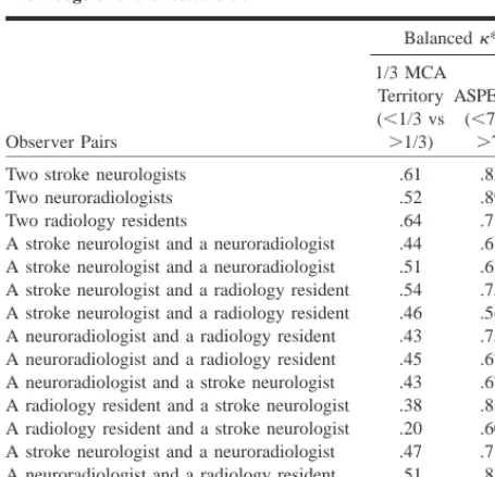
Interobserver Agreement
Table 2 gives the balancedk scores for interserver agreement among all the independent ob-servers when they used the 1/3 MCA rule and ASPECTS dichotomized between 7 or greater and less than 7. The interobserver agreement between pairs of stroke neurologists (k 5 .61 for the 1/3 MCA rule and .85 for ASPECTS), neuroradiolo-gists (k 5.52 and .89), and radiology residents (k
[image:6.612.46.529.77.334.2]TABLE 2: Pairwise interobserver agreement between independent observers for the 1/3 MCA territory rule and ASPECTS, with knowledge of the affected side
Observer Pairs
Balancedk*
1/3 MCA Territory (,1/3 vs .1/3)
ASPECTS (,7 vs
.7) Two stroke neurologists
Two neuroradiologists Two radiology residents
A stroke neurologist and a neuroradiologist A stroke neurologist and a neuroradiologist
.61 .52 .64 .44 .51 .85 .89 .71 .61 .63 A stroke neurologist and a radiology resident
A stroke neurologist and a radiology resident A neuroradiologist and a radiology resident A neuroradiologist and a radiology resident
.54 .46 .43 .45 .75 .56 .75 .67 A neuroradiologist and a stroke neurologist
A radiology resident and a stroke neurologist A radiology resident and a stroke neurologist A stroke neurologist and a neuroradiologist A neuroradiologist and a radiology resident A neuroradiologist and a radiology resident
.43 .38 .20 .47 .51 .39 .67 .83 .60 .71 .83 .63
* Balanced kappa:k ..80 implies excellent reliability; .61#k# .80 implies good reliability; .41# k# .60 implies moderate reli-ability; .21#k#.40 implies fair reliability;k#.20 implies poor reliability (26).
showed less than good reliability for ASPECTS, while all 11 of the 1/3 MCA rule results showed just fair reliability, with the remaining one imply-ing poor reliability.
Discussion
Early ischemic changes seen on CT scans ob-tained in the first few hours after stroke onset rep-resent early cytotoxic edema and perhaps the de-velopment of irreversible injury (19). Many authors have cited the potential superiority of diffusion-weighted MR imaging over CT, but to date MR imaging has not been able to discriminate salvage-able brain tissue from that which is irretrievably injured (20). Although diffusion-weighted imaging may become the method of choice, most physicians treating stroke will remain dependent on CT be-cause of its accessibility. However, the ability of physicians to correctly interpret early radiologic signs of acute stroke on CT scans is fraught with reservations and controversy (7). We believe that ASPECTS provides a solution to this problem.
ASPECTS is a robust clinical tool for several reasons. First, it has excellent reliability in the clin-ical setting, much superior to the 1/3 MCA territory rule. When the clinical situation is known, ASPECTS has proved reliable among physicians of different clinical backgrounds and experience. The agreement among physicians using ASPECTS was considerably better than when they applied the 1/3 MCA rule (the range of k improved from .20–.64 to .56–.89). Acute stroke therapy requires that the
treating clinician be comfortable in making an as-sessment of the severity of the CT findings at the bedside and that communication between col-leagues is consistent. We have shown that agree-ment between neuroradiologists and stroke neurol-ogists was good (k 5 .61–.71). This is essential both in facilitating the treatment process and in conducting clinical trials. Analysis of ECASS-1 CT scans found that 52 (8.4%) of 620 were misread locally. When three expert neuroradiologists reas-sessed the ECASS-1 CT scans, scoring them ac-cording to the 1/3 MCA rule, the chance adjusted pair-wise agreement was surprisingly low (k 5 .23–.51) despite 90% to 91% agreement (4). In a review of 50 CT scans from the Atlantis study (which used the 1/3 MCA rule) (21), agreement among the three neuroradiologist reviewers was moderate to good (pair-wisek coefficients of .44– .65) but consensus among reviewers could be achieved in 72% of cases. However, only neuro-radiologists interpreted the CT scans in the ECASS and Atlantis studies, and the scans were obtained within 6 hours of ictus. Second, ASPECTS is a systematic method. Wardlaw and Seller (22) showed that a systematic approach to assessing ce-rebral infarcts on CT scans produces excellent re-sults. In that study, the k statistics between two experienced neuroradiologists reading 119 brain CT scans for site and size without clinical infor-mation were good to excellent (k5.69–.87) (22). Our analysis showed that good to excellent reli-ability can be achieved with ASPECTS when the stroke symptom side is known, despite variations in the exact interpretation of the signs of early is-chemia and in the use of the two ASPECTS dia-grams (Fig 1A and B). One must conclude, there-fore, that the improvement stems from careful study of 10 specific areas on the initial CT scan, in which each area is compared with the opposite side.
It is interesting that the instructions for ASPECTS were interpreted differently by the neu-rologists and the neuroradiologists. Why does ASPECTS work when there is disparity concerning its exact interpretation? First, the two neurologists, who used only two ASPECTS sections to assess a score, missed only isolated infarcts near the vertex, and these by themselves are relatively rare. This would not have much impact on the statistics of 156 CT scans. The fact that one neurologist and one neuroradiologist initially read the CT scans blinded to clinical information reflects the speed and confidence with which these individuals worked. None of the three neurologists distin-guished between infarcts and volume averaging by reviewing the whole scan, as did the neuroradiol-ogists, but the neurologists still made limited com-parisons, which apparently helped them.
they would be contemplating giving a powerful thrombolytic drug, tPA, and they had already, in the research protocol, assessed the CT scan using the 1/3 MCA rule. They must, therefore, have read the whole CT scan, if only as a gestalt. The initial use of a CT scan is to exclude hemorrhage and tumor, and, in this context, to look for venous thrombosis.
Six observers were equally divided on the inter-pretation of what constituted the internal capsule for the purpose of ASPECTS. Surprisingly, this did not make as big an impact on the score as one would have expected. A possible explanation re-lates to the dichotomization at greater than 7 and 7 or less. Often, the caudate head, lentiform nucleus, and insula are infarcted at the same time, and so a maximum ASPECTS score would be 7, depending on how many, if any, cortical M areas were af-fected. Half our observers would give it an ASPECTS of 7 or less. Those who deducted a point for the involvement of the anterior half of the in-ternal capsule would score it 6. Because of the level of dichotomy, this would not affect the results. Questions 4 and 5 seem to indicate that in assessing the cortical M areas the precise combination of signs of ischemia is not significant. The important thing is that one look carefully at all areas. Eye-balling, or the gestalt method, of reading a CT scan does not work for the more subtle changes seen in acute infarcts less than 3 hours from onset.
Third, ASPECTS retains its utility with different CT techniques. Techniques have been fully dis-cussed by Graeb (23) and Russell (24), and optimal ones suggested, particularly the use of a large mAs. Lev et al (25) have discussed the benefit of work-stations, showing that the detection of ischemic brain parenchyma is facilitated with variable win-dow widths and center settings to accentuate the contrast between normal and edematous tissue. These authors initially used a center of 32 H with a width of 8 H. Reviewers then changed the set-tings to accentuate differences. Use of a narrow window width to review the CT scan on the work-station should improve the detection of early acute infarcts, just as it facilitated the diagnosis of isoat-tenuating subdural hematomas over a decade ago. The design of the original ASPECTS study (9) did not incorporate the use of workstations.
The ASPECTS system was used with three dif-ferent CT scan baselines. Early CT scans of the head were obtained with a superior OML baseline, but some centers have now changed to an OML or an inferior OML. There was at least a 1-cm differ-ence in the size of each M area between the min-imum and maxmin-imum chosen by each of our ob-servers; normal variations in the size of the MCA territory are probably greater than this (18). On the ganglionic level, the anatomic division of sections by the observers was always based on the position of the ends of the sylvian fissure. Therefore, a change of baseline would have no significant effect on the interpretation here. At the level of the roof
of the ventricles, the baseline changes made a ros-trocaudal-anteroposterior difference of 2 cm in the location of the M4 to M6 regions. The three vari-able factors are variations in observer interpreta-tions, baseline variainterpreta-tions, and vascular anatomic variations. As the latter is the most variable, we postulate this is the reason that ASPECTS can be used successfully on scans obtained on all axial baselines. This may not apply to the 1/3 MCA ter-ritory rule.
Conclusion
The availability and speed of CT scanners make them the instrument of choice for assessing acute ischemic stroke in many hospitals. While acute MR imaging provides fantastic pathophysiological in-formation, the utility and widespread applicability of diffusion-weighted MR imaging has yet to be proved within the first 3 hours of stroke ictus. ASPECTS is a CT-based system that provides a more accurate, robust, and practical method for as-sessing acute ischemic stroke than the 1/3 MCA rule. We encourage clinicians and radiologists to apply it in practice.
References
1. The National Institute of Neurological Disorders and Stroke rt-PA Stroke Study Group. Tissue plasminogen activator for acute hemisphere stroke. N Engl J Med 1995;333:1581–1587 2. Hacke W, Kaste M, Fieschi,et al. The European Cooperative
Acute Stroke Study (ECASS): safety and efficacy of intrave-nous thrombolysis with a recombinant tissue plasminogen ac-tivator in the treatment of acute hemisphere stroke. JAMA 1995;274:1017–1025
3. Hacke W, Kaste M, Fieschi C, for the ECASS II Group. Random-ized double blind placebo-controlled trial of thrombolytic ther-apy with intravenous alteplase in acute ischemic stroke. Lancet 1998;352:1245–1251
4. von Kummer R, Allen KL, Holle R, et al. Acute stroke: useful-ness of early CT findings before thrombolytic therapy.
Radi-ology 1997;205:327–333
5. Dippel DW, Du Ry van Beest Holle M, van Kooten F, Koudstaal PJ. The validity and reliability of signs of early infarction on CT in acute ischaemic stroke. Neuroradiology 2000;42:629–633 6. Schriger D, Kalafut M, Starkman S, et al. Cranial computed to-mography interpretation in acute stroke: physicians’ accuracy in determining eligibility for thrombolytic therapy. JAMA 1998;279:1293–1297
7. Grotta J, Chiu D, Lu M, et al. Agreement and variability in the interpretation of early CT changes in stroke patients qualify-ing for intravenous rtPA. Stroke 1999;30:1528–1533
8. Wardlaw JM, Dorman PJ, Lewis SC, Sandercock PAG. Can stroke physicians and neurologists identify signs of early ce-rebral infarction on CT? J Neurol Neurosurg Psychiatry 1999; 67:651–653
9. Barber PA, Demchuk AM, Zhang J, Buchan AM, for the ASPECTS Study Group. The validity and reliability of a novel quantitative CT score in predicting outcome in hyperacute stroke prior to thrombolytic therapy. Lancet 2000;355: 1670–1674
10. Scott JN, Buchan AM, Sevick RJ. Correlation of neurological dysfunction with CT findings in early acute stroke. Can J
Neurol Sci 1999;26:182–189
11. Grossman RJ, Yousem DM. Neuroradiology, the Requisites. St Louis: Mosby; 1994:1101
12. Sutton D. Textbook of Radiology and Imaging. New York: Chur-chill; 1998:1626
13. Tomura T, Uemura K, Inugami A, et al. Early CT findings in cerebral infarction: obscuration or the lentiform nucleus.
14. Truwit CL, Barkovich AJ, Gean-Marton A, et al. Loss of the insular ribbon: another early sign of acute middle cerebral artery infarction. Radiology 1990;176:801–806
15. Berman SA, Hayman LA, Hinck VC. Correlation of CT cerebral vascular territories with function: middle cerebral artery.
AJNR Am J Neuroradiol 1984;5:161–166
16. Stark DD, Bradley WG Jr. Magnetic Resonance Imaging. St Lou-is: Mosby; 1988:288–289
17. Osborn AG. Neuroradiology. St Louis: Mosby; 1995:138 18. van der Zwan A, Hillen B, Tulleken AF, Dujovny M, Dragovic L.
Variability of the territories or the major cerebral arteries. J
Neurosurg 1992;77:927–940
19. del Zoloppo GJ, von Kummer R, Hamanna GF. Ischemic damage of brain microvessels: inherent risks for thrombolytic treat-ment in stroke (editorial). J Neurol Neurosurg Psychiatry 1998; 65:1–9
20. Baird AE, Warach S. Magnetic resonance imaging of acute stroke. J Cereb Blood Flow Metab 1998;18:583–609
21. Marks M, Holmgren EB, Fox AJ, Patel S, von Kummer R, Froe-lich J. Evaluation of early computed tomography findings in acute ischemic stroke. Stroke 1999;30:389–392
22. Wardlaw JM, Seller R. A simple practical classification of ce-rebral infarcts on CT and its interobserver reliability. AJNR
Am J Neuroradiol 1994;15:1933–1939
23. Graeb DA. The challenge of imaging acute stroke in Canada (editorial). Can Assoc Radiol J 1999;50:365–369
24. Russell EJ. Diagnosis of hyperacute ischemic infarct with CT: key to improved clinical outcome after intravenous thrombol-ysis (editorial)? Radiology 1997;205:315–338
25. Lev MH, Farkas J, Gemmete J, et al. Acute stroke: improved nonenhanced CT detection: benefits of soft copy interpretation using variable window width and center level settings.
Radi-ology 1999;213:150–155