With 9 figures
Printed in Great Britain
DIEL CHANGES IN POTASSIUM ACTIVITY IN THE
HAEMOLYMPH OF THE COCKROACH
LEUCOPHAEA MADERAE
BY JANINE LETTAU,* W. A. FOSTER, J. E. HARKER
AND J. E. TREHERNEf
University of Cambridge, Department of Zoology, Downing Street, Cambridge CBz 3EJ, England
(Received 29 April 1977)
SUMMARY
1. A technique is described for continuous measurement, with ion-selective microelectrodes, of K+and Na+ activity in the blood of a free-walking cockroach Leucophaea maderae(Fab.). Measurements can be made for periods of up to 96 h.
2. In LD 16:8, there is a marked and consistent pattern in the diel variation of K+ activity, with minima 1 h before dusk and 2 h after dawn. The mean diel range in K+ activity covers a drop of 67 % below the maximum daily value. The maximum range in K+ activity for an individual cockroach in 24 h was from 4-5 to 25-0 mM-K+.
3. Simultaneous records of K+ activity and locomotor activity show that the first minimum in K+ activity occurs 1-2 h before the main, sharp peak in locomotor activity at dusk, and the second minimum 1-3 h after the subsidiary, broad peak in locomotor activity at dawn.
4. Diel fluctuations in whole-blood potassium concentration follow a roughly similar pattern to the K+ activity changes. Comparison of the concentration and activity changes allows some speculations about the diel movements of potassium within the blood.
5. Diel fluctuations in Na+ activity show no clear overall pattern.
INTRODUCTION
The functioning of many tissues is crucially dependent on the activity of a small number of inorganic ions in the body fluids. In insects the concentration of the major inorganic ions in the blood is highly variable, both within one individual and between individuals and species (e.g. Pichon, 1970; Florkin & Jeuniaux, 1974). Such variation in potassium and sodium activity is likely to be of most significance to the muscles and peripheral nerves, since these, unlike the CNS, are not sheathed from the blood (see Treherne, 1974). The observations of Hoyle (1954) and Ellis & Hoyle (1954) indicated that the level of activity of locusts could be controlled by the potassium concentration of the blood, although this was not supported by the field observations of Chapman (1958) or subsequent laboratory experiments of Moorhouse (1968).
• Present address: Botany School, Downing Street, Cambridge, England.
172 JANINE LETTAU AND OTHERS
Although this variability in blood ion concentration is well known, it is not clear whether it exhibits a consistent temporal pattern within one species. Treherne, Buchan & Bennett (1975) demonstrated a seasonal variation in Na+ concentration in Periplaneta, although the Na+ activity remained relatively constant. Brady (1968) established that Periplaneta blood is a changing ionic environment throughout the day: he observed in some animals a fall of about 10% in potassium concentration during the first hour of darkness.
The motivation of Brady's study was to establish whether potassium concentration, following Hoyle's (1954) observations, could act as a link between the circadian 'clock' and the overt expression of rhythmic locomotor activity. The present investigation was begun to improve and extend Brady's observations, using more modern techniques, in particular ion-selective electrodes. These have two important advantages over the whole-blood flame-photometry analysis used by Brady. First, they measure ionic activity of the plasma, not gross blood concentration, and therefore indicate the amount of ions freely available for physiological processes such as muscle contraction, whereas flame-photometry gives a measure of total ionic concentration, which includes, for example, ions that are bound. Second, ion-selective microelectrodes allow continuous measurements to be made on one particular cockroach; this eliminates the artefacts caused by repeatedly taking blood from one cockroach (Brady, 1968) and the great variability caused by sampling from different cockroaches.
The present study has three main aims. First, to develop methods for characterizing the ionic environment of cockroach blood using ion-selective microelectrodes. Second, to describe the diel changes in potassium and sodium activity in cockroach blood. Third, to relate such changes to known circadian rhythms, in particular locomotor activity.
MATERIALS AND METHODS 1. Insect cultures
We used Leucophaea maderae rather than Periplaneta americana because of the more precise and predictable locomotor activity of the former species (Harker, unpublished observations). Cultures of Leucophaea were maintained in large, light-tight boxes at a room temperature of 21-26 °C in a 16 h light:8 h dark cycle (LD 16:8). The light source was a 20 W fluorescent light producing 124 lux on the base of the box. Water was freely available. The cockroaches were fed ad libitum on a diet of the following composition: 350 g rolled oats, 350 g wheat feed, 50 g linseed cake, 120 g grass meal, 35 g casein, 25 g dried powdered yeast, 25 g sucrose, 50 ml linseed oil, 50 ml arachis oil and 11 g.W. salt mixture. In all experiments only adult males were used.
2. Measurement of potassium activity
Ion-selective microelectrodes based on the design of Khuri, Agulian & Wise (1971) were used. A small amount of potassium liquid ion-exchanger (Corning no. 47132) was injected into the tip (ID 0-3 /i) of a siliconized micropipette (Pyrex redrawn tubing: OD 1-2 mm, ID o-6 mm), which was then filled with o-i M-KC1. The mean resistance of the K+ selective electrodes was 5-8 + 0-45 x io9Q (mean
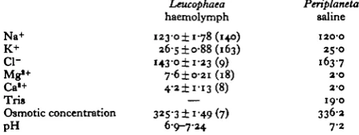
In pure KC1 the potassium electrodes gave a slope of between 52 and 55 mV per potassium activity decade. The electrodes were calibrated (immediately before im-plantation) in the cockroach saline designed by Treherne, Buchan & Bennett (1975). Although devised for Periplaneta, this saline has similar concentrations of major cations to those of Leucophaea blood (Table 1). The activity coefficient (y) of K+ and Na+ in this saline was calculated from a modified form of the Debye-Hiickel equation,
where z = valency of the ion, / = ionic strength of the solution, A and b are constants (A = 0-507 at 20 °C, b = o-i zi; Robinson & Stokes (1959)). y = 074 for K+ and Na+. The selectivity of individual electrodes for potassium over sodium was between 45 and 70 to 1 and varied with the age of the electrode. The electrodes showed a reduced performance in cockroach saline as compared with pure KC1, and calculations show (see Eisenman, 1962) that this reduced performance can be accounted for by inter-ference from sodium ions.
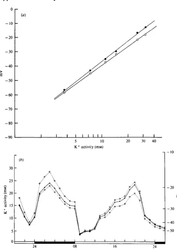
The stability of the electrodes is important, since they are intended for continuous use for at least 24 h whilst implanted in a living cockroach. The electrodes were always recalibrated in cockroach saline after removal from the experimental animal. The new calibration line had generally shifted a little and had a reduced slope (Fig. 1 a). Fig. 1 b shows the effect which this change in calibration line has on the calculated potassium activity in a typical experiment. The discrepancy between the two curves is most marked at the higher values, but the general pattern is similar. Potassium activities at intermediate stages of the experiment were in fact inferred by assuming that the calibration line changed linearly with time during the experiment (Fig. 1 b). There is no direct justification for this, but the calibration hne of electrodes in cockroach saline changed linearly with time.
The implanted electrodes eventually become encapsulated by haemocytes. The haemocytes do not appear to interfere with the behaviour of the electrode for about the first 24-60 h, the exact time depending on the individual electrodes. During this period the performance of an electrode in blood deteriorates in a broadly similar way to that of an electrode in cockroach saline. At some time after 24 h of implantation providing the electrodes do not fail for other reasons, the electrode response flattens out to give a stable potential record. Electrodes are invariably encapsulated at this point. When tested in cockroach salines of varying potassium concentrations, the encapsulated electrodes produce an unvarying potential reading. This encapsulation response of the cockroach sets a limit on the life of the electrode; in favourable cases, the electrodes may remain workable for up to 96 h.
3. Measurement of sodium activity and pH
JANINE LETTAU AND OTHERS
- 1 0
- 2 0
- 3 0
- 4 0
- 5 0
- 6 0
- 7 0
- 8 0
- 9 0 (a)
I I I I I I I I
-i - 1 0
[image:4.451.41.408.59.577.2]175
Silicone rubber
Insulated silver wires
Ion-selective and indifferent
electrodes
[image:5.451.36.357.61.355.2]Sticky wax
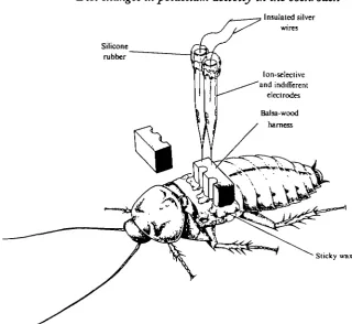
Fig. 2. Diagram showing technique for implantation of electrodes.
about 60 mV per activity decade. Given the composition of Leucophaea blood (Table 1), there is no danger of potassium interfering with the performance of the electrodes.
The sodium electrodes were not very stable, drifting by about 10 mV per 24 h. Intermediate values were inferred as for the potassium electrodes. The electrodes became encapsulated at about the same rate as did the potassium electrodes. The resistance of the sodium electrodes (3 x ioB~3 x io10 Q) increased by a factor of 10 during 24 h in cockroach haemolymph.
pH electrodes (Transidyne 805; tungsten carbide body with antimony film: supplied by Clark Electromedical Instruments) with a tip diameter of 5 fi and insulated in epoxy resin were calibrated in imidazole buffers.
4. Implantation of electrodes
Cockroaches were temporarily immobilized with a small dose of diethyl ether. Both pairs of wings were cut off near the base. The electrodes were inserted on either side of the midline of the metanotum through holes made with pins. The electrodes were held in position by a balsa wood harness (Fig. 2). The harness was glued to the thorax with sticky wax (Kerr Manufacturing Co., Detroit, Michigan).
176 JANINE LETTAU AND OTHERS
Table 1. Ionic composition ofhaemolymph o/Leucophaea maderae and
composition of experimental saline (from Treherne et al. 1975)
(Na+ and K+ are pooled night and day readings. Other readings taken 10.00-17.00 h. Values
are means ± 1 x S.H. with number of readings in parentheses. Ions in mM, osmotic concentra-tions in mmol.)
Leucophaea Periplaneta
haemolymph saline Na+ i23-o± 1-78 (140) 120-0 K+ 265 ±0-88(163) 25-0 Cl- 143'°±i-*3 (9) 1637
Mg*+ 7-6 ±0-21 (18) 2-0
Ca'+ 4-2±ri3(8) 2-0 Tris — 190 Osmotic concentration 32S"3 ±i"49 (7) 336-2 pH 6-9-7-24 7 2
a large light-tight box providing LD 16:8 at a temperature of 22-25 °C The light source was a 25 W tungsten light bulb, producing 66 lux on the base of the perspex box. The cockroach was not given food or water during the experiment.
Locomotor activity of the cockroach whilst in the box was measured with a light-activated switch (combined photodiode and monolithic integrated circuit).
The experimental treatment does not appear to have had a major effect on the timing of cockroach behaviour since the pattern of locomotor activity of individual cockroaches was broadly similar before, during and after the experimental treatment. The potential of the electrodes was measured with a Keithley Electrometer 600B (input impedance io14 Q.) and displayed on a Tekman dual-channel recorder. Loco-motor activity was also displayed on this recorder. The electrometer was used to measure electrode resistance.
5. Measurement of whole-blood ionic and osmotic concentrations and haematocrit values
Blood was sampled from the bases of the mid and hind coxae with 2 fi\ disposable micropipettes. Night-time samples were taken in the dark using a red torch. The 2 fil sample was diluted in 2 ml of distilled water, and K+, Na+, Mg*+ and Ca2+ concentra-tions were measured with an atomic absorption spectrophotometer (Unicam SP 90A Series 2). Chloride was measured by electrometric titration (Ramsay, Brown & Croghan, 1955). Osmotic pressure was measured with a Nanolitre Osmometer (Clifton Technical Physics, N.Y.).
For the measurement of haematocrit, haemolymph was sampled from heat-fixed cockroaches in 2 /il micropipettes and spun at about 3000 g for 15 min in a haematocrit centrifuge. The volume of the cell fraction was then measured under a microscope.
RESULTS
1. Characteristics of Leucophaea blood
7-3
7-2
71
70
6-9
6-8 L.
[image:7.451.57.383.71.353.2]24 16
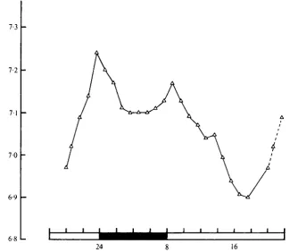
Fig. 3. Diel changes in pH in one male cockroach measured with an implanted pH electrode. LD 16:8. Dotted lines join points that have been plotted twice.
are broadly similar to those in the Periplaneta saline devised by Treherne et al. (1975). This saline was used for calibrating the electrodes in the present study.
The pH of the blood of one individual Leucophaea, measured with an implanted electrode, varied between 6-90 and 7-24 over a 22 h period (Fig. 3). There were two peaks, at dusk and dawn, corresponding to the periods of maximum locomotor activity, but it would be unwise to infer too much from the record of one individual.
Preliminary measurements of haematocrit indicate that between 2 and 6% of the haemolymph volume is occupied by haemocytes (14 measurements from 7 male Leucophaea); this compares with Brady's (19676) figure of 2-6% from 16 animals.
2. Diel changes in potassium activity
The diel changes in potassium activity of three adult male Leucophaea in LD 16:7 are shown in Fig. 4. There is a dramatic variation in K+ activity throughout the day: the maximum range for an individual cockroach in 24 h was from 4-5 to 25-0 mM. There is a consistent pattern in this diel change in potassium activity; a minimum about an hour before lights-off, rising to a peak during the hours of darkness, falling to a second low point about 2 h after dawn, and rising to a second peak during the day. A composite curve from 14 cockroaches, with activity expressed as a % change from the maximum for each individual, is shown in Fig. 5.
i78 JANINE LETTAU AND OTHERS tivi l « 30 25 20 15 10 5 0
A
/ 1/ ^ 1
/\ 1
/ \
/ V
-1 -1 -1 -1 -1
7 \
/ \
/A \
/A ^
/LA\
i i i i i
A
/ ^
\ / X*
\ / ^ N
?
\ / // ^
\ /
V^
?
;' y
V
r' i i '• \> '• \ o
Ff. i O
^^•Q Q ( it o
t *
U d ' ' 1
16 24 08 16 24 08
Fig. 4. Diel changes in K+ activity in 3 individual male cockroaches. Dotted lines join points that have been plotted twice.
The remaining cockroaches were sham-operated as though for measurement of potassium activity, but only locomotor activity was measured. There is a sharp peak in locomotor activity in the hour after lights-off. Activity remains relatively high during the remainder of the dark period with a small broad peak in the hours around lights-on. The major peak in locomotor activity occurs about 1 h after the first low point in K+ and the small dawn peak in locomotor activity some hours before the second low point in potassium activity.
The individual potassium activity records indicate that the observed pattern is probably not the result of some artefact of implantation (e.g. wounding). Individual cockroaches showed the same pattern of K+ activity no matter what time of day the electrodes were implanted (Fig. 4). Wounding causes an increase in haemocyte numbers in Periplaneta (Brady, 1967 A), and this leads to an increase in whole-blood K+ con-centration of the order of 2-0 mM (Brady, 19676). The effect of this on K+ activity would probably be of little importance.
3. Diel changes in sodium activity
179
JANINE LETTAU AND OTHERS
I I I I I I I I
16 24 08 16 24 08 Fig. 6. Diel changes in Na+ activity in 4 individual male cockroaches. Dotted lines join points
that have been plotted twice.
150r
no
1-1 -1 -1 1 1 1 1 1
16 24 08 16 24 08 Fig. 7. Diel changes in whole-blood Na+ concentration. Bars are ± 1 x s.E. of the mean. Dotted
30
25
-151
-10 24 08 16 24 08
Fig. 8. (a) Composite curve of diel changes in K+ activity from 14 cockroaches, expressed in
mM. Bars are ± 1 x s.E. of the mean. (6) Diel changes in mean whole-blood K+ concentration.
Points are means of 12-24 readings from 6—14 individual males. Bars are ± 1 x s.E. of the mean. L D 16:8. Dotted lines join points that have been plotted twice.
4. Diel changes in concentrations of potassium and sodium
182 JANINE LETTAU AND OTHERS
lights-off, but there is a considerable scatter about the mean for each value and a lessl consistent pattern than for K+.
Whole blood potassium concentration rises to a peak about 5 h before lights-off, falls a little an hour before lights-off, rises again at the beginning of the dark period and falls away to low values during the remainder of the dark period (Fig. 86). The fluctuations in potassium concentration and activity follow a roughly similar pattern (Fig. 8 a).
DISCUSSION
The present observations clearly emphasize the accepted view (e.g. Pichon, 1970) that insect blood is a highly variable ionic environment. The observations further demonstrate, for the first time, a marked and consistent diel rhythm in the activity of a major blood ion, potassium. This rhythm is probably of particular significance to the functioning of excitable tissues - nerve and muscle. Although the insect CNS and larger peripheral nerves are protected behind the so-called 'blood-brain' barrier (Treherne & Pichon, 1972; Treherne, 1974), the smaller peripheral nerves (Lane & Treherne, 1973) and the muscles and neuromuscular junctions (Osborne, 1970, 1975) appear not to be sheathed from the blood that bathes them. There is unfortunately no published information on the electrophysiology of Leucophaea nerve or muscle, but the present observations may be related to electrophysiological analysis of excitable
tissues in other insects.
Preliminary, day-time (12.00-18.00 h) measurements of potassium activity in 13 individual Periplaneta americana with ion-selective electrodes gave a mean value of 9-4 mM (range 7-7-11-5 mMJ (W. A. Foster, unpublished observations). A diel fluctua-tion in blood potassium activity in Periplaneta comparable to that shown by Leucophaea (say 60 %, Fig. 5 a) would cause severe disturbance of central nervous function, were it not for the existence of an effective blood-brain barrier (Thomas & Treherne, 1975). If the smaller peripheral nerves in Leucophaea are, by analogy with those of Periplaneta, freely accessible to small ions from the blood, then the observed diel variation in K+ activity could have a profound effect on axonal function.
Changes in blood potassium activity of the magnitude described here (e.g. 4-5-25*0 mM) might be expected to produce axonal depolarization that would induce significant inactivation of the inward current of the action potential (see, Hodgkin & Huxley, 1952). In desheathed cockroach giant axons, for example, elevation of the potassium level from the supposed extracellular concentration of 3-0 mM in intact connectives caused substantial reduction of the amplitude of the recorded action potential at 10 mM and conduction block at around 12-5 mM-K+ (Thomas & Treherne, 1975). It is possible, therefore, that any exposed small peripheral axons might be specialized to enable them to function at high potassium concentrations, either because their resting potential is unusually insensitive to external potassium concentrations in this physiological range, or because sodium-inactivation following large depolarizations is insufficient to block action potentials, as has been demonstrated in the giant axon of a serpulid worm (Carlson & Treherne, 1977).
IGbssina, step changes in external potassium concentration similar to the diel changes observed in the present investigation have been shown to depolarize the neurosecretory terminals, causing the release of diuretic hormone from the neurohaemal organs (Maddrell & Gee, 1974). If the neurohaemal areas in Leucophaea are accessible, as in Rhodnius, then it follows that the diel changes in potassium activity could profoundly affect the release of neurosecretory products into the blood and could, as a result, influence a variety of bodily functions. It is, of course, impossible to predict the extent of such an effect since it is likely to depend upon the rate of depolarization and the kinetics of calcium inactivation (see Baker & Rink, 1975; Nordmann, 1976).
The resting potentials of cockroach muscle (Usherwood, 1969; Wareham, Duncan & Bowler, 1974) and locust muscle (Hoyle, 1954) are sensitive to changes in external concentrations of potassium. Wareham et al. (1974) demonstrated that there is a linear relation between external potassium concentration and muscle resting potential in Periplaneta, with a slope of 43 mV per decade change in potassium concentration. In locusts, Hoyle (1954) showed that changes in potassium concentration of up to 60% could follow a short period of starvation. This could in turn account for an increase in the muscle resting potential of about 12 mV and an improvement in the mechanical responses of the muscles to nervous stimulation. In Leucophaea, changes of this magnitude apparently occur twice daily and will almost certainly influence muscle resting potential and also muscle performance.
JANINE LETTAU AND OTHERS
10 0-9 0-8 0-7 0-6 0-5 0-4 0-3 0-2 0 1 0
i i i
16 24 08 16 24 08
Fig. 9. Diel changes in apparent activity coefficient of potassium (T"K). See text for details. Bars are ± 1 x s.E. of the mean. Dotted lines connect points that have been plotted twice.
It is not clear what mechanisms directly control the variations in potassium activity. Broadly speaking, K+ activity could change either by movement of K+ between com-partments within the haemolymph or by movement in and out of the haemolymph. K+ may become associated with soluble blood proteins or with the haemocytes, which in cockroaches may contain a significant proportion (sometimes more than 50%) of the total blood potassium (Brady, 1967 a, b). It is clear from observations on the dehydration and rehydration of Periplaneta that there is considerable capacity for storage of Na+ and K+ in sites outside the haemolymph (Wall, 1970). An increased rate of Malpighian tubule secretion could reduce total K+ by the temporary accumula-tion of a potassium-rich fluid in the tubules during solute recycling.
A comparison of diel changes in total blood K+ (Fig. 8) and K+ activity (Fig. 8 a) gives some indication of the movement of K+ within the blood. A plot of the apparent activity coefficient yk (see Treherne et al. 1975), derived from values in Fig. 8, gives an idea of the diel changes in the proportion of freely available potassium in the blood (Fig. 9). Assuming that the blood is broadly similar, in terms of ionic strength and available water content, to the calibration saline (Table 1), y ^ should not exceed 0-74. The fact that it does suggests, for example, that at certain times of day the effective amount of solvent water in the blood is reduced by sequestration in some compartment. The haemocytes are unlikely to form a large part of this compartment since they occupied only some 2-6 % of the blood volume.
tend to make blood proteins more negative and hence attractive to K+. Confirmation of the pH change would be of great interest. Further measurements, particularly paired readings of K+ activity and whole-blood K+ in the same individual, are required before the trends outlined in Fig. 9 can be confirmed.
The fact that the baseline potassium activity of insect blood may fluctuate in a diel fashion is of considerable practical importance to those who study the physiology of insect tissues, in particular excitable tissues. For example, it may well be important to standardize the time of day of an experiment and to take diel variations into account in the elaboration of insect salines.
It is hoped that the present observations will provoke further work on the diel variations in insect blood ions, particularly in relation to the role of potassium in the expression of circadian locomotor activity.
This work was supported by a Science Research Council Grant to Dr J. E. Harker. We are indebted to Dr R. B. Moreton for helpful discussions. Mr P. C. Stearn gave valuable practical assistance. Mr J. W. Rodford prepared Fig. 2.
REFERENCES
ASPKREN, K. VAN & ESCH, I. VAN (1956). The chemical composition of the haemolymph in Periplaneta
americana with special reference to the mineral constituents. Archs nierl. Zool. iz, 342-360.
BAKER, P. F., RINK, T. J. (1975). Catecholamine release from bovine adrenal medulla in response to maintained depolarization. J. Physiol. 253, 593-620.
BRADY, J. (1967a). Haemocytes and the measurement of potassium in insect blood. Nature, Lond.
315.
96-97-BRADY, J. (19676). The relationship between blood ions and blood-cell density in insects. J. exp. Biol. 47. 3i3-3a6.
BRADY, J. (1968). Control of the circadian rhythm of activity in the cockroach. III. A possible role of the blood-electrolytes. J. exp. Biol. 49, 30-47.
CARLSON, A. D. & TREHERNE, J. E. (1977). Ionic basis of axonal excitability in an extreme euryhaline osmoconformer, the serpulid worm Mercierella etngmatica (Fauvel). J. exp. Biol. 67, 205-215. CHAPMAN, R. F. (1958). A field study of the potassium concentration in the blood of the red locust
Nomadacris septemfasciata (Serv.) in relation to its activity. Aram. Behaviour 6, 60—67.
CYMBOROWSKI, B. & BRADY, J. (1972). Insect circadian rhythms transmitted by parabiosis - a re-examination. Nature Netv Biol. Lond. 336, 221-222.
EISENMAN, G. (1962). Cation selective glass electrodes and their mode of operation. Biophyt.J. 2 (suppl.),
259-333-ELLIS, P. E. & HOYLE, G. (1954). A physiological interpretation of the marching of hoppers of the African migratory locust {Locusta migratoria migratorioidet R. & F.). J. exp. Biol. 31, 271-279. ESKIN, A. (1972). Phase shifting a circadian rhythm in the eye of Aplysia by high potassium pulses.
J. comp. physiol. 8o, 353-376.
FLORKIN, M. & JEUNIAUX, C. (1974). Hemolymph composition. In The Physiology of Insecta (ed. M. Rockstein), vol. 5, pp. 255-307. New York and London: Academic Press.
HARKER, J. E. (1956). Factors controlling the diurnal rhythm of activity of Periplaneta americana L.
J. exp. Biol. 33, 224-234.
HINCKE, J. A. M. (1959). Glass microelectrodes for measuring intracellular activities of sodium and potassium. Nature, Lond. 184, 1257-1258.
HODGKJN, A. L. & HUXLEY, A. F. (1952). The dual effect of membrane potential on sodium conductance in the giant axon of Loligo.J. Physiol. 116, 497-506.
HOYLE, G. (1954). Changes in the blood potassium of the African migratory locust (Locusta mtgratoria
mjgratorioides R. and F.) during food deprivation and the effect of neuromuscular activity, jf. exp. Biol. 31, 260-270.
KHURI, R. N., AGULIAN, S. K. & WISE, W. M. (1971). Potassium in the rat kidney proximal tubules
in situ: determination by K+-selective liquid ion-exchange microelectrodes. Pflugers Arch. ges. Physiol. 3**, 39-46.
186 JANINE LETTAU AND OTHERS
LANE, N. J. & TRBHBRNE, J. E. (1973). The ultrastructural organization of peripheral nerves in twoj insect species (Periplaneta americana and Schutocerca gregaria). Tissue and Cell 5, 703-714. MADDRBLL, S. H. P. & GEE, J. D. (1974). Potassium-induced release of the diuretic hormones of
Rhodmus prolixus and Glossina austeni: Ca dependence, time course and localization of neurohaemal
areas. J. exp. Biol. 61, 155-171.
MOORHOU8E, J. E. (1968). Locomotor activity and orientation in locusts. Ph.D. thesis, University of
London.
Njus, D.( SULZMAN, F. M. & HASTINCS, J. W. (1974). Membrane model for the circadian clock. Nature,
Land. 348, 116-120.
NOHDMANN, J. J. (1976). Evidence for calcium inactivation during hormone release in the rat neuro-hypophysis.J'. exp. Biol. 65, 669-683.
OSBORNE, M. P. (1970). Structure and function of neuromuscular junctions and stretch receptors.
Symp. R. ent. Soc. Land. 5, 77-100.
OSBORNE, M. P. (1975). The ultrastructure of nerve-muscle synapses. In Insect Muscle (ed. P. N. R. Usherwood), pp. 151-205. London and New York: Academic Press.
PICHON, Y. (1970). Ionic content of haemolymph in the cockroach, Periplaneta americana. A critical analysis. J. exp. Biol. 53, 195-209.
PICHON, Y. & BOISTEL, J. (1963). Modifications of the ionic content of the haemolymph and of the activity of Periplaneta americana in relation to diet. J. Insect Physiol. 9, 887-891.
RAMSAY, J. A., BROWN, R. H. J. & CROGHAN, P. C. (1955). Electrometric titration of chloride in small volumes. J. exp. Biol. 3a, 822-829.
ROBINSON, R. A. & STOKES, R. H. (1959). Electrolyte Solutions. 2nd ed. London: Butterworths. SATTER, R. L., GALSTON, A. W. & RACUSEN, R. (1975). Rhythmic changes in K+ content and electrical
properties of plant motor cells. Biophys.J. 15, 176a.
THOMAS, M. V. & TREHERNB, J. E. (1975). An electrophysiological analysis of extra-axonal sodium and potassium concentrations in the central nervous system of the cockroach {Periplaneta americana L.). J. exp. Biol. 63, 801-811.
TODD, M. E. (1958). Blood composition of the cockroach Leucophaea maderae Fabricius. J. N. York
ent. Soc. 66, 135-143.
TREHERNE, J. E. (1974). The environment and function of insect nerve cells. In Insect Neurobiology (ed. J. E. Treherne), pp. 187-244. Amsterdam: North Holland.
TREHERNB, J. E., BUCHAN, P. B. & BENNETT, R. R. (1975). Sodium activity of insect blood; physiological significance and relevance to the design of physiological saline. J. exp. Biol. 6a, 721-732.
TREHERNE, J. E. & PICHON, Y. (1972). The insect blood-brain barrier. Adv. Insect Physiol. 9, 257-313. USHERWOOD, P. N. R. (1969). Electrochemistry of insect muscle. Adv. Insect Physiol. 6, 205-278. WALL, B. J. (1970). Effects of dehydration and rehydration on Periplaneta americana. J. Insect Physiol.
16, 1027-1042.