572
In vivo Quantitation of Regional Cerebral Blood Flow in
Glioma and Cerebral Infarction: Validation of the
HIPDm-SPECT Method
Burton
Dray
er,"
2
Ronald Jaszczak,' Allan Friedman,
3
Robert Albright,
'·2
Hank Kung,
4
Kim Greer
,'
Mi
chael
Lischko
,'
Neil Petry
,'
and
Edward Coleman'
todine-123 labeled hydroxyiodopropyldiamine (HIPDm) is a diffusible indicator with an 85%-90% extraction fraction and stable retention in the brain for more than 2 hr. Equilibrium-phase imaging and quantitation using single-photon emission computed tomographic (SPECT) scanning defined a distribution of HIPDm in proportion to regional cerebral blood flow (rCBF). Studies in calves affirmed a close correspondence (r
=
0.97) in calculated rCBF between HIPDm and microspheres using the tissue deposition-arterial input function microsphere methodol-ogy. Using this same mathematical analysis in vivo, reproducible rCBF data within the expected range of normal were obtained on repeated studies in the same nonhuman primate. With a diffuse encephalopathy secondary to subarachnoid blood, a bilaterally symmetric decrease in rCBF was present. A prominent focal decrease in HIPDm accumulation and calculated rCBF was noted with cerebral infarction in the distribution of a ligated middle cerebral artery. Patient studies with glioma revealed diminished HIPDm accumulation due to decreased flow and/or pH in the region of the neoplasm as well as in the associated vasogenic edema and overlying gray matter.Since the initial application of xenon-133 s.tudies in man [1, 2], new techniques have been sought to characterize regional cerebral blood flow (rCBF) with improved morphologic specificity. The non-radioactive xenon-transmission computed tomographic (CT) method [3] provides excellent anatomic resolution but is somewhat cumbersome due to the stringent requirements of the gas-breathing apparatus, particularly if end-tidal xenon concentration is to accu-rately reflect the arterial input function. Oxygen-15 techniques using positron emission tomography (PET) require a costly on-site cyclotron and chemistry laboratory [4].
New approaches are therefore being explored to use the far less costly and more widely accessible technique of single-photon emis-sion computed tomography (SPECT) in conjunction with diffusible inert gasses [5] or new classes of iodine-123-labeled radiotracers [6-12]. This study describes the validation of rCBF measurements calculated using one of these iodine-123-labeled radi opharmaceu-ticals, hydroxyiodopropyldiamine (HIPDm) [13]. We are thus at-tempting to determine whether rCBF in ml 100 g-I min-I can be determined accurately and reproducibly in living subjects. Although
a visual or quantitative analysis of brain distribution is sufficient for defining an abnormality when a focal pathologic process is present, an absolute value for rCBF is necessary if studies are to be com-pared in different stages of disease or an analysis is to be made of diffuse, bilaterally symmetric pathologic processes (e.g., Alzheimer disease, metabolic encephalopathy).
Materials and Methods
Isotope Detection
The Duke-Siemens SPECT system [14, 15] consists of two o p-posing large-field-of-view (LFOV) gamma scintillation cameras mounted on a rotatable gantry with each head rotating through 3600
in 2-26 min. Our studies use low-energy, all-purpose parallel (LEAP) or medium-energy collimators. Thirty-two contiguous transaxial sections, each 6.4 mm thick, were reconstructed from projection data acquired during a single continuous 3600
rotation. The data were also reformatted into longitudinal sections. An inter-nal first-order attenuation correction was applied to the data during the reconstruction to compensate for the effects of gamma ray attenuation. In addition to a visual analysis of the hard film copy, regions of interest (ROI) were selected for computing counts/voxel and ultimately rCBF.
Isotope Preparation
N ,N, N '-trimethyl-N '-(2-hydroxyl-3-methyl-5-iodobenzyl)-1 ,3-pro-panediamine, HCI (HIPDm) (fig. 1) was synthesized and prepared in kit form [13]. High-purity, no-carrier-added iodine-1 23 (T'h = 13.3 hr) was made by the 1271 (P,5n) '23Xe_'231 reaction (Crocker Nuclear Laboratory, University of California at Davis). The radiochemical purity of the labeled HIPDm was greater than 93% and the dose of iodine-123 was 10-14 mCi (370-519 MBq) for animal studies and 5-6 mCi (185-222 MBq) for patients.
HIPDm Characterization
The first pass fraction of 1231_HIPDm extracted by brain tissue was estimated after a bolus infusion of the radiotracer into the internal carotid artery and the monitoring of the initial time activity
'Department of Radiology, Box 3808, Duke University Medical Center, Durham, NC 27710. Address reprint requests to B. P. Drayer.
'Department of Neurology, Duke University Medical Center, Durham, NC 27710. 'Department of Neurosurgery, Duke University Medical Center, Durham, NC 27710.
'Department of Nuclear Medicine, Veterans Administration Medical Center, Buffalo, NY 14215.
AJNR:4, May/June 1983
PET AND RADIONUCLIDE
STUDIES
5
7
3
curve from consecutive 1 sec collections using a gamma camera. The retention of 1231_HIPDm activity in the brain was analyzed using three 26 min SPECT scans obtained in the 8-34, 40-66, and 100-126 min intervals after the intracarotid or intravenous administration of thp radiotracer. All baboon (Papio cynocephalus) studies were performed in an intubaled animal using intravenous sodium pento-barbital anesthesia. The CO2 concentration in the arterial blood was in the 35-45 torr range.
Cerebral Blood Flow Analysis
If a close correspondence exists between the derived rCBF as determined by carbonized microspheres and HIPDm, the straight-forward flow equation based on organ deposition and arterial input sampling [16] can be used for in vivo HIPDm ("metabolic micro-sphere") studies: F = (WCi/Ca)
x
100, where F is the rCBF in ml 100 g-I min-1, W the constant withdrawal rate in ml min-1, Ci the
HO
Fig. 1 .-N,N,N'-trimethyl-N'-(2-hydroxyl-3-methyl-5-iodobenzyl)-1 ,3 -pro-panediamine· HCI(HIPDm).
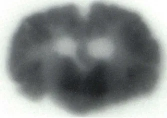
Fig. 2._ 1251_HIPDm autoradiography. A 12-mm-thick coronal autoradiographic section of cow brain confirms that >251_HIPDm distribution in brain (gray>
white> cerebrospinal fluid) is proportional to rCBF.
Fig. 3.-Cerebral infarction in a ba -boon 48 hr after surgical ligation of mid-dle cerebral artery (MCA) resulted in profound contralateral hemiparesis, hemianesthesia, and homonymous he m-ianopsia. A, CT. Large area of de-creased density in distribution of ligated MCA. B, SPECT. Decreased accumula -tion of HIPDm (arrows) in same MCA distribution consistent with decreased rCBF. C, Gross pathology. Gross sp eci-men of infarction (arrows) in MCA te rri-tory.
A
activity of 1231_HIPDm in a selected ROI from the SPECT scans in counts sec-I g-I, and Ca the total activity by well-counting in an arterial blood sample constantly withdrawn over the immediate 5 min of radiotracer infusion minus the activity in a 5 min sample begun 10 min after infusion in counts sec-I. A calibration factor to convert well-counter activity to SPECT unit activity is derived by well-counting and SPECT-scanning the blood. "Arterialized" ve-nous blood [17, 18] was drawn from a hand vein after warming to 44°C in a specially designed hand warmer for human studies. Brair activity from chosen ROls (Ci) was obtained from the 8-34, 40-66, and 100-126 min SPECT scans. In order to compensate for finite resolution and partial volume averaging affecting the mean activity in the selected gray or white matter ROI, we used a simulated brain phantom containing proportional concentrations of iodine-123.
To determine the reproducibility of the SPECT -HIPDm method, the same normal baboon was studied three times at weekly inte r-vals. This same animal was studied again 2 weeks later while diffusely encephalopathic following a retroorbital craniotomy and resultinq subarachnoid hemorrhage. After clinical recovery, a fifth study was performed on this animal 2 days after ligation of the left middlE. c:erebral artery producing focal cerebral infarction with n eu-rologic symptomatology.
In order to verify the use of the microsphere paradigm for HIPDm studies, scandium-46 microspheres and iodine-125 HIPDm were injected via a catheter into the left ventricle of the heart in the calf. The animals were killed at 30 min followed by excision of multiple small sections of gray and white matter for well-counting. In add i-tion, arterial blood was drawn at a constant rate over the 0-5 and 10-15 min time intervals. Thick-section autoradiographs were also prepared from 1 2-mm-thick coronal brain sections to correspond to the SPECT slice thickness.
Human Studies
Four patients with glioma were studied after the infusion of 5-6 mCi (185-222 MBq) of HIPDm. The Ca was determined from arterialized venous blood withdrawn at a constant rate and the Ci from chosen ROls (25
x
25 mm) on the SPECT scans. All patients had an intravenously enhanced CT study performed to determine tumor localization, blood-brain barrier integrity, and extent of va-sogenic edema. A SPECT scan was also obtained to analyze blood-brain barrier integrity using 99mTc-glucoheptonate and compared with the SPECT-HIPDm rCBF study.Results
Extraction and Retention
In addition to the brain, the major uptake of HIPDm occurs in the lungs. No significant activity was seen in the eyes. The first pass
[image:2.612.64.292.285.375.2] [image:2.612.94.261.428.546.2] [image:2.612.100.553.614.730.2]574
PET AND RADIONUCLIDE STUDIES
AJNR:4, May/June 1983A
B
Fig. 4.-Glioblastoma multiforme (GBM) in 53-year-old man. Decreased accumulation of HIPDm
(arrows) in GBM, associated vasogenic edema, and
adjacent parietal gray matter. Milder decrease in HIPDm accumulation (rCBF) in ipsilateral occipital
lobe, not involved by tumor or edema. Normal gr
ay-white distribution of HIPDm evident in contralateral
hemisphere.
Fig. 5.- Comparative distribution of HIPDm versus microspheres. In vivo comparison in two separate cows after left ventricular infusion of 99mTc microspheres (A) and 1231_HIPDm (B). Similarity of gray-white distribution is striking in visual and quantitative terms, further affirming mathematical
modeling of HIPDm as "metabolic microspheres."
extraction was 85%-90% at a mean CBF of 50-55 ml 100 g-1
min-I and pH of 7.35-7.40. The brain activity and the gray-white
distribution remained constant in all studies over the 2 hr of sca
n-ning. The gray-white distribution in the normal studies was 1.32
±
0.14 at an approximate spatial resolution of 15 mm FWHM. After
phantom compensation, the gray-white ratio was in the 4 to 1 range.
This coincided closely with the iodine-1 25 HIPDm autoradiographic
studies (fig. 2), which also demonstrated a distribution of activity in the gray and white matter in proportion to capillary density (i.e., about 4 to 1).
Equilibrium Image Analysis
A visual distinction was consistently present between the gray
and white matter. Focal regions of diminished distribution of HIPDm
were visually apparent with both cerebral infarction (fig. 3) and
glioblastoma multiforme (fig. 4). There was also a less prominent
decreased distribution of HIPDm in the occipital lobe (fig. 4) i
psilat-eral to a glioma involving the more anterior visual pathway. There
was a close correspondence between the SPECT scan appearance
using technetium-99m microspheres and iodine-123 HIPDm in-jected into the left ventricle of a calf (fig. 5). In both studies, there
was symmetric distribution of activity bilaterally and a distinction of
the increased activity in gray matter from the underlying white
matter.
Quantitative Analysis of rCBF
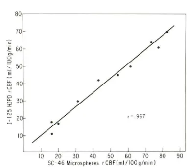
A close correlation was obtained when comparing rCBF
mea-sured using 1231_HIPDm and 46Sc-microspheres (fig. 6). A Spearman
rank correlation was used to determine an
r
of 0.97 on blood flowsfrom 15-90 ml 100 g-I min-I. Due to this close correspondence,
the mathematical modeling developed for use with microspheres
was applied to the in vivo HIPDm studies.
The results of serial rCBF estimations in the same baboon on the
same day and on different days are summarized in table 1. The
phantom-derived compensation factor was 1.73 for regions of co
r-80
70
c ~ 60
~
0 0
,
50E
u-40 OJ
u
C> "- 30
r
'"
~.
'
967..c
2010
10 20 30 40 50 60 70 80 90 SC-46 Microspheres r CSF I ml / 100q/min I
Fig. 6.-Verification of microsphere methodology for HIPDm rCBF es ti-mations. Tight correspondence between '6SC microspheres and 1251_HIPDm_ derived rCBF with flows of 10-90 ml 100 g-1 min-1 (pH 7.35-7.40). The ,sSc microspheres were infused into left ventricle of the cow immediately after left ventricular infusion of HIPDm.
tical gray matter and 0.59 for white matter locales. Using these compensation factors, the expected 4 to 1 ratio of gray to white
matter flow was obtained. A statistically significant difference in
gray matter flow in the frontal, temporal, parietal, and occipital lobes was not noted.
When the animal had a diffuse encephalopathy after subarach
-noid hemorrhage, a bilaterally symmetric decrease in gray and
white matter flow was noted. With cerebral infarction, a prominent,
well defined, focal area of diminished HIPDm accumulation was
apparent in the distribution of the ligated middle cerebral artery (fig.
3). The extent of severely diminished flow was slightly decreased
as compared with the abnormality noted on CT scanning. The
contralateral hemisphere showed no abnormality on non enhanced
[image:3.612.60.560.79.252.2] [image:3.612.341.532.338.509.2]AJNR:4, May/June 1983 PET AND RADIONUCLIDE STUDIES
575
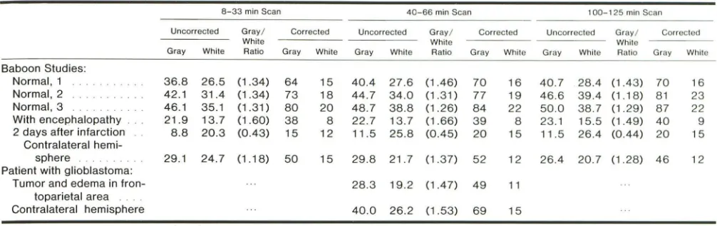
TABLE 1 : SPECT -HIPDm: Normal and Pathologic rCBF in a Baboon and Pathologic rCBF in a Patient
8-33 min Scan 40-66 min Scan 100-1 25 min Scan
Uncorrected Gray/ Corrected Uncorrected Gray/ Corrected Uncorrected Gray/ Corrected
White While White
Gray While Ratio Gray While Gray While Ratio Gray While Gray White Ratio Gray While
Baboon Studies:
Normal,1 36.8 26.5 (1.34) 64 15 40.4 27.6 (1.46) 70 16 40.7 28.4 (1.43) 70 16
Normal,2 42.1 31.4 (1.34) 73 18 44.7 34.0 (1.31) 77 19 46.6 39.4 (1.18) 81 23
Normal,3 46.1 35.1 (1.31) 80 20 48.7 38.8 (1.26) 84 22 50.0 38.7 (129) 87 22
With encephalopathy 21.9 13.7 (1.60) 38 8 22.7 13.7 (1.66) 39 8 23.1 15.5 (1.49) 40 9 2 days after infarction 8.8 20.3 (0.43) 15 12 11.5 25.8 (0.45) 20 15 11.5 26.4 (0.44) 20 15
Contralateral
hemi-sphere 29.1 24.7 (1.18) 50 15 29.8 21.7 (1.37) 52 12 26.4 20.7 (1.28) 46 12
Patient with glioblastoma:
Tumor and edema in fron- 28.3 19.2 (1.47) 49 11
toparietal area
Contralateral hemisphere 40.0 26.2 (1.53) 69 15
Note.-Mean (n = 5) rCBF in ml 100 9 1 min 1 calculated from 25 X 25 mm regions of interest. Coefficient of variation of derived SPECT counts is 5%-11 %. Predicted gray /while
ratio at 15 mm FWHM = 1.41 ± 0.42 as per Mazziotta et al. (19).
gray matter flow on the SPECT -HIPDm studies, consistent with
diaschisis.
In all four patients with high-grade astrocytomas, a decreased
accumulation of HIPDm was apparent on the SPECT study (fig. 4).
The region of decreased activity was larger than the area of blood
-brain barrier abnormality as determined using 99mTc-glucohept
on-ate. The SPECT -HIPDm abnormality coincided quite closely, how
-ever, with the area of contrast enhancement and associated vaso
-genic edema as seen on the CT scan. There was decreased uptake
of HIPDm not only in the neoplasm, but also in the overlying gray matter and uninvolved ipsilateral occipital lobe. The decrease in
calculated flow was less with glioma than with acute infarction (table
1).
Discussion
Studies in animals as well as initial studies in man strongly
suggest that the HIPDm-SPECT method will provide useful and
reproducible data concerning rCBF. HIPDm crosses the blood-brain barrier with a high first-pass extraction. The radiopharmaceutical is
retained in the brain for more than 2 hr at an equilibrium concentra
-tion, permitting the acquisition of high-quality images without time
restraint. Even though a large amount of the injected dose is trapped by the lungs, about 5%-6% of the injected dose enters the brain.
There has been no evidence of toxicity in either mammals or man.
The lipid solubility of HIPDm accounts for its ready permeability across the blood-brain barrier. The mechanism of brain retention is far more complex. Current theories suggest a pH gradient hypot
h-esis in which the non ionized diamine in the blood stream is lipophilic
and thus crosses the blood-brain barrier. However, with the lower
pH in the brain, the cationic radiotracer is no longer lipophilic and
thereby becomes metabolically trapped within the brain substance
[7]. Loberg [8] suggests that the pH gradient hypothesis may
account for the initial uptake of HIPDm in the brain, however, the
final distribution of the agent may be related to other types of
metabolism or binding. Additional studies would also be of value to
determine whether the small percentage of residual circulating
iodine-123 is free, bound to HIPDm, or bound to one of its met ab-olites.
The high degree of correspondence in postmortem studies using
simultaneously injected microspheres and HIPDm confirmed the utility of using the mathematical microsphere methodology for es
ti-mating rCBF in vivo using the SPECT -HIPDm paradigm. Due to the
excellent retention of HIPDm, blood flow can be calculated at any
time in the first 2 hr after intravenous infusion of the radiopharm
a-ceutical using the equilibrium SPECT image to quantitate brain
concentration. The 1.32 ± 0.14 ratio of gray-to-white activity is fully consistent with the computer-simulated data of Mazziotta et al.
[19] when the full-width-half-maximum is about 15 mm. The use of
a simulated brain phantom provides a useful compensation factor
for the underestimation of gray-matter flow and overestimation of
white-matter flow derived from the uncorrected SPECT scan data.
As expected, estimated gray-matter flows were decreased
sym-metrically and bilaterally in the diffusely encephalopathic baboon.
There was a prominent decrease in HIPDm accumulation with
cerebral infarction in the middle cerebrill artery distribution appar
-ent by both visual and quantitative analysis. However, the decrease
in flow in the contralateral hemisphere (diaschisis) was not apparent
on visual inspection. In this animal with a large cerebral infarction, crossed cerebellar diaschisis was not found. The mechanism of the
decrease in HIPDm accumulation needs elucidation as to whether
this is a pure flow phenomenon or related to the pH gradient changes.
The mechanism of the diminished accumulation of HIPDm with
gliomas is also not straightforward [20]. This decrease in tracer accumulation even in the presence of an abnormality in blood-brain
barrier permeability and hypervascularity as determined by angi
og-raphy suggests that the decreased brain activity may truly represent
a decrease in flow rather than or in addition to an abnormality in the pH gradient. Gliomas are known to have decreased metabolic rate
with associated anaerobic glycolysis with resulting decrease in oxygen consumption [21]. PET studies have also suggested a mild decreased flow with gliomas [22]. An additional factor is the de-creased flow in the surrounding vasogenic edema and overlying
cerebral cortex. The decline in rCBF in the region of vasogenic
edema may be related to a combination of increase in volume
(water) as well as decrease in flow due to compression of the
microcirculation.
The HIPDm-SPECT studies showed abnormalities in flow in
regions of brain where no structural abnormality was noted by CT
scan. The decrease in flow in the structurally uninvolved occipital
lobe in the glioma patient with a lesion involving the visual pathway
more anteriorly and the contralateral hemisphere with cerebral
infarction highlights the additional information concerning the
un-derstanding of disease that a blood flow study provides. As with
PET scanning, HIPDm-SPECT studies can thus be applied to the
[image:4.612.53.556.92.251.2]576
PET AND RADIONUCLIDE
STUDIES
AJNR:4, May/June 1983Although 1231-HIPDm is an extremely useful radiotracer to m
eas-ure rCBF, iodine-123 is cyclotron-produced. Its 13.3 hr half-life requires rapid transportion if an on-site cyclotron is not available. If
the measurement of brain function using SPECT scanning is to
achieve more widespread popularity, the development of a tech -netium-99m-labeled flow tracer is critical. In affirming the accuracy
and reproducibility of the SPECT quantitation of brain activity, the value of such a radiotracer becomes apparent, even at the
com-munity hospital level. Until the development of technetium-99m-labeled radiopharmaceuticals for evaluating brain metabolism, io -dine-123-labeled compounds can serve as a very useful alternative
and provide for a far greater degree of labeling flexibility.
REFERENCES
1. Mallett BL, Veall N. Measurement of regional cerebral clea
r-ance rates in man using xenon-133 inhalation and extracranial recording. Clin Sci 1965;29: 179-191
2. Obrist WD, Thompson HK, Wang HS, et al. Regional cerebral blood flow estimated by 133Xenon inhalation. Stroke 1975;6: 245-256
3. Drayer BP. Functional applications of CT of the central nervous system. AJNR 1981;2: 495-510
4. Frackowiak RS, Lenzi GL, Jones T, et al. Quantitative m
ea-surement of regional cerebral blood flow and oxygen metabo -lism in man using 150 and positron emission tomography: theory, procedure, and normal values. J Comput Assist Tomogr
1980;4: 727 -736
5. Kanno I, Lassen NA. Two methods for calculating cerebral
blood flow from emission computed tomography of inert gas concentrations. J Comput Assist Tomogr 1979;3: 71-76 6. Winchell HS, Baldwin RM, Lin TH. Development of 1-123-
la-beled amines for brain studies: localization of 1-123 iodophen-ylalkyl amines in rat brain. J Nucl Med 1980;21 :940-946
7. Kung HF, Blau M. Regional intracellular pH shift: a proposed new mechanism for radiopharmaceutical uptake in brain and
other tissues. J Nucl Med 1980;21 : 147 -152
8. Loberg MD. Radiotracers for cerebral functional imaging-a new class. J Nucl Med 1980;21: 183-186
9. Kuhl DE, Barrio JR, Huang SC, et al. Quantifying local cerebral
blood flow by N-isopropyl-p-('231)iodoamphetamine (IMP)
to-mography. J Nucl Med 1982;23: 196-203
10. Hill TC, Holman BL, Lovett R, et al. Initial experience with
SPECT (single photon computerized tomography) of the brain using N-isopropyl 1-123 p-iodoamphetamine: concise comm u-nication. J Nuc/ Med 1982;23: 191-195
11. Drayer B, Jaszczak R, Friedman A, et al. Tomographic m
eas-urement of regional brain perfusion using hydroxyiodopropy
l-diamine and single photon emission computed tomography. Stroke 1982;13:12
12. Coleman RE, Drayer BP, Jaszczak RJ. Studying regional brain
function: a challenge for SPECT. J Nucl Med 1982;23: 266
-270
13. Tramposch KM, Kung HF, Blau M. Brain imaging with 1-123 labeled diamines: a kit preparation suitable for routine clinical
use. J Nucl Med 1981 ;22: 1 2
14. Jaszczak RJ, Chang L T, Stein NA, et al. Whole-body single
-proton emission computed tomography using dual, large-fie ld-of-view scintillation cameras. Phys Med Bioi 1979;24: 112
3-1143
15. Jaszczak RJ, Coleman RE, Whitehead FR. Physical factors affecting quantitative measurements using camera-based sin -gle photon emission computed tomography (SPECT). IEEE
Trans Nucl Sci 1981 ;28: 69-80
16. Marcus M, Heistad 0, Ehrhardt J, et al. Total and regional cerebral blood flow measurement with 7-10,15,25, and 50 /lm microspheres. J Appl Physio/1976;40: 501-507
17. Goldschmidt S, Light AB. A method of obtaining from veins
blood similar to arterial blood in gaseous content. J Bioi Chem 1925;64: 53-58
18. Phelps ME, Huang SC, Hoffman EJ, et al. Tomographic
mea-surement of local cerebral flucose metabolic rate in humans
with (F-18)-2-fluoro-2-deoxy-D-glucose: validation of method. Ann Neuro/1979;6: 371-388
19. Mazziotta JC, Phelps ME, Plummer 0, et al. Quantitation in
positron emission computed tomography: 5. Physical-anatom -ical effects. J Comput Assist Tomogr 1981 ;5: 734-743 20. LaFrance NO, Wagner HN, Whitehouse P, et al. Decreased
accumulation of isopropyl-iodoamphetamine (1-123) in brain
tumors. J Nucl Med 1981 ;22: 1 081-1 083
21. Palpogyi R. Regional cerebral blood flow in patients with intr a-cranial tumors. J Neurosurg 1969;31 : 149-163
22. Ito M, Lammertsma AA, Wise RJS, et al. Measurement of
regional cerebral blood flow and oxygen utilization in patients
with cerebral tumors using 15