Reduction of oxygen provides one of the most important
sources of metabolic energy for living organisms, and the full
(four electron) reduction of dioxygen (O
2) to water has unusual
properties which are well suited to this role. First, the energy
available from reduction by biological substrates is large and,
second, energy barriers to the activation of dioxygen mean that
the reduction can be enzymatically controlled and that
relatively high concentrations of oxygen can be tolerated.
Following a major increase in oxygen production by
photosynthesis 1–2 billion years ago, atmospheric oxygen
concentrations have been very much greater than those
required for maximal rates of mitochondrial electron transfer,
and this has enabled multicellular organisms to utilize large
oxygen gradients for transfer purposes. Even bigger organisms
have evolved special transport systems (e.g. lungs, heart and
blood) to augment the potential of these gradients to support
oxygen transfer over large distances. The intricacy of the
anatomical organization underlying oxygen transfer by these
organs is striking, as is the microscopic organization of the
oxygen-consuming tissues. This is seen, for instance, in
analyses of blood capillary density, in the zonation of
metabolic enzyme activities and in the intracellular disposition
of organelles such as mitochondria (Weibel et al. 1992;
Jungermann, 1995). In fact, the whole of the macro- and
micro-anatomy of a large animal have to be constrained by the need
for adequate provision of oxygen.
Such processes must be dependent on mechanisms for the
sensing of oxygen and the regulation of gene expression. One
potential mechanism of sensing is through the adequacy of
high-energy phosphate supply from mitochrondrial oxidative
phosphorylation. Many processes are probably responsive to
such ‘metabolic’ oxygen sensing (Adair et al. 1990), but it has
long been clear that there are difficulties in proposing the
operation of such a system in isolation. First, it would require
a level of continuing metabolic compromise to generate an
‘error’ signal. Second, the multiple processes involved in
JEB1358
A great many aspects of the anatomy and physiology of
large animals are constrained by the need to match oxygen
supply to cellular metabolism and appear likely to involve the
regulation of gene expression by oxygen. Some insight into
possible underlying mechanisms has been provided by
studies of erythropoietin, a haemopoietic growth factor which
stimulates red cell production in response to hypoxia. Studies
of hypoxia-inducible cis-acting sequences from the
erythropoietin gene have led to the recognition of a
widespread transcriptional response to hypoxia based on the
activation of a DNA-binding complex termed
hypoxia-inducible factor-1 (HIF-1). Perturbation of the
transcriptional response by particular transition metal ions,
iron chelators and certain redox-active agents have suggested
a specific oxygen sensing mechanism, perhaps involving a
haem protein in a flavoprotein/cytochrome system. In
addition to erythropoietin, HIF-1-responsive genes include
examples with functions in cellular energy metabolism, iron
metabolism, catecholamine metabolism, vasomotor control
and angiogenesis, suggesting an important role in the
coordination of oxygen supply and cellular metabolism. In
support of this, we have demonstrated an important role for
HIF-1 in tumour angiogenesis. HIF-1 itself consists of a
heterodimer of two basic-helix–loop–helix proteins of the
PAS family, termed HIF-1
α
and HIF-1
β
, although other
closely related members of this family may also contribute to
the response to hypoxia. We have fused domains of HIF-1
genes to heterologous transcription factors to assay for
regulatory function. These experiments have defined several
domains in HIF-1
α
which can independently confer the
hypoxia-inducible property, and they suggest a mechanism
of HIF-1 activation in which post-translational
activation/derepression of HIF-1
α
is amplified by changes in
HIF-1
α
abundance most probably arising from suppression
of proteolytic breakdown. Pursuit of the mechanism(s)
underlying these processes should ultimately lead to better
definition of the oxygen-sensing process.
Key words: erythropoietin, hypoxia-inducible factor-1, transcription, oxygen, redox.
Summary
Introduction
OXYGEN SENSING, HYPOXIA-INDUCIBLE FACTOR-1 AND THE REGULATION OF
MAMMALIAN GENE EXPRESSION
PETER J. RATCLIFFE*, JOHN F. O’ROURKE, PATRICK H. MAXWELL
ANDCHRISTOPHER W. PUGH
Erythropoietin Group, Room 425, Institute of Molecular Medicine, John Radcliffe Hospital, Headington, Oxford
OX3 9DS, UK
*e-mail: peter.ratcliffe@hammer.imm.ox.ac.uk
oxygen delivery appear to be tightly coordinated so that
capacity at different points is well matched, and it is difficult
to understand how sensing at a single distal point could provide
the necessary control. Third, analyses of two systems long
known to be involved in the regulation of mammalian oxygen
supply (the control of ventilation by the carotid body and the
regulation of red cell production by erythropoietin) have
strongly suggested the operation of other processes. In the case
of erythropoietin production, the response to hypoxia cannot
be mimicked by inhibitors of oxidative phosphorylation (Necas
and Neuwirt, 1972). For the carotid body, it is clear that,
although some response to cyanide exposure is observed, the
operating P
O∑
is very much higher than that limiting
mitochondrial respiration (Gonzalez et al. 1994).
The rapid dynamic response and extreme sensitivity of these
systems has attracted great interest in the underlying
mechanism(s) of oxygen sensing. Though each had
traditionally been regarded as involving the operation of a
highly specialized and tissue-restricted ‘sensor’, recent
evidence indicates that at least some of the underlying
processes are much more widely distributed. Oxygen-sensitive
K
+channels, first recognized in the carotid body, have now
been described in a number of different vascular cells, and
oxygen sensitivity has been recognized for a variety of
different ion channels (Weir and Archer, 1995). Studies of
erythropoietin gene regulation have provided even more
powerful evidence of a general oxygen-sensing system with a
widely operative role in the regulation of gene expression
(Maxwell et al. 1993; Bunn and Poyton, 1996).
Oxygen sensing and the regulation of the erythropoietin
gene
Erythropoietin is the major regulator of erythropoiesis.
Production by the kidneys and liver is increased in response to
a reduction in haematocrit, arterial hypoxaemia or an increased
haemoglobin oxygen-affinity. In this way, a feedback loop is
completed in which a reduction in blood oxygen availability is
sensed and corrected by stimulating red cell production in the
bone marrow. Increased erythropoietin production parallels
increases in erythropoietin mRNA production, and both are
induced more than 100-fold by severe stimulation (Ratcliffe,
1993). In addition to this dramatic response to severe
perturbations of oxygen delivery, small increases in
erythropoietin production are induced by much more minor
stimuli, such as the donation of a unit of blood (Lorentz et al.
1991). Furthermore, the basal plasma level is suppressed in
cases of primary polycythaemia, demonstrating that regulation
is operative in the normal physiological range of oxygen
delivery (Cotes et al. 1986).
In enabling a molecular approach to the underlying
mechanism, an important step was made by Goldberg et al.
(1987), who demonstrated oxygen-regulated gene expression
and erythropoietin production by certain human hepatoma
cells. As had been observed in vivo, production was also
induced by particular transition metal ions (cobalt II, nickel II
and manganese II). When cells were exposed to carbon
monoxide, stimulation of erythropoietin production by
hypoxia, but not by cobaltous ions, was much reduced. This
led the authors to propose the existence a
haemoprotein-sensing molecule which (like haemoglobin) would respond to
the liganding of oxygen (or carbon monoxide) with a
conformational change. Arguing from knowledge that
cobaltous ions are a substrate for ferrochelatase and that cobalt
protoporphyrin does not bind oxygen, it was proposed that
cobalt could substitute for iron in such a sensor, leading to a
constitutive deoxy form which would stimulate gene
expression even in normoxic cells (Goldberg et al. 1988).
Other data have suggested the operation of a redox process
in the oxygen-sensing mechanism. For instance, induction of
erythropoietin gene expression is very sensitive to hydrogen
peroxide. Hydrogen peroxide production is itself
oxygen-dependent, being reduced during hypoxia. Fandrey et al.
(1994) have therefore proposed that this molecule is a signal
pathway intermediate and have provided supporting evidence
from the action of compounds with known effects on hydrogen
peroxide production. However, it is not yet known whether
these compounds act by perturbing hydrogen peroxide level
within the concentration range observed in the normal
normoxic to hypoxic transition, so that the evidence for
hydrogen peroxide level as a sensor is incomplete. Another
interesting characteristic is sensitivity to iodonium compounds
(Gleadle et al. 1995b). These compounds are believed to
operate as redox-activated flavoprotein inhibitors (O’Donnell
et al. 1993). Since flavoproteins are often linked to haem
proteins in electron transport systems, it might be that the
interaction of oxygen with such a system forms the basis of
sensing. Though such a model might accommodate the haem
protein hypothesis, a different mode of operation is required.
If it is proposed that a reduced electron flow to oxygen signals
hypoxia and somehow activates the system, why do iodonium
compounds and carbon monoxide (which should also act to
block electron flow) not activate the system? A possible
explanation is that the interaction of oxygen with a haem group
regulates or even diverts electron flow from a signalling redox
couple by providing an alternative electron acceptor.
Yet another characteristic requiring explanation is the
stimulation of erythropoietin gene expression by iron chelators
(Wang and Semenza, 1993a; Gleadle et al. 1995a). Here again,
two quite different explanations have been suggested. Haem
iron is not chelatable, but iron chelation might interfere with
the synthesis of a rapidly turning over haem protein or with the
function of a ferroprotein with a chelatable iron moiety.
Alternatively, iron chelation might interfere with the
generation of signalling active oxygen species through the
Haber–Weiss reaction.
new approach to the problem – from the gene outwards.
Isolation of the DNA control sequences which mediate
oxygen-regulated expression could be followed proximally
from the DNA-binding transcription factors to the signal
transduction molecules, and so on, to the regulator. In pursuit
of this, sequences from the Epo (erythropoietin) locus were
assayed for oxygen-regulated activity after transfection of the
Epo-producing cell lines Hep3B or HepG2. These studies
identified a powerful regulatory element lying 3
′
to both the
human and murine genes (the Epo 3
′
enhancer) (Beck et al.
1991; Pugh et al. 1991; Semenza et al. 1991). Detailed studies
of this enhancer defined several binding sites, one of which
was critical for hypoxia-inducible function and bound a
complex termed hypoxia-inducible factor-1 (HIF-1) (Semenza
and Wang, 1992). In keeping with an important regulatory role,
activation of HIF-1 was found to share several key
physiological characteristics with erythropoietin regulation:
progressive activation by hypoxia of graded severity,
activation by cobaltous ions and sensitivity to the protein
synthesis inhibitor cycloheximide. Subsequently,
affinity-purification of the complex enabled Semenza and colleagues
to identify encoding cDNAs and to demonstrate that the
DNA-binding complex was a heterodimer of two
basic-helix–loop–helix proteins of the PAS family, HIF-1
α
and
HIF-1
β
(Wang et al. 1995). HIF-1
α
was a newly defined member
of this family. However, HIF-1
β
had previously been identified
as a dimerization partner for another basic-helix–loop–helix
PAS protein, the aryl hydrocarbon receptor (AHR), where it is
essential for the xenobiotic response and is termed the aryl
hydrocarbon nuclear receptor translocator (ARNT) (Reyes et
al. 1992). The term PAS is an acronym derived from the names
of the first members of this gene family Per, AHR, ARNT and
Sim; Per and Sim are Drosophila genes involved in the control
of periodic and midline gene expression, respectively. The
defining characteristic is the PAS domain, an imperfectly
duplicated sequence of approximately 50 amino acids
containing the characteristic motif His-X-X-Asp (Huang et al.
1993). Recent data base searches and cloning experiments have
greatly expanded the family (Hogenesch et al. 1997; Tian et
al. 1997; Zhou et al. 1997). At least one other member,
endothelial PAS protein-1 (EPAS-1), appears to be directly
involved in hypoxic gene regulation, whilst several others
appear to be able to interact with the HIF-1 dimerization
partners.
A general system of gene regulation by oxygen
What was not expected when the analysis of erythropoietin
gene regulation began was that this would uncover a general
mechanism of cellular oxygen sensing and transcriptional
control. The first clear evidence that this system was operative
outside the context of erythropoietin regulation was provided
by transfection studies of the erythropoietin enhancer
(Maxwell et al. 1993). In contrast with the tight tissue
restriction of erythropoietin gene expression, transiently
transfected reporter genes linked to the erythropoietin enhancer
showed similar hypoxia-inducible activity in both the
hepatoma cells and a wide variety of
non-erythropoietin-producing cells. In keeping with this, HIF-1 was found to be
widely expressed (Wang and Semenza, 1993b), and
functionally critical HIF-1 sites were defined in other genes,
the first to be recognized encoding the glycolytic enzymes
phosphoglycerate kinase-1 and lactate dehydrogenase A (Firth
et al. 1994). The definition of a common regulatory mechanism
for genes with functions quite different from erythropoietin
suggested that many other HIF-1-responsive genes might exist,
and a large number have now been identified through
functional similarities with the induction of erythropoietin
gene expression, reduced expression in HIF-1-deficient cells or
the functional definition of HIF-1-binding sites in cis-acting
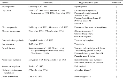
sequences. These include genes involved in various aspects of
energy metabolism (glucose transporters, glycolytic and
gluconeogenic enzymes), iron metabolism (transferrin),
catecholamine metabolism (tyrosine hydroxylase), vasomotor
control (nitric oxide synthases, endothelin-1) and angiogenesis
(vascular endothelial growth factor, platelet-derived growth
factor) (see Table 1 for references). Though these studies
strongly suggested a major role for HIF-1 in many
physiological and pathophysiological processes, this cannot be
immediately deduced from studies of gene expression in tissue
culture.
In the whole organism, hypoxia most commonly occurs in
the more complex setting of ischaemia and, in assessing the
importance of HIF-1, it is important to understand the
circumstances under which activation occurs in vivo, the
effects on gene expression in this setting and the
pathophysiological consequences. Such studies will ultimately
require the development of techniques for the assay of HIF-1
activation that can be applied to heterogeneous tissues in whole
animals. In the meantime, we have addressed this issue (at least
in the tumour setting) by analysis of tumour xenografts from
tissue culture cells in nude mice (Maxwell et al. 1997). We
examined a series of hepatoma (mouse hepa-1) cell lines which
were originally selected for a defective xenobiotic response
through resistance to benzo[a]pyrene (Hankinson, 1979). Cells
from one complementation group of resistant cells are deficient
in the common heterodimerization partner HIF-1
β
/ARNT
(Hoffman et al. 1991) and, in addition to manifesting a
deficient xenobiotic response, they are unable to form the
HIF-1 complex and show defective oxygen-regulated gene
expression in tissue culture (Wood et al. 1996). We therefore
compared gene expression, vascularization and growth in
tumours from wild-type cells, a HIF-1
β
/ARNT-deficient line
(c4), a revertant selected from c4 which has regained wild-type
levels of HIF-1
β
/ARNT (Rc4) and a line (c31) that has a
different defect in the xenobiotic response and is functional for
HIF-1.
often observed, and these were often close to necrotic regions.
This intense focal gene expression was not seen in tumours
derived from the mutant (c4) cells, but was observed in the
revertant line (Rc4) and in the c31 cells, both of which form
HIF-1 normally. This pattern of VEGF expression around
(presumably hypoxic) necrotic zones has been noted
previously and has led to the suggestion that hypoxia is a major
regulator of tumour angiogenesis (Plate et al. 1992; Shweiki et
al. 1992). Our experiments show that, at least in the context of
these hepatoma cells, activation of HIF-1 is largely responsible
for this pattern of gene expression within the tumour. When
vascularity and growth were examined, substantial differences
were observed which correlated with the presence or absence
of a functional HIF-1
β
/ARNT gene product. In particular,
tumours derived from the HIF-1
β
/ARNT-deficient (c4) cells
showed a striking reduction in vascularity (Fig. 1B).
These experiments strongly suggest that HIF-1 is activated
within solid tumours and provide evidence for an important
pathophysiological role in this setting. Although the evidence
linking hypoxia and erythropoietin regulation, through
HIF-1, to hypoxia and tumour angiogenesis is rather strong, it is
necessary to recognize the importance of other factors in
shaping the pattern of gene expression. The genes listed in
Table 1 are apparently linked to a common mechanism of
regulation by oxygen, yet the pattern of inducible expression
is in each case quite different. For instance, the VEGF gene
is also expressed in normal tissues – yet in many organs,
including kidney, it is induced modestly, if at all, by anaemic
stimulation sufficient to generate a massive increase in
erythropoietin production (Sandner et al. 1996). These
differences cannot easily be explained by cell-specific gene
expression and heterogeneous organ oxygenation. Rather,
A
[image:4.609.225.561.74.516.2]they suggest the operation of cooperative effects at the
cellular level, in which transcriptional activation by HIF-1
requires ancillary factors that may be cell-type-specific or
responsive to some other aspect of cellular physiology. For
example, VEGF expression is also increased by a number of
activated oncogenes, and it may be that an interaction with
such molecules constrains the circumstances under which the
gene is inducible by HIF-1 (Mazure et al. 1996). Other
insights into the mechanisms underlying the selective
responses to HIF-1 have come from studies of sequences
close to HIF-1 binding sites. For instance, the function of the
Epo enhancer is also dependent on at least two other sites
adjacent to the HIF-1-binding site (Blanchard et al. 1992;
Semenza and Wang, 1992; Pugh et al. 1994). One of these
has been shown to bind the tissue-specific transcription factor
hepatic nuclear factor (HNF-4) (Galson et al. 1995). In the
lactate dehydrogenase A gene promoter, a functional
interaction between an HIF-1 site and a cyclic AMP response
element has been defined (Firth et al. 1995). Thus, the
assembly of a functional transcriptional complex requires
factors other than HIF-1, and it appears likely that this
requirement for cooperative factors is used to define the
individual features of an inducible response.
Activation of HIF-1
[image:5.609.53.561.83.415.2]In pursuing the mechanism of oxygen sensing, a great deal
of attention has been turned towards analysis of the mechanism
of activation of HIF-1. In keeping with the demonstration of
HIF-1 DNA-binding activity in a wide variety of cell types,
mRNAs for HIF-1
α
and HIF-1
β
have been found in all cells
and tissues examined (Wenger et al. 1996; Wiener et al. 1996).
In the majority of studies, the mRNAs themselves were not
found to be inducible by hypoxic stimulation, indicating that
the activation of HIF-1 involves post-translational and/or
translational mechanisms (Huang et al. 1996; Wenger et al.
1996; Wood et al. 1996). We were unable to observe
translational regulation in fusions containing the HIF-1
promoter and 5
′
untranslated region fused to a luciferase
reporter and focused our attention on analyses likely to define
post-translational mechanisms of regulation. As with other
transcription factors, studies of regulatory mechanisms are
Table 1. Some processes involved in oxygen-regulated gene expression
Process References Oxygen-regulated gene Expression
Erythropoiesis Goldberg et al. 1991 Erythropoietin ↑
Glycolysis Firth et al. 1994, 1995; Marti et al. 1994; Lactate dehydrogenase A ↑ Semenza et al. 1994, 1996; Ebert et al. 1995 Phosphoglycerate kinase-1 ↑
Aldolase A and C ↑
Phosphofructokinase L and C ↑
Pyruvate kinase M ↑
Enolase A ↑
Gluconeogenesis Hellkamp et al. 1991; Kietzmann et al. 1993 Phosphoenolpyruvate carboxykinase ↓ Glucose transporters Ebert et al. 1995; O’Rourke et al. 1996 Glucose transporter 1 ↑ Glucose transporter 3 ↑ Glucose transporter 2 ↓ Catecholamine synthesis Czyzyk-Krzeska et al. 1992 Tyrosine hydroxylase ↑
Iron transport Rolfs et al. 1997 Transferrin ↑
Growth factors Kourembanas et al. 1990; Shweiki et al. Vascular endothelial growth factor ↑ 1992; Goldberg and Schneider, 1994; Transforming growth factor β ↑ Gleadle et al. 1995a Platelet-derived growth factor B ↑ Placental growth factor ↓ Nitric oxide synthesis McQuillan et al. 1994; Melillo et al. 1995 Inducible nitric oxide synthase ↑ Endothelial nitric oxide synthase ↓
Vasomotor regulator Bodi et al. 1995 Endothelin-1 ↑
High-energy phosphate O’Rourke et al. 1996 Adenylate kinase-3 ↑ metabolism
Haem metabolism Lee et al. 1997 Haem oxygenase 1 ↑
Evidence for a common mechanism of regulation for the genes listed is provided by functional similarities in the response (e.g. induction by hypoxia, Co2+ions and iron chelators) and/or the definition of functionally critical HIF-1 sites in cis-acting sequences and/or altered regulation in mutant cells which do not produce HIF-1.
potentially confounded by dependence on a series of
interrelated events (e.g. dimerization, DNA binding,
interaction with transcriptional activators) for an assayable
response. For this reason, we have sought to define regions of
HIF-1 genes that can independently confer oxygen-regulated
activity on heterologous transcription factors. In this work, we
have used two types of chimeric gene: those in which the
heterologous transcription factor encoded a DNA-binding
activity but lacked trans-activation; and others in which an
activation domain was either included with the heterologous
DNA binding domain or added from a second heterologous
gene. This allowed for the analysis of regulatory domains from
HIF-1 genes which did not necessarily contain intrinsic
activation potential. In these experiments, we found that
sequences from HIF-1
α
but not from HIF-1
β
could convey the
hypoxia-inducible property, indicating a regulatory role for the
α
subunit (Pugh et al. 1997).
An example of such an experiment is shown in Fig. 2. The
fusion of the powerful activation domain (amino acids
410–490) from the herpes simplex virus protein VP16 to a
DNA-binding domain derived from amino acids 1–147 of the
yeast transcription factor Gal4 creates a powerful transcription
factor which activates expression from promoters containing a
Gal4 binding site. When co-transfected into Hep3B cells with
a gene containing such a promoter linked to a luciferase
reporter, this Gal4/VP16 fusion is highly active. When
particular sequences derived from the HIF-1
α
gene are
encoded between the Gal4 and the VP16 sequences, two
striking effects are observed. First, in normoxic cells, the
overall level of activity is much reduced. Second, when cells
are exposed to hypoxia, cobaltous ions or desferrioxamine, the
apparent repression is greatly relieved, producing a regulated
activity which reflects that of HIF-1. The experiment illustrated
in Fig. 2B demonstrates a regulatory domain of HIF-1
α
between amino acids 530 and 652. In other work, we have
defined sequences lying further towards the amino terminus of
HIF-1
α
that also have this property (J. F. O’Rourke et al.,
unpublished work).
As a first analysis of the mechanisms by which such
regulatory domains operate, we assayed levels of protein
product in cells transfected with similar fusion genes. Studies
so far have demonstrated that, at least with the regulatory
sequences between amino acids 530 and 652, a major effect is
observed on the level of expressed fusion protein. In normoxic
cells, the fusion protein levels are much reduced by the
inclusion of this sequence, with this reduction being relieved
not only by exposure to hypoxia, cobaltous ions and
desferrioxamine, but also by a variety of inhibitors of
proteosomally mediated proteolysis. This suggests that one
mechanism of regulation might be mediated by proteolysis of
HIF-1
α
, with activation involving an inducible blockade of a
constitutively high rate of degradation targetted through these
sequences.
However, it is unlikely that this is the only mechanism of
regulation. In our chimeric transcription factor assays,
regulated activity was also observed which appeared to be
independent of the level of expressed protein. This was
particularly the case with the most C-terminal HIF-1
α
sequences. When fused to the Gal4 DNA-binding domain,
C-terminal sequences from HIF-1
α
demonstrated
oxygen-regulated trans-activation. Detailed analysis of these
sequences showed that, while HIF-1
α
amino acids 786–826
showed constitutive trans-activation, amino acids lying
immediately N-terminal to position 786 suppressed total
Activator plasmid
Reporter plasmid
Gal4 aa 1–147
HIF-1α sequence
VP16 transactivator CMV P
Gal4 UAS tk promoter LUCIFERASE
A
B
Gal VP16
Gal/VP16 530 652
HIF-1α
9 16 27
1.1 1.0 1.0
N H Co DFO N H Co DFO
Relative luciferase activity
0
[image:6.609.202.555.86.338.2]activity and conferred inducibility (J. F. O’Rourke et al.,
unpublished work). Thus, a Gal4 fusion bearing amino acids
775–826 showed reduced activity in normoxic cells which was
relieved by hypoxia. In this case, the fusion protein appeared
to be expressed at a similar level in normoxic and stimulated
cells.
Overall, these findings suggest a model involving at least
two mechanisms of HIF-1 activation, with induction or
derepression of activation domains being amplified by
regulation of transcription factor abundance, occurring through
changes in protein stability (Pugh et al. 1997). Such a model
would be consistent with assays of endogenous HIF-1
immunoactivity. These demonstrate rapid nuclear
accumulation of HIF-1
α
during hypoxia from very low levels
in unstimulated cells (Wang et al. 1995; Huang et al. 1996).
Upon reoxygenation, levels decline within minutes, indicating
a very short half-life in normoxic cells. Similar studies of
HIF-1
β
/ARNT in whole-cell extracts have shown modest or even
absent induction by hypoxia, with considerable levels being
present in unstimulated cells (Huang et al. 1996). Consistent
with a constitutive excess of the
β
subunit, forced
overexpression of the
α
subunit in normoxic cells is itself
sufficient to drive HIF-1-dependent reporter gene expression.
However, in keeping with a dual mechanism of activation,
hypoxic stimulation of these cells further enhances activity
(Jiang et al. 1996).
The definition of regulatory and activation domains for
HIF-1
α
now points the way to defining the next steps in regulation.
Quite possibly, this will involve regulated cofactors. For
instance, the proteosome is constitutively active, and any
regulated activity on HIF-1
α
could be modulated by a cofactor
or by a modification of the target. Similarly, the known
activation domains possess no clear homology with classical
activator sequences, and it may be that they operate to recruit
a cofactor. Interestingly, one known transcriptional
coactivator, p300, is known to affect the activity of the system
(Arany et al. 1996).
Evolutionary origins of HIF-1
One important question regarding a system which appears
to play such a central role in mammalian gene regulation by
oxygen is what its role might be in non-mammalian cells – in
particular, in simpler organisms which might facilitate genetic
analysis of the sensing mechanism. To address this, we have
studied the binding of nuclear extracts from non-mammalian
cells to an oligonucleotide from the mouse erythropoietin
enhancer (Nagao et al. 1996). So far, our clearest results have
been from Drosophila melanogaster SL2 cells and are shown
in Fig. 3. These cells contain a sequence-specific
hypoxia-inducible DNA-binding activity which has a similar
electrophoretic mobility to HIF-1, making it highly likely that
a homologous system is operative in insects. In ongoing
experiments, we are exploring the nature of this activity, but it
is clearly of interest that the first basic-helix–loop–helix PAS
proteins were defined in Drosophila and that more recent
studies have isolated new genes, sima (Nambu et al. 1996) and
trachealess (Wilk et al. 1996), which show even better
sequence similarity and (at least in the case of trachealess)
have a clear role in the organization of oxygen-delivery
systems in the fruit fly. Thus, it seems likely that the
Epo-wt
Epo-wt Epo-mut
N
− −
H N H N H N H
HeLa SL2
Probe:
Extract:
Competitor:
Condition:
1 2 3 4 5 6 7 8
I
[image:7.609.320.563.77.542.2]C
Fig. 3. DNA-binding assay demonstrating HIF-1-like activity in
uncovering of this widespread system of oxygen-regulated
gene expression in mammalian cells will soon also lead to the
recognition of a connection with oxygen-regulated gene
expression in non-mammalian systems.
The work conducted in the authors’ laboratory was
supported by the Wellcome Trust and the Medical Research
Council, UK.
References
ADAIR, T. H., GAY, W. J. AND MONTANI, J.-P. (1990). Growth regulation of the vascular system: evidence for a metabolic hypothesis. Am. J. Physiol. 259, 393–404.
ARANY, Z., HUANG, L. E., ECKNER, R., BHATTACHARYA, S., JIANG, C., GOLDBERG, M. A., BUNN, H. F. ANDLIVINGSTON, D. M. (1996). An essential role for p300/CBP in the cellular response to hypoxia.
Proc. natn. Acad. Sci. U.S.A. 93, 12969–12973.
BECK, I., RAMIREZ, S., WEINMANN, R. ANDCARO, J. (1991). Enhancer element at the 3′-flanking region controls transcriptional response to hypoxia in the human erythropoietin gene. J. biol. Chem. 266, 15563–15566.
BLANCHARD, K. L., ACQUAVIVA, A. M., GALSON, D. L. ANDBUNN, H. F. (1992). Hypoxic induction of the human erythropoietin gene: cooperation between the promoter and enhancer, each of which contains steroid receptor response elements. Molec. cell. Biol. 12, 5373–5385.
BODI, I., BISHOPRIC, N. H., DISCHER, D. J., WU, X. ANDWEBSTER, K. A. (1995). Cell-specificity and signaling pathway of endothelin-1 gene regulation by hypoxia. Cardiovasc. Res. 30, 975–984. BUNN, H. F. AND POYTON, R. O. (1996). Oxygen sensing and
molecular adaptation to hypoxia. Physiol. Rev. 76, 839–885. COTES, P. M., DORÉ, C. J., LIUYIN, J. A., LEWIS, S. M., MESSINEZY,
M., PEARSON, T. C. ANDREID, C. (1986). Determination of serum immunoreactive erythropoietin in the investigation of erythrocytosis. New Engl. J. Med. 315, 283–287.
CZYZYK-KRZESKA, M. F., BAYLISS, D. A., LAWSON, E. E. AND MILLHORN, D. E. (1992). Regulation of tyrosine hydroxylase gene expression in the rat carotid body by hypoxia. J. Neurochem. 58, 1538–1546.
EBERT, B. L., GLEADLE, J. M., O’ROURKE, J. F., BARTLETT, S. M., POULTON, J. AND RATCLIFFE, P. J. (1995). Isoenzyme specific regulation of genes involved in energy metabolism by hypoxia, cobalt and desferrioxamine: similarities with the regulation of erythropoietin. Biochem. J. 313, 809–814.
FANDREY, J., FREDE, S. ANDJELKMANN, W. (1994). Role of hydrogen peroxide in hypoxia-induced erythropoietin production. Biochem.
J. 303, 507–510.
FIRTH, J. D., EBERT, B. L., PUGH, C. W. ANDRATCLIFFE, P. J. (1994). Oxygen-regulated control elements in the phosphoglycerate kinase 1 and lactate dehydrogenase A genes: similarities with the erythropoeitin 3′ enhancer. Proc. natn. Acad. Sci. U.S.A. 91, 6496–6500.
FIRTH, J. D., EBERT, B. L. AND RATCLIFFE, P. J. (1995). Hypoxic regulation of lactate dehydrogenase A: interaction between hypoxia inducible factor 1 and cyclic AMP response elements. J. biol.
Chem. 270, 21021–21027.
GALSON, D. L., TSUCHIYA, T., TENDLER, D. S., HUANG, E., REN, Y., OGURA, T. ANDBUNN, H. F. (1995). The orphan receptor hepatic nuclear factor 4 functions as a transcriptional activator for
tissue-specific and hypoxia-tissue-specific erythropoietin gene expression and is antagonized by EAR3/COUP-TF1. Molec. cell. Biol. 15, 2135–2144.
GLEADLE, J. M., EBERT, B. L., FIRTH, J. D. AND RATCLIFFE, P. J. (1995a). Regulation of angiogenic growth factor expression by hypoxia, transition metals and chelating agents. Am. J. Physiol. 268, C1362–C1368.
GLEADLE, J. M., EBERT, B. L. AND RATCLIFFE, P. J. (1995b). Diphenylene iodonium inhibits the induction of erythropoietin and other mammalian genes by hypoxia: implications for the mechanism of oxygen sensing. Eur. J. Biochem. 234, 92–99. GOLDBERG, M. A., DUNNING, S. P. AND BUNN, H. F. (1988).
Regulation of the erythropoietin gene: evidence that the oxygen sensor is a heme protein. Science 242, 1412–1415.
GOLDBERG, M. A., GAUT, C. C. AND BUNN, H. F. (1991). Erythropoietin mRNA levels are governed by both the rate of gene transcription and posttranscriptional events. Blood 77, 271–277. GOLDBERG, M. A., GLASS, G. A., CUNNINGHAM, J. M. ANDBUNN, H.
F. (1987). The regulated expression of erythropoietin by two human hepatoma cell lines. Proc. natn. Acad. Sci. U.S.A. 84, 7972–7976. GOLDBERG, M. A. ANDSCHNEIDER, T. J. (1994). Similarities between the oxygen-sensing mechanisms regulating the expression of vascular endothelial growth factor and erythropoietin. J. biol.
Chem. 269, 4355–4359.
GONZALEZ, C., ALMARAZ, L., OBESO, A. AND RIGUAL, R. (1994). Carotid body chemoreceptors: from natural stimuli to sensory discharges. Physiol. Rev. 74, 829–877.
HANKINSON, O. (1979). Single-step selection of clones of a mouse hepatoma line deficient in aryl hydrocarbon hydroxylase. Proc.
natn. Acad. Sci. U.S.A. 76, 373–376.
HELLKAMP, J., CHRIST, B., BASTIAN, H. ANDJUNGERMANN, K. (1991). Modulation by oxygen of the glucagon-dependent activation of the phosphoenolpyruvate carboxykinase gene in rat hepatocyte cultures. Eur. J. Biochem. 198, 635–639.
HOFFMAN, E. C., REYES, H., CHU, F.-F., SANDER, F., CONLEY, L. H., BROOKS, B. A. AND HANKINSON, O. (1991). Cloning of a factor required for activity of the Ah (Dioxin) receptor. Science 252, 954–958.
HOGENESCH, J. B., CHAN, W. K., JACKIW, V. H., BROWN, R. C., GU, Y.-Z., PRAY-GRANT, M., PERDEW, G. H. AND BRADFIELD, C. A. (1997). Characterization of a subset of the basic-helix–loop–helix-PAS superfamily that interacts with components of the dioxin signaling pathway. J. biol. Chem. 272, 8581–8593.
HUANG, L. E., ARANY, Z., LIVINGSTON, D. M. ANDBUNN, H. F. (1996). Activation of hypoxia-inducible transcription factor depends primarily on redox-sensitive stabilization of its α subunit. J. biol.
Chem. 271, 32253–32259.
HUANG, Z. J., EDERY, I. AND ROSBASH, M. (1993). PAS is a dimerization domain common to Drosophila period and several transcription factors. Nature 364, 259–262.
JIANG, B.-H., RUE, E., WANG, G. L., ROE, R. ANDSEMENZA, G. L. (1996). Dimerization, DNA binding and transactivation properties of hypoxia-inducible factor 1. J. biol. Chem. 271, 17771–17778. JUNGERMANN, K. (1995). Zonation of metabolism and gene expression
in liver. Histochemistry 103, 81–91.
KIETZMANN, T., SCHMIDT, H., UNTHAN, F. K., PROBST, I. AND JUNGERMANN, K. (1993). A ferro-heme protein senses oxygen levels, which modulate the glucagon-dependent activation of the phosphoenolpyruvate carboxykinase gene in rat hepatocyte cultures. Biochem. biophys. Res. Commun. 195, 792–798.
tension regulates the expression of the platelet-derived growth factor – β chain gene in human endothelial cells. J. clin. Invest. 86, 670–674.
LEE, P. J., JIANG, B.-H., CHIN, B. Y., IYER, N. V., ALAM, J., SEMENZA, G. L. AND CHOI, A. M. K. (1997). Hypoxia-inducible factor-1 mediates transcriptional activation of the heme oxygenase-1 gene in response to hypoxia. J. biol. Chem. 272, 5375–5381.
LORENTZ, A., JENDRISSEK, A., ECKARDT, K.-U., SCHIPPLICK, M., OSSWALD, P. M. AND KURTZ, A. (1991). Serial immunoreactive erythropoietin levels in autologous blood donors. Transfusion 31, 650–654.
MARTI, H. H., JUNG, H. H., PFEILSCHIFTER, J. ANDBAUER, C. (1994). Hypoxia and cobalt stimulate lactate dehydrogenase (LDH) activity in vascular smooth muscle cells. Pflügers Arch. 429, 216–222. MAXWELL, P. H., DACHS, G. U., GLEADLE, J. M., NICHOLLS, L. G.,
HARRIS, A. L., STRATFORD, I. J., HANKINSON, O., PUGH, C. W. AND RATCLIFFE, P. J. (1997). Hypoxia inducible factor-1 modulates gene expression in solid tumors and influences both angiogenesis and tumor growth. Proc. natn. Acad. Sci. U.S.A. 94, 8104–8109. MAXWELL, P. H., PUGH, C. W. ANDRATCLIFFE, P. J. (1993). Inducible
operation of the erythropoietin 3′ enhancer in multiple cell lines: evidence for a widespread oxygen sensing mechanism. Proc. natn.
Acad. Sci. U.S.A. 90, 2423–2427.
MAZURE, N. M., CHEN, E. Y., YEH, P., LADEROUTE, K. R. AND GIACCIA, A. J. (1996). Oncogenic transformation and hypoxia synergistically act to modulate vascular endothelial growth factor expression. Cancer Res. 56, 3436–3440.
MCQUILLAN, L. P., LEUNG, G. K., MARSDEN, P. A., KOSTYK, S. K. AND KOUREMBANAS, S. (1994). Hypoxia inhibits expression of eNOS via transcriptional and posttranscriptional mechanisms. Am.
J. Physiol. 267, H1921–H1927.
MELILLO, G., MUSSO, T., SICA, A., TAYLOR, L. S., COX, G. W. AND VARESIO, L. (1995). A hypoxia-responsive element mediates a novel pathway of activation of the inducible nitric oxide synthase promoter. J. exp. Med. 182, 1683–1693.
NAGAO, M., EBERT, B. L., RATCLIFFE, P. J. ANDPUGH, C. W. (1996).
Drosophila melanogaster SL2 cells contain a hypoxically inducible
DNA binding complex which recognises mammalian HIF-1 binding sites. FEBS Lett. 387, 161–166.
NAMBU, J. R., CHEN, W., HU, S. AND CREWS, S. T. (1996). The
Drosophila melanogaster similar bHLH-PAS gene encodes a
protein related to human hypoxia-inducible factor 1α and
Drosophila single-minded. Gene (in press).
NECAS, E. ANDNEUWIRT, J. (1972). The effect of inhibitors of energy metabolism on erythropoietin production. J. Lab. clin. Med. 79, 388–396.
O’DONNELL, V. B., TEW, D. G., JONES, O. T. G. ANDENGLAND, P. J. (1993). Studies on the inhibitory mechanism of iodonium compounds with special reference to neutrophil NADPH oxidase.
Biochem. J. 290, 41–49.
O’ROURKE, J. F., PUGH, C. W., BARTLETT, S. M. ANDRATCLIFFE, P. J. (1996). Identification of hypoxically inducible mRNAs in Hela cells using differential display PCR. Eur. J. Biochem. 241, 403–410. PLATE, K. H., BREIER, G., WEICH, H. A. AND RISAU, W. (1992).
Vascular endothelial growth factor is a potential tumour angiogenesis factor in human gliomas in vivo. Nature 359, 845–848.
PUGH, C. W., EBERT, B. L., EBRAHIM, O. ANDRATCLIFFE, P. J. (1994). Characterisation of functional domains within the mouse erythropoietin 3′enhancer conveying oxygen-regulated responses in different cell lines. Biochim. biophys. Acta 1217, 297–306.
PUGH, C. W., O’ROURKE, J. F., NAGAO, M., GLEADLE, J. M. AND RATCLIFFE, P. J. (1997). Activation of hypoxia inducible factor-1; Definition of regulatory domains within the α subunit. J. biol.
Chem. 272, 11205–11214.
PUGH, C. W., TAN, C. C., JONES, R. W. ANDRATCLIFFE, P. J. (1991). Functional analysis of an oxygen-related transcriptional enhancer lying 3′to the mouse erythropoietin gene. Proc. natn. Acad. Sci.
U.S.A. 88, 10553–10557.
RATCLIFFE, P. J. (1993). Molecular biology of erythropoietin. Kidney
Int. 44, 887–904.
REYES, H., REISZ-PORSZASZ, S. AND HANKINSON, O. (1992). Identification of the Ah receptor nuclear translocator protein (Arnt) as a component of the DNA binding form of the Ah receptor.
Science 256, 1193–1195.
ROLFS, A., KVIETIKOVA, I., GASSMANN, M. ANDWENGER, R. H. (1997). Oxygen-regulated transferrin expression is mediated by hypoxia-inducible factor-1. J. biol. Chem. (in press).
SANDNER, P., GESS, B., WOLF, K. ANDKURTZ, A. (1996). Divergent regulation of vascular endothelial growth factor and of erythropoietin gene expression in vivo. Pflügers Arch. 431, 905–912.
SEMENZA, G. L., JIAN, B.-H., LEUNG, S. W., PASSANTINO, R., CONCORDET, J.-P., MAIRE, P. ANDGIALLONGO, A. (1996). Hypoxia response elements in the aldolase A, enolase 1 and lactate dehydrogenase A gene promoters contain essential binding sites for hypoxia-inducible factor 1. J. biol. Chem. 271, 32529–32537. SEMENZA, G. L., NEJFELT, M. K., CHI, S. M. ANDANTONARAKIS, S. E.
(1991). Hypoxia-inducible nuclear factors bind to an enhancer element located 3′to the human erythropoietin gene. Proc. natn.
Acad. Sci. U.S.A. 88, 5680–5684.
SEMENZA, G. L., ROTH, P. H., FANG, H.-M. AND WANG, G. L. (1994). Transcriptional regulation of genes encoding glycolytic enzymes by hypoxia-inducible factor 1. J. biol. Chem. 269, 23757–23763.
SEMENZA, G. L. ANDWANG, G. L. (1992). A nuclear factor induced by hypoxia via de novo protein synthesis binds to the human erythropoietin gene enhancer at a site required for transcriptional activation. Molec. cell. Biol. 12, 5447–5454.
SHWEIKI, D., ITIN, A., SOFFER, D. ANDKESHET, E. (1992). Vascular endothelial growth factor induced by hypoxia may mediate hypoxia-initiated angiogenesis. Nature 359, 843–845.
TIAN, H., MCKNIGHT, S. L. ANDRUSSELL, D. W. (1997). Endothelial PAS domain protein 1 (EPAS1), a transcription factor selectively expressed in endothelial cells. Genes Dev. 11, 72–82.
WANG, G. L., JIANG, B.-H., RUE, E. A. ANDSEMENZA, G. L. (1995). Hypoxia-inducible factor 1 is a basic-helix–loop–helix-PAS heterodimer regulated by cellular O2tension. Proc. natn. Acad. Sci.
U.S.A. 92, 5510–5514.
WANG, G. L. ANDSEMENZA, G. L. (1993a). Desferrioxamine induces erythropoietin gene expression and hypoxia-inducible factor 1 DNA-binding activity: implications for models of hypoxia signal transduction. Blood 82, 3610–3615.
WANG, G. L. ANDSEMENZA, G. L. (1993b). General involvement of hypoxia-inducible factor 1 in transcriptional response to hypoxia.
Proc. natn. Acad. Sci. U.S.A. 90, 4304–4308.
WEIBEL, E. R., TAYLOR, C. R. ANDHOPPELER, H. (1992). Variations in function and design: Testing symmorphosis in the respiratory system. Respir. Physiol. 87, 325–348.
WEIR, E. K. ANDARCHER, S. L. (1995). The mechanisms of acute hypoxic pulmonary vasoconstriction: the tale of two channels.
WENGER, R. H., ROLFS, A., MARTI, H. H., GUÉNET, J.-L. AND GASSMANN, M. (1996). Nucleotide sequence, chromosomal assignment and mRNA expression of mouse hypoxia-inducible factor-1α. Biochem. biophys. Res. Commun. 223, 54–59.
WIENER, C. M., BOOTH, G. AND SEMENZA, G. L. (1996). In vivo expression of mRNAs encoding hypoxia-inducible factor 1.
Biochem. biophys. Res. Commun. 225, 485–488.
WILK, R., WEIZMAN, I. ANDSHILO, B.-Z. (1996). trachealess encodes a bHLH-PAS protein that is an inducer of tracheal cell fates in
Drosophila. Genes Dev. 10, 93–102.
WOOD, S. M., GLEADLE, J. M., PUGH, C. W., HANKINSON, O. AND RATCLIFFE, P. J. (1996). The role of aryl hydrocarbon receptor nuclear translocator (ARNT) in hypoxic induction of gene expression: studies in ARNT deficient cells. J. biol. Chem. 271, 15117–15123.