CELL RECOGNITION AND PATTERN FORMATION IN THE DEVELOPING NERVOUS SYSTEM
BY DAVID TRISLER
Laboratory of Biochemical Genetics, National Heart, Lung and Blood Institute, National Institutes of Health, Bethesda, MD 20892, USA
Summary
The topographic map of cell position in the avian retina is conserved and inverted when retinal ganglion neurons synapse with neurons in the optic tectum. Developmental mechanisms based on molecular gradients that specify positional information and pattern formation have been postulated in the establishment of these topographic maps of cells in retina and optic tectum. Two cell surface proteins in retina, T O P D V a nd TOPAp, are distributed in dorsoventral and anteroposterior topographic gradients, respectively. Corresponding gradients of TOP molecules present in the tectum are inverted with respect to the retinal gradients. These orthogonal gradients of TOPD V and T O P A P molecules provide a possible Cartesian coordinate system for designation of cell position at all points in the retinotectal map.
Introduction
12 D. TRISLER
Fig. 1. Topographic projection of retinal ganglion cell axons to the optic tectum in lower vertebrates. (A) Dorsal retinal neurons innervate ventral tectum and ventral retina innervates dorsal tectum. (B) Anterior retina projects to posterior tectum and posterior retina projects to anterior tectum. (A, B after Attardi and Sperry, 1963; DeLong and Coulombre, 1965). (C) Continuous point-to-point topographic map of retinal projection onto tectum demonstrated by electrophysiological data (after Gaze
et al. 1963; Gaze and Sharma, 1970; Yoon, 1971; Chung and Cooke, 1975; Schmidt et al. 1978). (D) Ganglion cell axons misrouted after disruption in retina and optic nerve
can reorient on the tectum and project to the correct target site (after Thanos et al. 1984). D, dorsal; V, ventral; A, anterior (nasal); P, posterior (temporal/caudal).
to fill the available synaptic field of tectum after ablation of part of the retina or tectum, yet maintain the topographic order of the map (Gaze et al. 1963; Gaze and Sharma, 1970; Yoon, 1971; Chung and Cooke, 1975; Schmidt etal. 1978). The orderly retinotectal projection apparently relies, in part, on the ability of retinal axons to read tectal cues. In the developing chick retina, misrouted ganglion cell axons, disrupted in the retina and optic nerve, can reorient on the tectal surface and project to the correct target site (Fig. ID; Thanos et al. 1984). Some misrouted growth cones migrate to the appropriate anterior-posterior tectal longitude, make a 90° turn and project to the correct position along the dorsoventral axis of the tectum.
and optic tectum (Trisler etal. 1981; Constantine-Paton etal. 1986; Rabacchi and Drager, 1987; Trisler and Collins, 1987; Muller et al. 1990).
Topographically graded molecules in retina and optic tectum
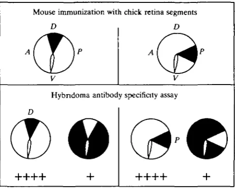
Topographically graded molecules in retina were detected with monoclonal antibodies generated by the fusion of spleen cells from mice immunized with a small portion of dorsal or posterior retina to P3X63Ag8 mouse myeloma cells (Fig. 2). One antibody to a cell surface molecule bound more abundantly to cells from dorsal retina than to cells from the remainder of the retina. A second antibody bound preferentially to cells of posterior retina. These molecules, termed TOP for toponymic (i.e. a marker of position), are present throughout the retina but are distributed in topographic gradients (Fig. 3). TOPD V is graded dorsoven-trally and TOPA P is graded anteroposteriorly. Bilaterally symmetrical gradients of TOP molecules are present in the retinas of both right and left eyes.
A 35-fold gradient of TOPD V was found extending from the dorsal to the ventral . margins of the retina aligned parallel to the long axis of the choroid fissure (Fig. 4),
and a 16-fold gradient of TO PAP is present from the anterior retinal margin to the posterior margin perpendicular to TOPD V. The concentration of TOP molecules detected varied continuously and logarithmically with the logarithm of distance along the circumference of retina.
Mouse immunization with chick retina segments
D D
d>
[image:3.451.103.346.353.547.2]Hybndoma antibody specificity assay
14 D . TRISLER
u.
[image:4.451.85.371.49.295.2]7 8 1 Retina section
Fig. 3. Topographic gradients of (A) TOPDV ( • ) and (B) TOPAP ( • ) molecules in E14 chick retina. Molecular gradients detected with mouse monoclonal anti-TOPov and anti-TOPAP antibodies. Values shown are picomoles of [125I]-F(ab')2 fragment of rabbit IgG directed against mouse IgG specifically bound to retinal cells exposed to anti-TOP antibody per milligram of retina protein (A after. Trisler et at. 1981).
20
10
A TOPD V in retina
D
50 100
Ventral Dorsal Anterior Percentage of maximal distance
50 100 Posterior
[image:4.451.84.369.383.598.2]Indirect immunofluorescence of anti-TOPDv antibody binding to cells in tissue sections taken along the dorsoventral axis of retina of E5 embryos revealed TOPD V was most abundant in dorsal retina, intermediate in middle, and least abundant in ventral retina (Fig. 5). No obvious heterogeneity was observed in the cell population from each location. Most or all cells across the thickness of retina from the vitreal surface to the pigmented epithelium stained. Visual scanning of fluorescently stained transverse sections of whole retina showed that TOPD V was continuously graded. This indicates that the dorsoventral gradient is due to differences in the amount of TOPD V per cell rather than to differences in the number of cells expressing TOPDV- The ring fluorescence pattern around each cell is consistent with the fact that TOPD V is a cell surface molecule. In older embryos and in adult retina, TOPD V is most abundant in the synaptic layers and in the ganglion cell axon layer (Fig. 5G).
Similarly, TOPAp is distributed in a graded pattern along the anteroposterior axis (Fig. 5). The ring fluorescence of cells from anterior retina was weakest, that of middle cells was intermediate and that of cells from posterior retina exhibited the greatest binding of anti-TOPAP in E5 embryos. Autoradiography of anti-TOPAp binding along the anterior-posterior equatorial meridian of the entire E8 retina revealed the TOPAp gradient. Since TOPD V and TOPA P molecules are graded on the basis of number of molecules per cell, TOPD V can be used to identify cell position along the dorsoventral axis, while TOPA P identified cell position along the anteroposterior axis of retina.
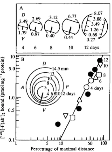
Gradients of TOP molecules were present in retina at all developmental ages tested from E4 to E18 and in hatchling chicks (Fig. 6). The orientation of the gradients remained constant throughout development; however, the magnitude of the TOPD V gradient increased 10 times from a threefold gradient at E4 to a 35-fold gradient at E12. The TOPA P gradient increased fivefold from E5 to E18. Thus, the TOP gradients are present early during blast cell proliferation and persist throughout development when both neurogenesis and gliogenesis occur and tissue organization is established.
Both TOPD V and TOPA P are cell surface proteins. TOPD V has an apparent relative molecular mass of 47xlO3 (Moskal et al. 1986) and TOPA P of 40X103. Topographic differences in T O P A P distribution were found in immunoblots of retina cell proteins resolved by sodium dodecyl sulfate-polyacrylamide gel electrophoresis. Approximately 16-fold more TOPA P was detected in cells from the posterior margin of retina than from the anterior margin (Fig. 7). Anal-ogously, more T O P A P w as present in the posterior half-retina than in the anterior half-retina. These results suggest that the graded nature of TOPA P demonstrated by radioimmuno-binding assay and by indirect immunofluorescence reflects the distribution of the antigen itself and not that of a masking molecule or of antigen accessibility. However, a gradient of post-translational modification of evenly distributed TOPA P molecules has not been ruled out.
16
D . TRISLERAxons from peripheral dorsal retina cells of E12 embryos were shown by fluorescent dye-tracing to be present in dorsal nerve. Indirect immunofluor-escence revealed a greater abundance of TOPD V in dorsal nerve than in ventral
50 100
Ventral Dorsal Anterior Percentage of maximal distance
50 100 Posterior
Fig. 6. TOP antigen gradients in chick retina as a function of developmental age. (A) TOPDV; (B) TOPAP in ( • ) , E4; (O), E5; ( • ) , E8; ( • ) , E10; ( • ) , E12; (A), E14;
(A), E18 retinas.
nerve. Ganglion cell axons from posterior retina were in anterior nerve, as were higher levels of TOPA P. Although the distribution of TOP molecules reiterates the organization of ganglion cell axons in the nerve, the question of whether TOP molecules in the nerve are associated with axons, with glia or with both is not resolved. In E12 embryos, at the age when retinal axons have covered the tectal surface, TOPAp was most abundant on the anterior medial portion of tectum (Fig. 8) where posterior retinal axons project (DeLong and Coulombre, 1965).
TOP molecules are present on optic tectum cells as well as in retina (Trisler and Collins, 1987). They are distributed in gradients in E5 embryos 1 day before ganglion cell axons arrive at the tectum (Fig. 9). TOPD V and TOPA P gradients in tectum are inverted with respect to the retinal gradients. TOPD V is 10-fold higher in ventral than in dorsal tectum and TOPA P is eightfold higher in anterior than in posterior tectum. The quantities of TOPD V and TOPA P detected per cell by indirect immunofluorescence varied continuously along the axes of the tectal gradients (Fig. 5). Little or no heterogeneity of TOPD V or TOPA P expression was found among cells from ventricular to pial surfaces at a given position along the respective gradients. Thus, as in retina, TOP molecules can be used to mark cell position in tectum.
18 D . T R I S L E R
7 A M
rx 1 0 "
31
3
5
7
9 1 1 1 3 1 5 1 7 1 9
18-B
Fig. 7. Demonstration of TOPAP topographic distribution in retina and determination of TOPAp relative molecular masses by sodium dodecyl sulfate-polyacrylamide gel electrophoresis and immunoblot analysis. (A) Lanes 1 and 11, relative molecular mass standards; lanes 2-10, serial twofold dilutions of 25 fig of E8 protein from the anterior retinal margin; lanes 12-20, serial twofold dilutions of 25 fig of E8 protein from the posterior retinal margin; (B) detail of autoradiogram of anti-TOPAp binding to serial dilutions of 25 fig of retina protein from anterior half-retina (lanes 2-10) and posterior half-retina (lanes 12-20).
suggests a possible role for the molecules in orienting the dorsoventral and anteroposterior axes of the retinal projection onto the tectum.
Cell lineage and regulation of TOP expression
The retina grows by accretion of rings of cells in the proliferative zone at the peripheral margin (Coulombre, 1955; Kahn, 1974). Expression of the TOP gradient during retinal growth was examined by comparison of the amount of
T O P D V in the proliferative zone at the poles of the gradient in E4-E10 retinas with the amount of TOPDv at the corresponding distances along the gradient axis in E12 retina (Fig. 11). The cells in the proliferative zone at the dorsal margin of retina expressed progressively more TOPDv with retinal growth, while those in the proliferative zone at the ventral margin expressed progressively less TOPDv, thereby increasing the magnitude of the gradient (Trisler, 1987). There was close agreement in the amount of TOPD V detected on cells at a given distance along the gradient axis from the fundus, the oldest portion of the retina, throughout the developmental period tested. Thus, each position along the axis has a constant, TOPDv value through these developmental ages.
Fig. 8. TOP molecules in optic nerve and optic tectum. (A,B) Anterogradely transported rhodamine isothiocyanate from dorsal and posterior retina in E12 optic nerve from within 1 mm of the nerve head, respectively; (C,D) image-enhanced video micrographs of T O PD V and T O PA P binding in E12 optic nerve, respectively; (E) dorsal
view of a whole mount of E12 optic tectal lobes stained with anti-TOPAp antibody and
horseradish-peroxidase-labeled rabbit antibody to mouse IgG.
10 50 100
Ventral Dorsal Anterior Percentage of maximal distance
50 100 Posterior
[image:9.451.67.357.399.618.2]20 D . TRISLER
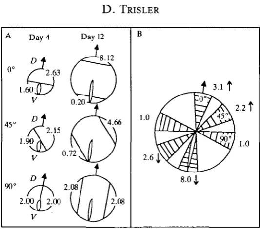
to the axis of the antigen gradient was determined by comparing TOPD V expression in cells of the proliferative zone of E4 and E12 retinas at different positions around the peripheral margin (Fig. 12). Both the magnitude and the sign of change in TOPD V expression during development varied depending on the position of the parental cells. Progeny cells in dorsal retina expressed more TOPD V than parental cells while those in ventral retina expressed less. The greatest magnitude of change in expression was along the axis of the gradient (0°). Little or no change in TOPD V expression occurred in cells with retinal growth along the perpendicular axis (90°). Intermediate rates of change in TOPD V expression were found during cell division at 45° to the gradient axis.
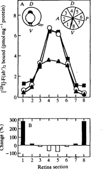
Ablation of parental cells in the proliferative zone at the dorsal and ventral poles of the gradient in embryos 60 h after fertilization altered the level of TOPD V expression in dorsal and ventral retina later in development (Fig. 13). Progeny cells that replaced the ablated dorsal region expressed 60% less TOPD V than normal in E16 retina. After ventral ablation, cells in ventral retina expressed 300 % more TOPDV- These values are similar to that expected for the progeny of parental cells from regions neighboring the ablated portion. This suggests that by 60 h of development the level of TOPD V expressed by the retina cells and their progeny is already determined.
Function of TOPDy in retinal development
The role of molecular markers of cell position in the development of the nervous system was examined using a monoclonal antibody to TOPD V (Trisler, 1983; Trisler et al. 1986). Antibodies provide a means of blocking molecular function (Levi-:Montalcini and Booker, 1960; Crawford et al. 1982; Schwartz and Spirma, 1982; Warner et al. 1984). Several groups have demonstrated effects of antibodies against nervous system molecules on growth cone behavior, neurite outgrowth and tissue organization in vivo and in vitro. Antibodies against chicken cognin and N-CAM inhibit cell-cell adhesion (Hausman and Moscona, 1979; Thiery et al. 1977, respectively) and anti-N-CAM disrupts axonal fasciculation (Thanos et al. 1984; Fraser et al. 1984). Antibody T61/3/12 blocks neurite outgrowth of chick retina cells in vitro (Henke-Fahle and Bonhoeffer, 1983), whereas antibody to Thy-1 stimulates neurite outgrowth of rat retinal ganglion cells in vitro (Leifer et al. 1984). Antibody LI inhibits granular cell migration in rat cerebellar explants (Lindner et al. 1983). Our objectives were to determine the accessibility of TOPD V to antibody in the in vivo retina, to determine the persistence of antibody in retina after injection into the embryo, and to identify changes in the development of retina continuously exposed to anti-TOPDV antibody.
D
2 a. 7 00 "o S 3 •o a
1
10 5.0 1.0 0.5 0.1. A D 2.49 V 4: B
L
'
A[
-2.69 3^12 ^ 7 7
0-97 1 5 / ^ /
6 8 10
D
y^- -14.5 mm
\k\jj 6 81012 days
V / I
V
i i i 1 1 1 1 / i
^ 8 . 0 7 / T 3.88 A f Z/3.49 -/\
I f 126 VJ
V 0.68 1 /0.27 ^ 12 days
^ 1 2
^vio
• / Q 8
O 4 days
)
1 1 1 1 1 nl
5 10 50 Percentage of maximal distance
[image:13.451.130.325.71.335.2]100
Fig. 11. Increase in TOPDV gradient with retinal growth. (A) TOPDV detected at the poles of the gradient of E4-E10 retinas and along the axis of the gradient of E12 retina. (B) TOPDV values at these positions for each age as a function of distance along the axis of the gradient in E12 retina. Circumferential distances from dorsal to ventral poles for each age are shown: Q, E4; A, E6; • , E8; V, E10; • . E12 (after Trisler, 1987).
ventral gradient of A b T O PD V complexes of the same magnitude and orientation detected by in vitro binding studies was present 24 h after intraocular injection of antibody.
A gradient of Ab-TOPDV complexes in retina persisted for 4 days after intraocular injection of mouse ascites fluid containing anti-TOPov antibody (Fig. 15). The Ab-TOPD V gradient was maintained for 10 days when hybridoma cells, that synthesize anti-TOPDV antibody, were injected. The hybridoma cells provide a continuous source of antibody for long-term maintenance of Ab-TOPDV complexes in the retina. The decrease in Ab-TOPDV complexes in retina after E18 represents a loss in accessibility of TOPD V to intraocular antibody, perhaps due to developmental changes in permeability of the inner limiting membrane of the retina.
22
D. TRISLERFig. 12. Comparison of TOPDV expression in parental cells (E4) and progeny cells (E12) during embryonic development as a function of the angle of growth from the axis of the TOPDV gradient in retina. (A) TOPDV detected at the peripheral margins of E4 and E12 retina along the axis (0°) and at 45° and 90* to the axis. (B) Change of TOPDV expression at each angle from E4 to E12 retinal growth (after Trisler, 1987).
If
1
a ie
~s
B a. •o c 3B
r JD a?f
&
(Jha i 8 6 4 2 0 300 200 100 0 -100 A •(( -Wa
T
li
- 1 I D V i 2 i B i 22
Q
Bff
\\
//
/ / +r
i i 3 4 i i"-
1 11U
[image:15.451.153.322.78.353.2]3 4 Retina t A\ -A. I 5 1
u
1 5k
\
i 6 i 6 section Dw
V1
\
K
7 8 1 ^I
1 i 7 8Fig. 13. TOPDV gradients in E16 retinas after ablation of parental cells at the dorsal or ventral poles of the gradient in the cell proliferative marginal zone 60 h after fertilization. (A) TOPDV in O, normal retina; A, retina after dorsal pole ablation; • , retina after ventral pole ablation. (B) Percentage change of TOPDV expression for each region of retina after ablation.
Conclusions
The topographic map of cell position in retina is conserved and inverted when retinal axons project to and synapse with neurons of the optic tectum. Develop-mental mechanisms based on molecular gradients that specify positional infor-mation and pattern forinfor-mation have been postulated in the establishment of the topographic map of cells in avian retina and optic tectum. Orthogonal gradients of toponymic (TOP) cell membrane molecules are present in the retina and optic tectum. T O P D V molecules are distributed dorsoventrally and TOPA P molecules are graded anteroposteriorly. The polarities of the gradients are inverted in tectum with respect to retina. TOPDv is most abundant in dorsal retina and ventral tectum and TOPAp is most abundant in posterior retina and anterior tectum. The quantities of TOPD V and TOPAp detected per cell vary continuously along the axis of the respective gradient. Thus, TOP molecules can be used to identify cell position along the dorsoventral and anteroposterior axes of the developing retina and optic tectum.
consti-24
D. TRISLER [image:16.451.79.372.48.294.2]Dorsal
Fig. 14. Anti-TOPDV distribution in retina after in ovo injection. (A) Anti-TOPDV in E3 retina 1 day after injection of antibody into the amniotic cavity. (B) Anti-TOPDV in E12 retina 1 day after intraocular injection of antibody (after Trisler et al. 1986).
•S 7
0>
s
-°- 6
I 00
1 5
D-1
A Antibody B Antibody and hybridoma cells
D
5 2 3 4 5 6 Days after injection
10
[image:16.451.80.370.356.560.2]4 5 6 7 8 Days after injection
Fig. 16. Time course of synapse formation in the inner syriaptic layer of retina after intraocular injection of hybridoma cells producing anti-TOPDV antibody. (A) Growth cone and (B) synapse density as a function of developmental age after exposure to anti-TOPDV ( • and • , respectively) and exposure to no antibody and to control antibodies P3X63Ag8 and 57D8 (O and D, respectively) (after Trisler et al. 1986).
tute a possible Cartesian coordinate system that can be used to identify cell position in the topographic map of retina and optic tectum. Synapse formation in retina was inhibited in the presence of anti-TOPDv antibody. This suggests that T O P D V niay be involved in the recognition of cell position that is required for normal synapse formation. The inverted configuration of the gradients in retina and tectum corresponds to the inverted topographic map of cell position in the retinotectal projection. Thus, TOP molecules may be involved in orienting the retinotectal map.
References
ATTARDI, D. G. AND SPERRY, R. W. (1963). Preferential selection of central pathways by regenerating optic fibers. Exptl Neurol. 7, 46-64.
BOVERI, T. (1901). Uber die Polaritat des Seeigeleies. Eisverb. Phys. Med. Ges. (Wurzburg) 34, 145-175.
CHILD, C. M. (1941). Patterns and Problems of Development. Chicago: University of Chicago Press.
CHUNG, S. H. AND COOKE, J. (1975). Polarity of structure and ordered nerve connections in the developing amphibian brain. Nature 258, 126-132.
CONSTANTINE-PATON, M., BLUM, A. S., MENDEZ-OTERO, R. AND BARNSTABLE, C. J. (1986). A cell surface molecule distributed in a dorso-ventral gradient in the perinatal rat retina. Nature
324, 459-462.
26 D . TRISLER
CRAWFORD, G., SLEMMON, J. R. AND SALVATERRA, P. M. (1982). Monoclonal antibodies selective for Drosophila melanogaster choline acetlytransferase. J. biol. Chem. 257, 3853-3856. DALCQ, A. AND PASTEELS, J. (1938). Potentiel morphog6n£tique regulation et 'axial gradients'
de Child. Bull. Acad. r. Med. Belg. 3, 261-308.
DANIELS, M. P. AND VOGEL, Z. (1970). Localization of a--bungarotoxin binding sites in synapses of developing chick retina. Brain Res. 201, 45-56.
DEL CERRO, M. P. AND SNIDER, R. S. (1968). Studies on the developing cerebellum: ultrastructure of growth cones. J. comp. Neurol. 133, 341-361.
DELONG, G. R. AND COULOMBRE, A. J. (1965). Development of the retino-tectal topographic projection in the chick embryo. Exptl Neurol. 13, 350-363.
FRASER, S. E. AND HUNT, R. K. (1980). Retinotectal specificity: Models and experiments in search of a mapping function. A. Rev. Neurosci. 3, 319-352.
FRASER, S. E., MURRAY, B. A., CHUONG, C.-M. AND EDELMAN, G. M. (1984). Alteration of the retinotectal map in Xenopus by antibodies to neural cell adhesion molecules. Proc. natn.
Acad. Sci. U.S.A. 81, 4222-4226.
GAZE, R. M., JACOBSON, M. AND SZEKELY, G. (1963).' The retino-tectal projection in Xenopus with compound eyes. J. Physiol., Lond. 165, 484-499.
GAZE, R. M. AND SHARMA, S. C. (1970). Axial differences in the reinnervation of the goldfish optic tectum by regenerating optic nerve fibers. Expl Brain Res. 10, 171-181.
GRIERER, A. AND MEINHARDT, H. (1972). A theory of biological pattern formation. Kybernetik
12, 30-39.
HAUSMAN, R. AND MOSCONA, A. (1979). Immunological detection of retina cognin on the surface of embryonic cells. Expl Cell Res. 119, 191-204.
HENKE-FAHLE, S. AND BONHOEFFER, F. (1983). Inhibition of axonal growth by a monoclonal antibody. Nature 303, 65-67.
HUGHES, W. F. AND LAVELLE, A. (1974). On the synaptogenic sequence in the chick retina.
Anat. Rec. 179, 297-301.
KAHN, A. J. (1974). An autoradiographic analysis of the times of appearance of neurons in the developing chick neural retina, Devi Biol. 38, 30-40.
KAWANA, E., SANDRI, C. AND AKERT, K. (1971). Ultrastructure of growth cones in the cerebellar cortex of the neonatal rat and cat. Z. Zellforsch. mikrosk. Anat. 115, 284-298.
LEIFER, D., LIPTON, S. A., BARNSTABLE, C. J. AND MASHLAND, R. H. (1984). Monoclonal antibody to Thy-1 enhances regeneration of processes by rat retinal ganglion cells in culture.
Science 224, 303-306.
LEVI-MONTALCINI, R. AND BOOKER, B. (1960). Destruction of the sympathetic ganglion in mammals by an antiserum to a nerve growth protein. Proc. natn. Acad. Sci. U.S.A. 46, 384-391.
LINDNER, J., RATHJEN, F. G. AND SCHACHNER, M. (1983). LI mono- and polyclonal antibodies modify cell migration in early postnatal mouse cerebellum. Nature 305, 427-430.
MOSKAL, J. R., TRISLER, D., SCHNEIDER, M. D. AND NIRENBERG, M. (1986). Purification of a membrane protein distributed in a topographic gradient in chicken retina. Proc. natn. Acad.
Sci. U.S.A. 83, 4730-4733.
MOLLER, B., STAHL, B. AND BONHOEFFER, F. (1990). In vitro experiments on axonal guidance and growth-cone collapse. J. exp. Biol. 153, 29-46.
RABACCHI, S. A. AND DRAGER, U. C. (1987). A positional marker for the embryonic mouse retina. Neurosci. Abstsr. 13, 590.
SCHMIDT, J. T., CICERONE, C. M. AND EASTER, S. S. (1978). Expansion of the half retinal projection to the tectum in goldfish: An electrophysiological and anatomical study. J. comp.
Neurol. 177, 257-278.
SCHWARTZ, M. AND SPIRMA, N. (1982). Sprouting from chicken dorsal root ganglia induced by nerve growth factor is specifically inhibited by affinity-purified antiganglioside antibodies.
Proc. natn. Acad. Sci. U.S.A. 79, 6080-6083.
SHEFFIELD, J. B. AND FISHMAN, D. A. (1970). Intercellular junctions in the developing neural retina of the chick embryo. Z. Zellforsch. mikrosk. Anat. 104, 405-418.
SPERRY, R. W. (1963). Chemoaffinity in the orderly growth of nerve fiber patterns and^ connections. Proc. natn. Acad. Sci. U.S.A. 50, 703-710.
cues influence the development of the chicken retinotectal projection. Proc. natn. Acad. Sci. U.S.A. 81, 1906-1910.
THIERY, J.-P., BRACKENBURY, R., RUTISHAUSER, U. AND EDELMAN, E. M. (1977). Adhesion among neural cells of the chick embryo. II. Purification and characterization of a cell adhesion molecule from neural retina. /. biol. Chem. 252, 6841-6845.
TRISLER, D. (1982). Are molecular markers of cell position involved in the formation of neural circuits? Trends Neurosci. 5, 1-5.
TRISLER, D. (1983). Approaches to in vivo alteration of neuronal development with monoclonal antibodies. In Molecular Approaches to the Nervous System. Soc. Neurosci. Short Course Syllabus, pp. 63-72.
TRISLER, D. (1987). Synapse formation in retina as influenced by molecules that identify cell position. Curr. Topics dev. Biol. 21, 277-308.
TRISLER, D., BEKENSTEIN, J. AND DANIELS, M. P. (1986). Antibody to a molecular marker of cell position inhibits synapse formation in retina. Proc. natn. Acad. Sci. U.S.A. 83, 4194-4198. TRISLER, D. AND COLLINS, F. (1987). Corresponding spatial gradients of TOP molecules in the
developing retina and optic tectum. Science 237, 1208-1209.
TRISLER, D., SCHNEIDER, M. D. AND NIRENBERG, M. (1981). A topographic gradient of molecules in retina can be used to identify cell position. Proc. natn. Acad. Sci. U.S.A. 78, 2145-2149.
WARNER, A. E., GUTHRLE, S. C. AND GILULA, N. B. (1984). Antibodies to gap junction protein selectively disrupt junctional communication in the early amphibian embryo. Nature 311, 127-131.
WHITELAW, V. A. AND COWAN, J. D. (1981). Specificity and plasticity of retinotectal connections: a computational model. J. Neurosci. 12, 1369-1387.