Michael A. Kuharik 1 Mary K. Edwards 1
Martin R. Farlow2 Gary J. Becker 1 Biagio Azzarelli3 Eugene C. Klatte1
Gary T. Augustyn1
R.
Gerald Dreesen 1Received September 30, 1987; accepted after revision December 26,1987.
, Department of Radiology, Indiana University Medical Center, University Hospital X64, 926 W. Michigan St., Indianapolis, IN 46223. Address re-print requests to M. K. Edwards.
2 Department of Neurology, Indiana University Medical Center, Indianapolis, IN 46223.
3 Department of Pathology, Indiana University Medical Center, Indianapolis, IN 46223.
AJNR 9:643-648, July/August 1988 0195-6108/88/0904-0643
© American Society of Neuroradiology
Gd-Enhanced
MR Imaging of
Acute and
Chronic Experimental
Demyelinating
Lesions
Experimental allergic encephalomyelitis, a demyelinating disease with marked simi-larity to multiple sclerosis, was produced in two of 12 dogs. All dogs were studied with serial MR imaging. T1- and T2-weighted MR images were obtained both before and after IV Gd-DTPA. Multiple, new periventricular white matter demyelinating lesions were observed after each clinical episode of the disease. Like multiple sclerosis, the acute lesions of experimental allergic encephalomyelitis on T2-weighted MR images were indistinguishable from the older, more chronic lesions. However, after Gd-DTPA, there was bright paramagnetic enhancement of the acute lesions and, in one animal, no enhancement of the chronic lesions on T1-weighted MR images. At necropsy, the differences in the MR paramagnetic enhancement correlated well with the relative histologic age of the demyelinating lesions.
Our results suggest that MR with Gd-DTPA may be used to differentiate acute, active demyelinating lesions from the more chronic, inactive lesions in this animal model.
Experimental allergic encephalomyelitis (EAE) is a well established animal model for human demyelinating diseases, particularly multiple sclerosis (MS) [1-8]. The disease is produced by inducing a cell-mediated hypersensitivity response to the myelin of the CNS with subsequent demyelination. The distribution, gross and histopathologic appearance of the CNS lesions, as well as the clinical course of EAE closely resembles MS [1-8].
MR imaging has dramatically improved the diagnostic evaluation in patients with MS [9, 10]. With MR, one can assess the number, size, and location of the MS lesions. However, MR has not previously been proved to reliably differentiate acute active MS lesions from chronic plaques. Recently, Grossman et al. [10] described paramagnetic enhancement of MS lesions present in patients with new clinical symptoms, thus suggesting a means by which active MS lesions may be distin-guished.
The purpose of this project was to (1) determine if the canine form of EAE is a useful animal model for human demyelinating diseases in relation to its neuropath-ologic and MR appearance; (2) observe the changes in the demyelinating lesions over the course of the disease with MR; and (3) determine if Gd-DTPA can be used with MR to differentiate acute from chronic demyelinating lesions as assessed by specimen correlation.
Methods and Materials
Experimental Model
644 KUHARIK ET AL. AJNR:9, July/August 1988
five different sites with the spinal cord-adjuvant mixture for a total of 1 ml. The injections were repeated every 15 days or until the disease developed. Once signs of the disease occurred, no additional injec-tions were given. The dogs were observed daily for neurologic signs typical of EAE: gait ataxia, progressive paralysis, and blindness. Dogs were imaged within 24 hr of such signs, and were otherwise scanned at approximately 10-day intervals.
MR Imaging
A 1.5-T superconducting Picker MR imager was used with a 20-cm field of view, a 192 x 192 matrix with two repetitions, and a 5-mm section thickness. Images were obtained in the axial plane with the dog lying supine after sedation with 5 ml of ketamine intramus-cularly. The head was placed within a knee surface coil for improved resolution. T1-weighted SE pulse sequences were 600/20 (TRITE), and T2-weighted SE pulse sequences were 2000/90.
The MR examinations were obtained first without and then with Gd-DTPA, which was administered as an IV bolus injection at a dose of 0.2 mmols/kg. The dogs were scanned 10 min after the injection. All dogs in the study had MR scans prior to EAE induction to exclude any previously existing cranial pathology.
Neuropathology
The dogs that acquired EAE were to be sacrificed during either (1) the acute phase of EAE, which was defined as 0-5 days after the onset of neurologic signs, or (2) the chronic phase of EAE, defined as more than 4 weeks after the onset of neurologic signs and after partial or complete remission of those signs. The animals were killed with an overdose of thiopental. The brains were removed intact and fixed in 10% buffered formalin. They were cut axially at 5-mm sections in approximately the same plane as the MR images. These sections were inspected for gross lesions. Histologic whole brain sections were cut at 10 ILm and stained with Hand E and with Smith's myelin stain.
Results
Experimental Model
We attempted to induce EAE in 12 dogs, of which most received multiple injections and were observed for at least 30 days (range, 30-114 days). Of the 12 dogs studied, only two
(dogs 3 and 6) developed EAE (17%). Both animals showed
clinical signs of EAE 12 days after the first injection. No additional injections were given. The initial sign was gait ataxia, which progressed rapidly to extremity paralysis or weakness. In dog 6, the neurologic deficit was maximal for 10 days and resolved completely over the next 14 days. The dog did well over the next 36 days, both in appearance and behavior, until 72 days after EAE induction. At this time, the animal again developed acute, diffuse neurologic signs,
rep-resenting a second acute episode of EAE.
Dog 6 was sacrificed on day 73 postinduction, which was considered to be during both an acute phase of EAE (1 day after the onset of the second episode of neurologic signs) and a chronic phase of EAE (60 days after the onset of the initial neurologic signs). Dog 3 died during the acute phase of EAE on day 12 postinduction (day 1 of neurologic signs).
MR Imaging
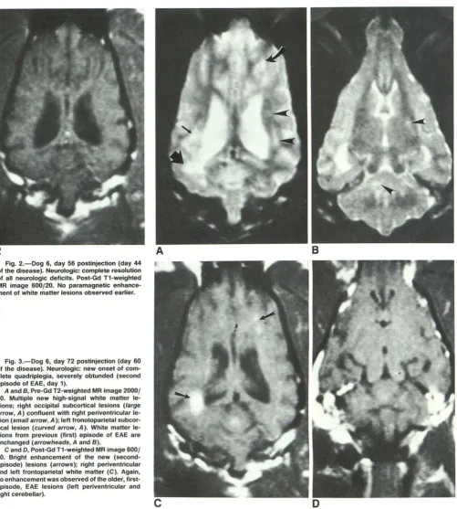
MR examinations were performed within 12 hr of the acute neurologic signs (day 12 postinduction for dogs 3 and 6). The pre-Gd T1-weighted MR images did not reveal any abnor-malities. However, pre-Gd T2-weighted images did show multiple small, focal regions of high signal in the periventricular and deep white matter of the cerebrum and cerebellum (Figs. 1 A and 1 B). After the administration of Gd-DTPA these same lesions were found to exhibit bright paramagnetic enhance-ment on the T1-weighted images (Figs. 1 C and 1 D). On day 30 postinduction (day 18 of the disease), dog 6 was observed to have persistent but slowly resolving paralysis. The white matter lesions on pre-Gd T2-weighted images appeared slightly larger and better defined. Faint paramagnetic en-hancement was noted on post-Gd T1-weighted images. By day 56 postinduction (day 44 of the disease) all neurologic deficits had completely resolved. MR images at this time demonstrated no change in the appearance of the multiple white matter abnormalities on pre-Gd T1- and T2-weighted images. On post-Gd T1-weighted images, however, no en-hancement was observed (Fig. 2). This animal continued to be neurologically intact until day 72 postinduction (day 60 of the disease) when the dog was found to be completely quadriplegic and severely obtunded. This exacerbation was considered the second episode of EAE. MR examination performed at that time revealed multiple, new high-signal subcortical and periventricular white matter lesions on the pre-Gd T2-weighted images (Figs. 3A and 3B, arrow). The white matter lesions from the previous (first) episode of EAE appeared unchanged and indistingushable in appearance from the new-onset (second-episode) lesions on pre-Gd T2-weighted images (Figs. 3A and 3B, arrowheads). Post-Gd T1-weighted images demonstrated bright enhancement of the new, second-episode lesions (Fig. 3C, arrows). Again, no enhancement of the older, first-episode lesions was observed.
Neuropathology
Dog 3 died during the acute phase of EAE (day 12 postin-duction, day 1 of the disease), and dog 6 was sacrificed 24 hr after the onset of the second episode of EAE (acute phase, second episode). At the time of necropsy, dog 6 was also clinically fully recovered from the first episode of EAE (day 61 of the disease). These lesions were stable, clinically silent, and considered chronic-phase EAE.
Fig. 1.-00g 6, day 12 postinjection (day 1 of the disease). Neurologic: hind-leg paralysis, foreleg weakness, mildly obtunded.
A and B, Pre-Gd T2-weighted MR image 2000/ 90. Multiple small, indistinct high-intensity le-sions (arrowheads) in left periventricular (A) and right cerebellar (B) white matter.
C and 0, Post-Gd T1-weighted MR image 600/ 20. Paramagnetic enhancement (arrowheads) of white matter lesions seen on T2-weighted im-ages in A and B; left periventricular (C) and right cerebellar (0) white matter.
c
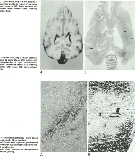
stained sections of these lesions showed the characteristic perivascular demyelination (Fig. 6B). These foci were there-fore characterized as acute demyelinating lesions.
Histologic examination of the chronic-phase (>60 days old) lesions revealed a disorganized diffuse cellular infiltration con-sisting of macrophages and plasma cells with the persistence of some lymphocytes. These lesions were believed to be subacute to early chronic demyelinating lesions. Truly chronic lesions in which gliosis had become marked were not seen.
This process may take many months to occur.
o
Discussion
646
KUHARIK ET AL. AJNR:9, July/August 19882
Fig. 2.-00g 6, day 56 postinjection (day 44 of the disease). Neurologic: complete resolution of all neurologic deficits. Post-Gd T1-weighted MR image 600/20. No paramagnetic enhance-ment of white matter lesions observed earlier.
Fig. 3.-00g 6, day 72 postinjection (day 60 of the disease). Neurologic: new onset of com-plete quadriplegia, severely obtunded (second episode of EAE, day 1).
A and B, Pre-Gd T2-weighted MR image 2000/ 90. Multiple new high-signal white matter le-sions; right occipital subcortical lesions (large arrow, A) confluent with right periventricular le-sion (small arrow, A); left fronotoparietal subcor-tical lesion (curved arrow, A). White matter le-sions from previous (first) episode of EAE are unchanged (arrowheads, A and B).
C and D, Post-Gd T1-weighted MR image 600/ 20. Bright enhancement of the new (second-episode) lesions (arrows); right peri ventricular and left frontoparietal white matter (C). Again, no enhancement was observed of the older, first-episode, EAE lesions (left periventricular and right cerebellar).
A
encountering MBP in the myelin sheaths of the CNS. A hypersensitivity response is induced with the accumulation of acute inflammatory cells in the neural perivascular spaces. Demyelination then proceeds either by direct cytotoxic effects on the myelin-producing cells or by localized production of cytotoxic antibodies, or both [2-4].
B
D
[image:4.613.63.564.83.640.2]Fig. 4.-Gross brain, dog 6. 5-mm axial sec-tions. Arrowhead points to region of abnormal white matter seen on MR. Focal necrosis left periventricular white matter after relatively chronic-phase EAE.
Fig. 5.-Myelin stain, dog 6. 10-l'm sections. Arrow points to acute-phase EAE lesions with
patchy demyelination of right periventricular
white matter. Arrowhead points to a relatively chronic-phase EAE lesion, left periventricular white matter.
Fig. 6.-Neurohistopathology acute-phase EAE lesions, dog 6. 10-l'm sections.
A, Hand E stain. Well organized, perivascular cellular infiltration by polymorphonuclear neutro-phils and lymphocytes.
B, Myelin stain. Perivascular demyelination about a venule (arrow).
4
A
disease was chosen for its morphologic/pathologic
resem-blance to human MS and for the experimental animal's large
brain size, which allowed for reproducible lesion imaging in the living animal.
MR is exquisitely sensitive in detecting white matter abnor-malities, particularly on the T2-weighted images. These high signal abnormalities are nonspecific, but in MS patients, the
-5
B
peri ventricular lesions in conjunction with a typical clinical history and spinal fluid abnormalities allow definitive diagnosis of the disease [12]. Unfortunately, MR examinations have not been able to differentiate acute, active MS lesions from chronic, inactive plaques [10].
[image:5.614.82.556.88.643.2]648 KUHARIK ET AL. AJNR:9, July/August 1988
the disease in dog 6 were correlated with the appearance of multiple, new periventricular white matter lesions that were generally not visualized on the pre-Gd T1-weighted images
and appeared as high signal foci on the T2-weighted images.
Like MS, the appearance of these lesions on MR during the
acute phase was indistinguishable from the relatively
chronic-phase lesions. This situation is illustrated in Figs. 3A and 3B,
where the acute lesions (arrows) cannot be differentiated
from the more chronic, 60-day-old lesions (arrowheads) on
pre-Gd T2-weighted images.
MR contrast enhancement is obtained by the use of
para-magnetic agents, such as Gd-DTPA. The Gd+3
ions possess
seven unpaired electrons that impart a relatively strong
mag-netic moment on the ion. This produces a local magnetic field
that shortens the relaxation times, T1 and T2, of the
neigh-boring hydrogen nuclei [13-20]. IV Gd-DTPA is exclusively
extracellular and does not normally cross the blood-brain
barrier [13-15, 18-20]. It is these properties that make
Gd-DTPA useful in assessing the blood-brain barrier breakdown,
which commonly occurs in the acute or active lesions of EAE
and MS [6, 10, 21]. After Gd-DTPA, the histologically
classi-fied acute-phase demyelinating lesions in our dog
demon-strated bright paramagnetic enhancement on the T1-weighted
images (Fig. 3C, arrows). These same lesions were thought
to be clinically active, causing the animals neurologic deficits.
Over the next 3-4 weeks, the neurologic deficits resolved
and the white matter lesions began to lose their paramagnetic
enhancement. Histologically classified
subacutejearly-chronic-phase demyelinating lesions, which occurred more than 60 days after the onset of the disease, did not exhibit
enhancement on the post-Gd T1-weighted images (Figs. 3C
and 3D). These lesions were considered clinically silent with an apparent reestablishment of the blood-brain barrier. This
difference in the paramagnetic enhancement characteristics
may allow the separation between the acute, active
demyeli-nating lesions and the relatively chronic, inactive lesions.
Our research complements the work of Grossman et al.
[10] in which paramagnetic enhancement was observed in
MS lesions thought to be active. Tissue specimens of these
lesions were, however, not available for gross and histologic
analysis of lesion age or activity. Our study suggests a
relationship between the relative age of the EAE
demyelinat-ing lesions and the presence of paramagnetic enhancement.
More specifically, we have demonstrated in one animal that
the demyelinating lesions of EAE evolve over time from acute
paramagnetic enhancing foci to chronic nonenhancing
plaques. Because of the marked similarities between EAE
and MS, we believe a similar phenomenon can be expected
during MS lesion evolution.
In conclusion, the results from our current investigation
suggest that MR with Gd-DTPA may be used to differentiate
acute from chronic demyelinating lesions in this animal model.
We believe the EAE animal model closely resembles MS in
its MR appearance and provides a unique opportunity to
observe the MR evolution of demyelinating lesions with gross
and histologic specimen correlation. Finally, these findings
may assist in the interpretation of MR with paramagnetic contrast in human demyelinating diseases.
ACKNOWLEDGMENTS
We thank Barry Owens and Edward Rhodes for technical support,
and Terri Kuharik and Barbara Smith for editorial and transcription assistance.
REFERENCES
1. McFarlin DE, McFarland HF. Multiple sclerosis. N Engl J Med
1982;307:1183-1188,1246-1251
2. Rowe MJ. Experimental allergic encephalomyelitis: a review. Bull Los Angeles Neural Soc 1969;34(1):55-66
3. Lisak RP, Behan PO. Experimental autoimmune demyelinating diseases:
experimental allergic encephalomyelitis and experimental allergic neuritis.
Biomed Pharmacother 1975;22: 81-87
4. Summers BA, Greisen HA, Appel MJ. Canine distemper and experimental
allergiC encephalomyelitis in the dog: comparative patterns of
demyelina-tion. J Comp Patho/1984;94:575-589
5. Ferraro A, Cazzullo CL. Chronic experimental allergic encephalomyelitis in
monkeys. J Neuropathol Exp Neuro/1948;7:235-260
6. Alvord EC. Summary and concluding remarks. In: Kiies MW, Alvord EC, eds. "Allergic" encephalomyelitis: experimental allergic encephalomyelitis and its relation to other diseases of man and animals. Chicago: Thomas,
1959:518-557
7. Wisniewski HM, Keith AB. Chronic relapsing experimental allergic
enceph-alomyelitis: an experimental model of multiple sclerosis. Ann Neurol
1977;1 :144-148
8. Wolf A, Kabat EA, Bezer AE. The pathology of acute disseminated
en-cephalomyelitis produced experimentally in the rhesus monkey and its
resemblance to human demyelinating disease. J Neurapathol Exp Neural
1947;6: 333-357
9. Sheldon JJ, Siddharthan R, Tobias J, Sheremata WA, Soil a K, Viamonte
M. MR imaging of multiple sclerosis: comparison with clinical and CT
examinations in 74 patients. AJR 1985;145:957-964
10. Grossman RI, Gonzalez-Scarano F, Atlas SW, Galetta S, Silberg DH.
Multiple sclerosis: gadolinium enhancement in MR imaging. Radiology
1986;161 :721-725
11. Rivers TM, Schwentker FF. Encephalomyelitis accompanied by myelin
destruction experimentally produced in monkeys. J Exp Med
1935;61 :689-703
12. Carr DH, Brown J, Bydder GM, et al. Gadolinium-DTPA as a contrast
agent in MRI: initial clinical experience in 20 patients. AJR 1984;143:2
15-224
13. Weinmann HJ, Brasch RC, Press WR, Wesbey GE. Characteristics of
gadolinium-DTPA complex: a potential NMR contrast agent. AJR
1984;142:619-624
14. Brasch RC, Weinmann H-J, Wesbey GE. Contrast-enhanced NMR
imag-ing: animal studies using gadolinium-DTPA complex. AJR
1984;142:625-630
15. Goldstein EJ, Burnett KR, Hansell JR, et al. Gadolinium-DTPA (an NMR
proton imaging contrast agent): chemical structure, paramagnetic
prop-erties and pharmacokinetics. Physiol Chem Phys Med NMR 1984;16:
97-104
16. Runge VM, Clanton JA, Lukehart CM, Partain CL, James AE. Paramagnetic
agents for contrast-enhanced NMR imaging: a review. AJR
1983;141 :1209-1215
17. Runge VM, Clanton JA, Price AC, et al. The use of Gd-DTPA as a perfusion
agent and marker of blood-brain barrier disruption. Magn Reson Imaging
1985;3:43-55
18. Grossman RI, Joseph PM, Wolf G, et al. Experimental intracranial septic
infarction: magnetic resonance enhancement. Radiology 1985;
155:649-653
19. Runge VM, Clanton JA, Price AC, et al. Evaluation of contrast-enhanced
MR imaging in a brain-abscess model. AJNR 1985;6: 139-147
20. Farlow MR, Markand ON, Edwards MK, Stevens JC, Kolar OJ. Multiple
sclerosis: magnetic resonance imaging, evoked responses, and spinal fluid
electrophoresis. Neuralogy 1986;36: 828-831
21. Vinuela FV, Fox AJ, Debrun GM, Feasby TE, Ebers GC. New perspectives
in computed tomography of multiple sclerosis. AJNR 1982;3:277-281,