Original Article
Analysis of the prognostic risk factors in middle-aged
and elderly patients with acute cerebral hemorrhage
undergoing minimally invasive surgery
Jiming Zou
Department of Health Care, Yantaishan Hospital, Yantai, Shandong, China
Received October 14, 2019; Accepted December 9, 2019; Epub January 15, 2020; Published January 30, 2020
Abstract: Objective: To evaluate the prognostic risk factors in middle-aged and elderly patients with acute cerebral hemorrhage undergoing minimally invasive surgery. Methods: From Mar 2017 to Jan 2018, 70 middle-aged and elderly patients with acute cerebral hemorrhage who received minimally invasive surgery were selected. There were 36 patients with good prognosis and 34 patients with poor prognosis judged by the attending physician. Risk fac-tors for prognosis were analyzed by univariate logistic regression and multivariate logistic regression analysis. Re-sults: According to univariate logistic regression analysis, statistical differences existed between patients with good prognosis and with poor prognosis in age, history of hypertension, preoperative systolic blood pressure, amount of bleeding, bleeding site, IVH, pulmonary infection, reoperation, preoperative blood glucose, preoperative GCS scores and NIHSS scores (P<0.05). According to multivariate logistic regression analysis, older age, greater amount of bleeding, bleeding region (basal ganglia), IVH, pulmonary infection, reoperation, preoperative blood glucose >7.8
mmol/L, preoperative GCS scores <9 and NIHSS scores ≥6 were independent risk factors related to the prognosis
of minimally invasive surgery. Conclusion: The conditions of patients before and after surgery affect the prognosis of patients treated with minimally invasive surgery, while older age, greater amount of bleeding, bleeding in the basal ganglia, IVH, pulmonary infection, reoperation, preoperative blood glucose >7.8 mmol/L, preoperative GCS scores
<9 and NIHSS scores ≥6 are independent risk factors.
Keywords: Minimally invasive surgery, acute cerebral hemorrhage, prognosis, risk factors
Introduction
A heavy burden is exerted on the intracranial
blood vessels, sometimes resulting in blood
vessels bursting, that’s intracranial
rahge [1]. Statistically, acute cerebral
hemor-rhage is one of the most common emergenci-
es and is a severe disease due to the high
incidence and mortality [2]. Acute cerebral
hemorrhage is characterized by acute onset,
rapid progression and poor prognosis, affecting
mostly middle-aged and elderly patients [3].
Surgical treatment, especially minimally
inva-sive surgery, reduces the mortality and
disabil-ity of patients [4]. Related studies revealed that
the proportion of patients suffering from poor
prognosis and recurrence after minimally
inva-sive surgery was high [5]. Postoperative
recur-rence of acute cerebral hemorrhage not only
aggravates the original disease of the patients,
but also leads to complications [6].
Materials and methods
Patient selection
From Mar 2017 to Jan 2018, 70 middle-aged
and elderly patients with acute cerebral he-
morrhage were treated with minimally invasive
surgery in our hospital. There were 36 pati-
ents with good prognosis and 34 patients with
poor prognosis (The prognosis was determin-
ed by the attending physician with self-made
questionnaires from the hospital after
minimal-ly invasive surgery) [9, 10].
Inclusion criteria: patients who were aged over
40 years and met the international diagnos-
tic criteria for cerebral hemorrhage [11]. All
patients were treated with minimal invasive
surgery, with no history of infectious diseases,
infections, anticoagulants, mental illness,
cog-nitive or consciousness impairment.
Exclusion criteria were as follows: patients with
history of blood disease, liver and renal
dys-function, or other tumor diseases.
The subjects and their families included in the
study signed informed consent in advance. This
study was approved by the ethics committee
of our hospital.
Information extraction
The baseline data were shown in Table 2.
Patients with acute cerebral hemorrhage were
treated with unilateral or bilateral ventricular
frontal puncture catheterization and external
ventricular drainage, among which, hematomas
Statistical methods
The data were analyzed by SPSS 22.0 (Beijing
Bizinsight Information Technology Co., Ltd.)
software. The count data were expressed by
[n (%)] and compared by X
2between the two
groups. The measurement data were shown as
mean ± standard deviation (X ± s) and
com-pared with independent t test. Variables for
prognostic factors are obtained through
for-ward selection, backfor-ward selection, and
step-wise selection. Univariate logistic regression
was used to select the significant variables,
then multivariate logistic regression was
per-formed to select the significant independent
risk factors. Table 1 shows the variables and
assignment values.
For multivariate logistic regression model,
bina-ry logistic regression was used with LR me-
thod.
P<0.05 indicated statistically significant
differences.
Results
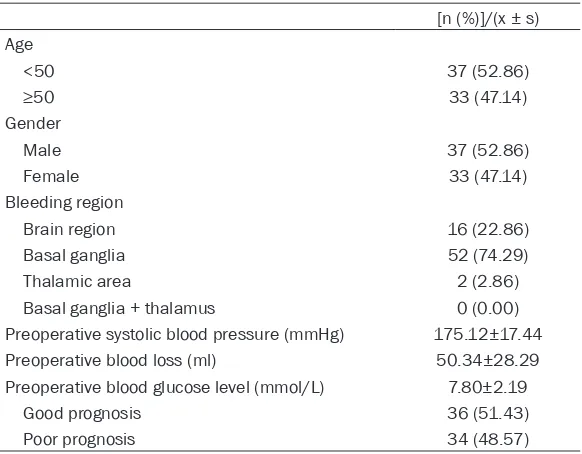
General information
[image:2.612.91.354.84.271.2]Seventy elderly patients with acute cerebral
hemorrhage who were admitted in our hospital
were selected. There were 37 males and 33
females, aged from 49 to 79 years old. Age,
gender, bleeding region, preoperative systolic
blood pressure (mmHg), preoperative blood
loss (ml), preoperative blood glucose level
(mmol/L) and other prognostic details are sh-
own in Table 2. In all, 36 patients showed good
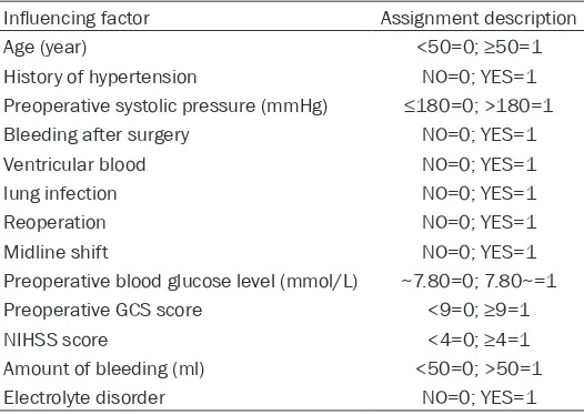
Table 1. Main research factors and variable assignment value
Influencing factor Assignment description
Age (year) <50=0; ≥50=1
History of hypertension NO=0; YES=1
Preoperative systolic pressure (mmHg) ≤180=0; >180=1
Bleeding after surgery NO=0; YES=1
Ventricular blood NO=0; YES=1
lung infection NO=0; YES=1
Reoperation NO=0; YES=1
Midline shift NO=0; YES=1
Preoperative blood glucose level (mmol/L) ~7.80=0; 7.80~=1 Preoperative GCS score <9=0; ≥9=1
NIHSS score <4=0; ≥4=1
Amount of bleeding (ml) <50=0; >50=1
Electrolyte disorder NO=0; YES=1
with volume below 70 ml were
treated with the temporal incision
and line of keyhole approach for
removal of the hematoma, and he-
matomas with volume below 70
ml were treated with craniotomy
accompanied by cranioplasty. All
patients received comprehensive
treatment measures such as
anti-hypertensive drugs, infection
con-trol and neurotrophic support.
Outcome measurement
prognosis while 34 patients exhibited poor
prognosis.
Univariate analysis of prognosis and related
factors after minimally invasive surgery
According to univariate logistic regression
anal-ysis, differences existed between patients with
good prognosis and patients with poor
progno-sis in age, history of hypertension, preoperative
systolic blood pressure, amount of bleeding,
bleeding site, IVH, pulmonary infection,
reoper-ation, preoperative blood glucose, preopera-
tive GCS sores and NIHSS scores (
P
<0.05).
Patients’ age, history of hypertension,
preoper-ative systolic blood pressure, amount of
[image:3.612.90.381.83.310.2]bleed-tory of hypertension, preoperative systolic
blood pressure, amount of bleeding, bleeding
site, IVH, pulmonary infection, reoperation,
pre-operative blood glucose, prepre-operative GCS
scores and NIHSS scores [12]. The prognosis
of patients with acute cerebral hemorrhage
after minimally invasive surgery is positively
correlated with the age of the patients. The
higher the age, the worse the prognosis [13].
Although the promotion of minimally invasive
treatment has greatly shortened the operation
time, reduced the size of the wound and
reduced the chance of complications [14].
Related studies indicated that middle-aged and
elderly patients with acute cerebral
hemor-rhage were vulnerable to surgery due to the
Table 2. General information
[n (%)]/(x ± s) Age
<50 37 (52.86)
≥50 33 (47.14)
Gender
Male 37 (52.86)
Female 33 (47.14)
Bleeding region
Brain region 16 (22.86)
Basal ganglia 52 (74.29)
Thalamic area 2 (2.86)
Basal ganglia + thalamus 0 (0.00)
Preoperative systolic blood pressure (mmHg) 175.12±17.44
Preoperative blood loss (ml) 50.34±28.29
Preoperative blood glucose level (mmol/L) 7.80±2.19
Good prognosis 36 (51.43)
[image:3.612.92.378.357.513.2]Poor prognosis 34 (48.57)
Table 3. Prognosis of different ages, history of hypertension, GCS
and NIHSS scores
Group Good prognosis (n=36) Poor prognosis (n=34) X2/t P
Age 8.184 0.004
<50 25 (69.44) 12 (35.29)
≥50 11 (30.56) 22 (64.71)
History of hypertension 9.715 0.002
Yes 12 (33.33) 24 (70.59)
No 24 (66.67) 10 (29.41)
Preoperative GCS score 28.600 <0.001
~9 3 (8.33) 24 (70.59)
9 to 15 33 (91.67) 10 (29.41)
NIHSS score 3.47±1.03 12.67±2.89 17.940 <0.001
ing, bleeding site, IVH,
pul-monary infection, reopera-
tion, preoperative blood
glu-cose, preoperative GCS sc-
ores and NIHSS scores were
risk factors related to poor
prognosis of minimally
inva-sive surgery (Tables 3-7).
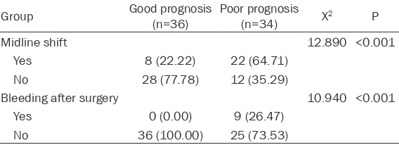
Comparisons of midline shift
situation, recurrent bleeding
after surgery were detailed in
Figures 1 and 2.
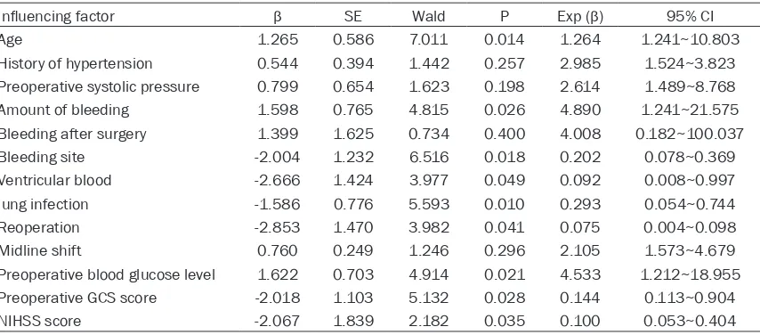
Multivariate analysis of
prognosis and related
fac-tors after minimally invasive
surgery
According to multivariate lo-
gistic regression analysis,
age, older age, greater am-
ount of bleeding, bleeding
site, IVH, pulmonary infecti-
on, reoperation, preoperative
blood glucose >7.8 mmol/L,
preoperative GCS scores <9
and NIHSS scores ≥6 were
independent risk factors re-
lated to the prognosis of
min-imally invasive surgery (Table
8).
Discussion
his-degradation of their organs [14]. Blood glucose,
systolic blood pressure and changes in amount
of bleeding have a great impact on prognosis of
patients with cerebral hemorrhage [15]. The
severity of the midline shift is often affected by
the amount of bleeding in patients with
cere-bral hemorrhage. More cerecere-bral hemorrhag-
ing in patients before operation leads to an
increase in intracranial pressure and greater
[image:4.612.91.523.84.232.2]mal range will is related to higher death rates
of patients [17]. A large number of studies on
prognostic factors of cerebral hemorrhage
suggested that the prognosis of patients with
cerebral hemorrhage was closely related to
factors such as age, history of hypertension,
preoperative systolic blood pressure, amount
of bleeding, bleeding site, IVH, pulmonary
infec-tion and preoperative blood glucose. They ex-
Table 4. Comparison of systolic blood pressure, blood glucose and blood loss before surgery
Group Good prognosis (n=36) Poor prognosis (n=34) X2 P
Preoperative systolic blood pressure (mmHg) 8.184 0.004
≤180 25 (69.44) 12 (35.29)
>180 11 (30.56) 22 (64.71)
Preoperative blood glucose level (mmol/L) 9.715 0.002
~7.80 12 (33.33) 24 (70.59)
7.80~ 24 (66.67) 10 (29.41)
Amount of bleeding (ml) 28.490 <0.001
<30 20 (55.56) 2 (5.88)
30~50 14 (38.89) 13 (38.24)
>50 2 (5.56) 19 (55.88)
Table 6. Comparison of the presence or absence of midline shift
and the prognosis of recurrent bleeding after surgery
Group Good prognosis (n=36) Poor prognosis (n=34) X2 P
Midline shift 12.890 <0.001
Yes 8 (22.22) 22 (64.71)
No 28 (77.78) 12 (35.29)
Bleeding after surgery 10.940 <0.001
Yes 0 (0.00) 9 (26.47)
[image:4.612.95.522.278.436.2]No 36 (100.00) 25 (73.53)
Table 5. Comparison of prognosis between different bleeding sites, hematoma morphology and
pres-ence or abspres-ence of ventricular hemorrhage
Group Good prognosis (n=36) Poor prognosis (n=34) X2 P
Bleeding region 6.256 0.044
Brain region 12 (33.33) 4 (11.76)
Basal ganglia 24 (66.67) 28 (82.35)
Thalamic area 0 (0.00) 2 (5.88)
Basal ganglia + thalamus 0 (0.00) 0 (0.00)
Hematoma morphology 0.061 0.806
rule 18 (50.00) 18 (52.94)
irregular 18 (50.00) 16 (47.06)
Ventricular blood 35.020 <0.001
Yes 1 (2.78) 24 (70.59)
No 35 (97.22) 10 (29.41)
[image:4.612.92.376.484.587.2]nor-plored the relationship bet-
ween age and postoperative
pulmonary infection and found
that the older the patient, the
higher the probability of lung
infection and worse prognosis
[18]. Hypertension,
character-ized by increased intracranial
pressure near the bleeding
site, may lead to enlargement
of cerebral hematoma or se-
vere cerebral hemorrhage [14,
19]. Data from multivariate
logistic regression analysis sh-
owed that age, amount of
bleeding, bleeding site, IVH,
Table 7. Comparison of prognosis of electrolyte imbalance,
pulmo-nary infection and reoperation
Group Good prognosis (n=36) Poor prognosis (n=34) X2 P
Electrolyte disorder 0.412 0.522
Yes 1 (2.78) 2 (5.88)
No 35 (97.22) 32 (94.12)
lung infection 28.600 <0.001
Yes 3 (8.33) 24 (70.59)
No 33 (91.67) 10 (29.41)
Reoperation 12.350 <0.001
Yes 0 (0.00) 10 (29.41)
No 36 (100.00) 24 (70.59)
Figure 1. Midline shift situation. *indicated that the
middle line shift is significantly higher in people in
this group (P<0.001).
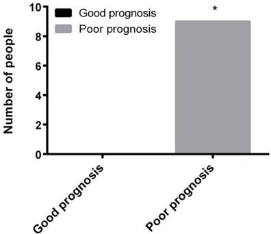
Figure 2. Bleeding after surgery situation. *indicated that the number of patients with recurrent bleeding
after surgery was significantly higher (P<0.001).
pulmonary infection, reoperation, preoperative
blood glucose, preoperative GCS scores and
NIHSS scores were independent risk factors
related to the prognosis of minimally invasive
surgery.
The amount and location of bleeding are the
decisive factors affecting the prognosis of pa-
tients with cerebral hemorrhage. The greater
the amount of bleeding, the worse the
progno-sis of patients [19]; the prognoprogno-sis of patients
with cerebral hemorrhage was directly affected
by different bleeding sites. Patients with
cere-bral hemorrhage in the posterior limb,
thala-mus and under the sac showed poor clinical
prognosis [20]. The preoperative GCS score
and NIHSS score have been used in the eva-
luation of patients with cerebral hemorrhage to
assess the degree of coma and neurological
deficits. GCS score and NIHSS score were posi
-tively correlated with the severity of cerebral
hemorrhage. The higher the NIHSS score, the
worse the clinical outcome of minimally
inva-sive surgery and the worse prognosis of pa-
tients [21]. Long-term accumulation of blood
in the ventricles can lead to hematoma and
necrosis of brain tissue around the hematoma,
which seriously affected the prognosis [13].
[image:5.612.89.287.497.668.2]intraventricu-lar hemorrhaging will affect the prognosis of
patients with acute cerebral hemorrhage [24].
The death of patients with cerebral
hemor-rhage is mostly caused by complications such
as pulmonary infection. Pulmonary infection
may be related to the brain stem respiratory
and circulatory center failure, indicating that
the patient is dying [25]. This supported the
idea that pulmonary infection is an important
prognostic risk factor for the prognosis of
cere-bral hemorrhage after minimally invasive
sur-gery. The relationship between reoperation of
hypertensive cerebral hemorrhage and prog-
nosis showed that patients with hypertensive
cerebral hemorrhage undergo reoperation. The
purpose of this treatment is to reduce ischemic
edema and restore the brain function of the
patient as much as possible. However, during
the reoperation, the primary brain injury will be
aggravated. In severe cases, the brain tissue
experiences irreversible pathological changes
[26].
This study did not explore the influence of dif
-ferent geographical environments and the level
of medical as well as the patients’ habits such
as smoking and drinking, which could affect
the prognosis and result in the contingency of
the experimental results [27, 28]. This study
did not explore the mechanism of the risk
fac-tors. The study will be improved based on the
above considerations in the future.
In conclusion, older age, greater amount of
bleeding, bleeding site, IVH, pulmonary
infec-tion, reoperainfec-tion, preoperative blood glucose
>7.8 mmol/L, preoperative GCS scores <9 and
NIHSS scores ≥6 are independent risk factors
for the prognosis of minimally invasive surgery.
Disclosure of conflict of interest
None.
Address correspondence to: Jiming Zou, Depart- ment of Health Care, Yantaishan Hospital, No. 91 Jiefang Road, Zhifu District, Yantai 264008, Shandong, China. Tel: +86-13505351660; E-mail: evagk1md@163.com
References
[1] Qureshi AI, Palesch YY and Suarez JI. Intensive blood-pressure lowering in cerebral hemor-rhage. N Engl J Med 2016; 375: e48.
[2] Yang J, Arima H, Wu G, Heeley E, Delcourt C, Zhou J, Chen G, Wang X, Zhang S, Yu S, Chalm-ers J and AndChalm-erson CS; INTERACT
Investiga-tors. Prognostic significance of perihematomal
edema in acute intracerebral hemorrhage: pooled analysis from the intensive blood pres-sure reduction in acute cerebral hemorrhage trial studies. Stroke 2015; 46: 1009-1013. [3] Lattanzi S, Cagnetti C, Provinciali L and
Silves-trini M. Neutrophil-to-lymphocyte ratio and neurological deterioration following acute ce-rebral hemorrhage. Oncotarget 2017; 8: 57489-57494.
[4] Chan E, Anderson CS, Wang X, Arima H, Saxe-na A, Moullaali TJ, Heeley E, Delcourt C, Wu G, Wang J, Chen G, Lavados PM, Stapf C, Robin-son T, Chalmers J and Huang Y; INTERACT2
In-vestigators. Significance of intraventricular
[image:6.612.94.521.83.271.2]hemorrhage in acute intracerebral hemor-rhage: intensive blood pressure reduction in
Table 8. Multivariate analysis of prognosis and related factors after minimally invasive surgery
Influencing factor β SE Wald P Exp (β) 95% CI
Age 1.265 0.586 7.011 0.014 1.264 1.241~10.803
History of hypertension 0.544 0.394 1.442 0.257 2.985 1.524~3.823 Preoperative systolic pressure 0.799 0.654 1.623 0.198 2.614 1.489~8.768
Amount of bleeding 1.598 0.765 4.815 0.026 4.890 1.241~21.575
Bleeding after surgery 1.399 1.625 0.734 0.400 4.008 0.182~100.037
Bleeding site -2.004 1.232 6.516 0.018 0.202 0.078~0.369
Ventricular blood -2.666 1.424 3.977 0.049 0.092 0.008~0.997
lung infection -1.586 0.776 5.593 0.010 0.293 0.054~0.744
Reoperation -2.853 1.470 3.982 0.041 0.075 0.004~0.098
Midline shift 0.760 0.249 1.246 0.296 2.105 1.573~4.679
Preoperative blood glucose level 1.622 0.703 4.914 0.021 4.533 1.212~18.955 Preoperative GCS score -2.018 1.103 5.132 0.028 0.144 0.113~0.904
acute cerebral hemorrhage trial results. Stroke 2015; 46: 653-658.
[5] Berkhemer OA, Fransen PS, Beumer D, van den Berg LA, Lingsma HF, Yoo AJ, Schonewille WJ, Vos JA, Nederkoorn PJ, Wermer MJ, van Walderveen MA, Staals J, Hofmeijer J, van Oostayen JA, Lycklama à Nijeholt GJ, Boiten J, Brouwer PA, Emmer BJ, de Bruijn SF, van Dijk LC, Kappelle LJ, Lo RH, van Dijk EJ, de Vries J, de Kort PL, van Rooij WJ, van den Berg JS, van Hasselt BA, Aerden LA, Dallinga RJ, Visser MC, Bot JC, Vroomen PC, Eshghi O, Schreuder TH, Heijboer RJ, Keizer K, Tielbeek AV, den Hertog HM, Gerrits DG, van den Berg-Vos RM, Karas GB, Steyerberg EW, Flach HZ, Marquering HA, Sprengers ME, Jenniskens SF, Beenen LF, van den Berg R, Koudstaal PJ, van Zwam WH, Roos YB, van der Lugt A, van Oostenbrugge RJ, Ma-joie CB and Dippel DW; MR CLEAN Investiga-tors. A randomized trial of intraarterial treat-ment for acute ischemic stroke. N Engl J Med 2015; 372: 11-20.
[6] Reinink H, de Jonge JC, Bath PM, van de Beek D, Berge E, Borregaard S, Ciccone A, Csiba L, Demotes J, Dippel DW, Korv J, Kurkowska-Jas-trzebska I, Lees KR, Macleod MR, Ntaios G, Randall G, Thomalla G and van der Worp HB. PRECIOUS: PREvention of complications to im-prove outcome in elderly patients with acute stroke. Rationale and design of a randomised, open, phase III, clinical trial with blinded out-come assessment. Eur Stroke J 2018; 3: 291-298.
[7] Hanley DF, Thompson RE, Muschelli J, Rosen-blum M, McBee N, Lane K, Bistran-Hall AJ, Mayo SW, Keyl P, Gandhi D, Morgan TC, Ullman N, Mould WA, Carhuapoma JR, Kase C, Ziai W, Thompson CB, Yenokyan G, Huang E, Broad-dus WC, Graham RS, Aldrich EF, Dodd R, Wij-man C, Caron JL, Huang J, Camarata P, Mende-low AD, Gregson B, Janis S, Vespa P, Martin N, Awad I and Zuccarello M; MISTIE Investigators.
Safety and efficacy of minimally invasive sur -gery plus alteplase in intracerebral haemor-rhage evacuation (MISTIE): a randomised, con-trolled, open-label, phase 2 trial. Lancet Neurol 2016; 15: 1228-1237.
[8] Saver JL, Goyal M, van der Lugt A, Menon BK, Majoie CB, Dippel DW, Campbell BC, Nogueira RG, Demchuk AM, Tomasello A, Cardona P, Devlin TG, Frei DF, du Mesnil de Rochemont R, Berkhemer OA, Jovin TG, Siddiqui AH, van Zwam WH, Davis SM, Castano C, Sapkota BL, Fransen PS, Molina C, van Oostenbrugge RJ, Chamorro A, Lingsma H, Silver FL, Donnan GA, Shuaib A, Brown S, Stouch B, Mitchell PJ, Davalos A, Roos YB and Hill MD; HERMES Col-laborators. Time to treatment with endovascu-lar thrombectomy and outcomes from
isch-emic stroke: a meta-analysis. JAMA 2016; 316: 1279-1288.
[9] Masotti L, Lorenzini G, Di Napoli M and Godoy DA. Prognostic ability of four clinical grading scores in spontaneous intracerebral hemor-rhage. Acta Neurol Belg 2017; 117: 325-327. [10] Benedictus MR, Hochart A, Rossi C, Boulouis
G, Henon H, van der Flier WM and Cordonnier C. Prognostic factors for cognitive decline after intracerebral hemorrhage. Stroke 2015; 46: 2773-2778.
[11] Fam MD, Hanley D, Stadnik A, Zeineddine HA, Girard R, Jesselson M, Cao Y, Money L, McBee N, Bistran-Hall AJ, Mould WA, Lane K, Cama-rata PJ, Zuccarello M and Awad IA. Surgical performance in minimally invasive surgery plus recombinant tissue plasminogen activa-tor for intracerebral hemorrhage evacuation phase III clinical trial. Neurosurgery 2017; 81: 860-866.
[12] Goto T, Ohata T, Shijo T, Yoshioka D and Kaneko M. Emergency valve surgery for infective endo-carditis complicated by acute intracranial hem-orrhage: a case report. Int J Surg Case Rep 2017; 32: 32-35.
[13] Baharoglu MI, Cordonnier C, Al-Shahi Salman R, de Gans K, Koopman MM, Brand A, Majoie CB, Beenen LF, Marquering HA, Vermeulen M, Nederkoorn PJ, de Haan RJ and Roos YB; PATCH Investigators. Platelet transfusion ver-sus standard care after acute stroke due to spontaneous cerebral haemorrhage associat-ed with antiplatelet therapy (PATCH): a ran-domised, open-label, phase 3 trial. Lancet 2016; 387: 2605-2613.
[14] Feng Y, He J, Liu B, Yang L and Wang Y. Endo-scope-assisted keyhole technique for hyper-tensive cerebral hemorrhage in elderly pa-tients: a randomized controlled study in 184 patients. Turk Neurosurg 2016; 26: 84-89. [15] Saxena A, Anderson CS, Wang X, Sato S, Arima
H, Chan E, Munoz-Venturelli P, Delcourt C, Rob-inson T, Stapf C, Lavados PM, Wang J, Neal B, Chalmers J and Heeley E; INTERACT2
Investi-gators. Prognostic significance of hyperglyce -mia in acute intracerebral hemorrhage: the INTERACT2 study. Stroke 2016; 47: 682-688. [16] Thigpen JL, Dillon C, Forster KB, Henault L,
Quinn EK, Tripodis Y, Berger PB, Hylek EM and
Limdi NA. Validity of international classification
of disease codes to identify ischemic stroke and intracranial hemorrhage among
individu-als with associated diagnosis of atrial fibrilla -tion. Circ Cardiovasc Qual Outcomes 2015; 8: 8-14.
Mine-matsu K and Toyoda K; SAMURAI Study Investi-gators. Blood glucose levels during the initial 72 h and 3-month functional outcomes in acute intracerebral hemorrhage: the SAMU-RAI-ICH study. J Neurol Sci 2015; 350: 75-78. [18] Yang N, Lin M, Wang BG, Zeng WY, He YF, Peng
HY, Zeng J, Wu ZY and Zhong Y. Low level of low-density lipoprotein cholesterol is related with increased hemorrhagic transformation af-ter acute ischemic cerebral infarction. Eur Rev Med Pharmacol Sci 2016; 20: 673-678. [19] Kazdal H, Kanat A, Findik H, Sen A, Ozdemir B,
Batcik OE, Yavasi O and Inecikli MF. Transor-bital ultrasonographic measurement of optic nerve sheath diameter for intracranial midline shift in patients with head trauma. World Neu-rosurg 2016; 85: 292-297.
[20] Gofir A, Mulyono B and Sutarni S. Hyperglyce -mia as a prognosis predictor of length of stay and functional outcomes in patients with acute ischemic stroke. Int J Neurosci 2017; 127: 923-929.
[21] Yuan MZ, Li F, Tian X, Wang W, Jia M, Wang XF and Liu GW. Risk factors for lung infection in stroke patients: a meta-analysis of observa-tional studies. Expert Rev Anti Infect Ther 2015; 13: 1289-1298.
[22] Koivunen RJ, Haapaniemi E, Satopaa J, Nieme-la M, Tatlisumak T and PutaaNieme-la J. Medical acute complications of intracerebral hemor-rhage in young adults. Stroke Res Treat 2015; 2015: 357696.
[23] Stanley D, Mason LJ, Mackin KE, Srikhanta YN, Lyras D, Prakash MD, Nurgali K, Venegas A, Hill MD, Moore RJ and Wong CH. Translocation and dissemination of commensal bacteria in post-stroke infection. Nat Med 2016; 22: 1277-1284.
[24] Matis GK and Birbilis TA. Poor relation between Glasgow coma scale and survival after head injury. Med Sci Monit 2009; 15: CR62-65. [25] Bhatt A, Lesko A, Lucas L, Kansara A and
Bara-ban E. Patients with low national institutes of health stroke scale scores have longer door-to-needle times: analysis of a telestroke network. J Stroke Cerebrovasc Dis 2016; 25: 2253-2258.
[26] Iso T, Yanagawa Y, Takeuchi I and Suwa S. Con-comitance acute cerebral infarction and re-mote intra-cerebral hemorrhaging on arrival. J Emerg Trauma Shock 2018; 11: 149-150. [27] Shitara K, Matsuo K, Hatooka S, Ura T,
Taka-hari D, Yokota T, Abe T, Kawai H, Tajika M and Kodaira T. Heavy smoking history interacts with chemoradiotherapy for esophageal can-cer prognosis: a retrospective study. Cancan-cer Sci 2010; 101: 1001-1006.