G PROTEIN-COUPLED RECEPTOR KINASE 3 (GRK3) REGULATES G PROTEIN- COUPLED RECEPTORS ON MURINE BONE MARROW NICHE MESENCHYMAL
STEM CELLS AND HEMATOPOIETIC STEM-PROGENITOR CELLS
Jaime Marie Brozowski
A dissertation submitted to the faculty at the University of North Carolina at Chapel Hill in partial fulfillment of the requirements for the degree of Doctor of Philosophy in the
Department of Microbiology and Immunology in the School of Medicine.
Chapel Hill 2017
Approved by:
Teresa K. Tarrant Jason Whitmire Kathleen Caron Glenn Matsushima Roland Tisch
ii
©2017
Jaime Marie Brozowski ALL RIGHTS RESERVED
iii ABSTRACT
Jaime Marie Brozowski: G protein-coupled receptor kinase 3 (GRK3) regulates G protein- coupled receptors on murine bone marrow niche mesenchymal stem cells and hematopoietic
stem-progenitor cells
(Under the direction of Teresa K. Tarrant)
The bone marrow microenvironment, termed niche, supports hematopoietic cell development and thus, is vital for establishment of the immune system. Within the niche reside bone marrow mesenchymal stem cells (BmMSCs) that surround the hematopoietic stem-progenitor cells (HSPCs) to support their development, maintenance, and function;
however, the intracellular regulatory mechanisms of BmMSCs and HSPCs are still being defined. The goal of this dissertation work is to provide further insight into the regulatory mechanisms that modulate functionality of BmMSCs and HSPCs. Our data suggest G protein-coupled receptor kinase 3 (GRK3) functions as a negative regulator of G protein- coupled receptors (GPCRs) on BmMSCs and HSPCs.
BmMSCs isolated from GRK3-deficient (Grk3-/-) mice have enhanced proliferation and osteogenic differentiation ex vivo compared to wildtype (WT) BmMSCs. Grk3-/- BmMSC cultures also have higher levels of CXCL12, an essential chemokine for HSPC development, and interestingly, Grk3-/- mice have increased hematopoietic cell numbers isolated from the bone marrow. Both Grk3-/- BmMSC proliferation and osteogenic
differentiation were reduced to WT level upon reduction of sphingosine-1-phosphate (S1P), and Grk3-/- BmMSCs have sustained ERK1/2 signaling upon stimulation of sphingosine-1- phosphate receptor (S1PR) with S1P in comparison to WT BmMSCs. In addition, we report
iv
GRK3 recruits β-arrestin, a protein necessary for receptor internalization, to the C-terminus of S1PR1, and we demonstrate BmMSCs lacking GRK3 regulation have impaired S1PR1 internalization. Our findings suggest GRK3 regulates GPCR S1PR on BmMSCs.
Grk3-/- mice have increased bone marrow lineage negative (Lin-) Sca1+ c-Kit+ (LSK) HSPC and oligopotent progenitor numbers, as well as increased total leukocytes in the peripheral blood. Since increased stem cell numbers and function potentiate cellular
engraftment and hematopoiesis, we tested whether GRK3 deficiency enhances hematopoietic cell function in vivo after short-term transplantation, termed colony forming unit-spleen (CFU-S) assay. Transplanted Grk3-/- LSK HSPCs or Grk3-/- whole bone marrow increases colony counts on the explanted spleen in comparison to WT controls, suggesting
hematopoiesis of Grk3-/- HSPC is enhanced. Further, both Grk3-/- hematopoietic myeloid granulocytic and monocytic (CFU-GM) and lymphoid (CFU-Pre-B) colony counts increased ex vivo upon CXCR4 ligand stimulation (CXCL12), and Grk3-/- myeloid colony counts reduced to WT levels with CXCR4 antagonist treatment (AMD3100). Taken together, in vivo and ex vivo CFU data suggest GRK3 regulates bone marrow HSPC numbers, and this is, at least in part, mediated through CXCL12/CXCR4 stimulation.
Herein, we describe a newly elucidated pathway of regulation on two niche cells, BmMSCs and HSPCs. Specifically, our data suggest GRK3 functions as a negative regulator of GPCRs on both BmMSCs and HSPCs and can modulate stem cell function.
v
To my unborn child, everything I aim to accomplish in life is now for you.
vi
ACKNOWLEDGEMENTS
Since joining Dr. Teresa K. Tarrant’s lab in 2013, I have been able to combine two long-standing passions of mine: stem cell and immunological research, which both ignite my scientific curiosity each day. I am thankful for Dr. Tarrant’s scientific mentorship over the duration of my dissertation projects, as she always welcomed dissemination of our work by supporting my fellowship applications, encouraging my participation in conferences, and giving the freedom to initiate collaborations. I have enjoyed (and will miss) our scientific brainstorming dialogs filled with “well, what if…” and “what about…”. Thank you for respecting my insight. Somewhere along the way under her guidance, I also learned to balance scientific dedication and focus with carving out time for fun (two-time champions of the Training Initiatives in Biomedical & Biological Sciences-TIBBS’ lab pumpkin carving contest!). It is with sincere gratitude that I am able to admit I have thoroughly enjoyed my Ph.D. studies. In addition to serving as my academic advisor, Dr. Tarrant has also served as a woman in science mentor. She has eloquently demonstrated and encouraged how one can maintain a devoted scientific career with balance of family life. For all of these things, I would like to sincerely thank Dr. Tarrant for her professional mentorship.
I would like to thank my dissertation committee, Drs. Jason Whitmire, Kathleen Caron, Glenn Matsushima, and Roland Tisch, for their time and continued guidance that aided my scientific development. In particular, I would like to acknowledge Dr. Whitmire, who served as Chair of my dissertation committee, for his dedicated involvement throughout
vii
my academic studies and interest in my professional development. I am blessed to have so many devoted mentors at the University of North Carolina at Chapel Hill (UNC).
I would like to acknowledge Dr. Matthew Billard (post-doc) with his much-
appreciated lab expertise and valuable mentoring during our day-to-day interactions. I would also like to acknowledge Roman Timoshchenko’s (lab technician) and Jessica Koontz’s (undergraduate thesis student) role in initiating the BmMSC studies; D. Stephen Serafin (lab technician) for his involvement in the modified TANGO assay; Brittney Allyn
(undergraduate student/lab technician) for her assistance in the CXCL12 ELISA assay; and Daniel Mattox (undergraduate student) for his invaluable weekly autoclaving of lab supplies and his interest in bioinformatics that aided our RNA-seq database query of human
BmMSCs.
I would also like to acknowledge Dr. Janet Rubin (PI; UNC) for her expertise that aided initiation of our BmMSC cultures before my arrival into lab, as well as our micro- computed tomography collaborations with Dr. Clinton T. Rubin (PI; Stonybrook), Dr.
Matthew J. Hilton (PI; Duke), and Dr. Yinshi Ren (post-doc; Duke); sphingosine kinase activity collaborations with Dr. Nancy L. Allbritton (PI; UNC), Taylor F. Harris (graduate student; UNC), and David Abraham (graduate student; UNC); and statistical collaboration with Dr. Amanda M. Eudy (post-doc; Duke). I would also like to thank Dr. Bryan Roth (UNC) for the TANGO constructs and Dr. Robert Lefkowitz (Duke) for the Grk3-/- mice.
I would like to acknowledge our use of the UNC Flow Cytometry Core Facility (supported in part by P30 CA016086 Cancer Center Core Support Grant to the UNC Lineberger Comprehensive Cancer Center), (former) Michael Hooker Microscopy Core at UNC, (new) UNC Hooker Imaging Core, UNC Animal Studies Core, and the ENCODE
viii
Consortium using Dr. Thomas Gingeras’ (Cold Spring Harbor Labs) RNA-seq database of human BmMSCs.
Finally, I would like to acknowledge fellowships, travel awards, training programs, and departments that have supported my scientific training and dissertation work. I am honored to be a recipient of the Ruth L. Kirschstein Predoctoral Individual National Research Service Award F31 Parent Fellowship (3 years, 2015 – 2018) through the National Heart, Lung, and Blood Institute of the National Institutes of Health under award number
F31HL128029. I would also like to acknowledge other laboratory funding through the National Institute of Arthritis and Musculoskeletal and Skin Diseases under award number R03AR059286 and National Institute of Allergy and Infectious Diseases under award
number K08AI070684. Note: The content herein is solely the responsibility of the author and does not necessarily represent the official views of the National Institutes of Health. In addition, I would like to express my gratitude toward the Howard Hughes Medical Institute (HHMI) Med into Grad training program (2013 – Current) that has furthered my education in translational medicine through exposure to both clinical practice and basic science research at UNC. I would like to acknowledge each mentor within the HHMI Translational Medicine program at UNC: Drs. Tarrant and Caron (PI and Co-Mentor), Dr. William Coleman (Program Director), and Dr. Patrick Brandt (Program Manager), as well as my HHMI fellowship stipend support (1 year, 2013) and travel award (2014). I would also like to acknowledge the Thurston Arthritis Research Center (TARC) and their support of my scientific education, and lastly, though certainly not least, I would like to acknowledge my graduate studies department: Microbiology and Immunology (M&I) at UNC, particularly Dr.
William Goldman (Department Chair) and Dr. Robert Bourret (Graduate Studies Director)
ix
for their scientific and professional development guidance, as well as administrative support by Dixie Flannery (Student Services Specialist) and Natalie Nesbitt (Executive Assistant to Dr. Goldman). The Department of M&I at UNC has also supported dissemination of my work through travel awards.
In summary, thank you to all the mentors, committee members, laboratory teammates, collaborators, Core Facility staff members, and departmental faculty and administrators. It has been an honor to interact with each of you and pursue my Ph.D.
graduate studies at Carolina.
x
TABLE OF CONTENTS
LIST OF FIGURES……….…xv
LIST OF ABBREVIATIONS AND SYMBOLS………...…xvi
CHAPTER 1: INTRODUCTION—THE BONE MARROW NICHE………..…1
Overview………..………..1
Healing Properties of Hematopoietic Cell Transplantation………...…1
Historical Relevance of the Bone Marrow Niche………..2
Cellular Components of the Bone Marrow Niche……….3
Hematopoietic stem cells………...…3
Mesenchymal stem cells………...….4
Osteoblasts……….5
CXCL12-abundant reticular (CAR) cells………..…6
Regions of the Bone Marrow Niche………..…7
G Protein-Coupled Receptor (GPCR) Signaling within the Bone Marrow Niche……8
Background on GPCRs………..…8
GPCR regulation: G protein-coupled receptor kinases (GRKs) and β-arrestin……….8
Ligand-induced GPCR signaling within the bone marrow niche………..9
PTH/PTHR signaling………...……...9
CXCL12/CXCR4 signaling……….…10
S1P/S1PR signaling……….11
xi
Targeting GPCRs as Therapeutic Strategies in the Bone Marrow………..…12
Granulocyte-colony stimulating factor (G-CSF)…...………..………12
AMD3100………12
FTY720 and SEW2871………13
Conclusion and Research Goals………...……13
CHAPTER 2: G PROTEIN-COUPLED RECEPTOR KINASE 3 MODULATES MESENCHYMAL STEM CELL PROLIFERATION AND DIFFERENTIATION THROUGH SPHINGOSINE-1-PHOSPHATE RECEPTOR REGULATION…………...…17
Overview………..…17
Significance Statement……….…18
Introduction………..…19
Materials and Methods……….…20
Animals………20
Bone marrow-derived mesenchymal stem cell (BmMSC) isolations……..…20
Chondrogenic, adipogenic, osteogenic differentiation………21
Bone marker mRNA expression (qRT-PCR)………..…22
ELISA………..…22
Micro-computed tomography (µCT)………...…22
Cellular proliferation………23
Immunoblotting………23
β-arrestin recruitment assay……….………25
S1PR1 internalization assay……….………25
Statistical analyses………..….26
Results………..………27
Grk3-/- BmMSCs have enhanced osteogenic differentiation ………..….27
xii
Grk3-/- BmMSC cultures have higher levels of CXCL12………...….28
GRK3 deficiency increases proliferation ex vivo……….….29
GRK3 deficiency does not affect mature bone formation in vivo………..29
Inhibition of S1P reduces the enhanced osteogenic differentiation and proliferation phenotype of Grk3-/- BmMSCs……….31
Grk3-/- BmMSCs have enhanced ERK1/2 signaling after S1P stimulation…..32
GRK3 recruits β-arrestin to the C-terminus of S1PR1 and affects S1PR1 internalization……….…..33
Discussion………...….35
Conclusion………...…39
CHAPTER 3: G PROTEIN-COUPLED RECEPTOR KINASE 3 REGULATES HEMATOPOIETIC STEM-PROGENITOR CELL FUNCTION MEDIATED THROUGH CXCL12/CXCR4………...….41
Overview………..…41
Significance Statement……….…43
Introduction………..…43
Materials and Methods……….…45
Animals………45
Flow cytometry bone marrow lineage analyses………...……45
In vivo colony forming unit-spleen (CFU-S) assays………46
Ex vivo colony forming unit-granulocytic and monocytic (CFU-GM) progenitor cell assay…...47
Ex vivo colony forming unit-pre-B (CFU-Pre-B) progenitor cell assay………..……47
Statistical analyses………...…47
Results………..…48
xiii
Grk3-/- HSPC and oligopotent progenitor cell numbers are increased
within the bone marrow………...…48
Transplantation of Grk3-/- LSK HSPCs increases hematopoietic stem cell colonies on the spleen………..….50
Transplantation of Grk3-/- bone marrow cells increases hematopoietic progenitor colony counts on the spleen………...……52
CXCL12 stimulation increases hematopoietic granulocytic and monocytic-GM progenitor colony formation (CFU-GM) of Grk3-/- bone marrow cells………...53
CXCL12 stimulation increases hematopoietic lymphoid pre-B cell colony formation (CFU-Pre-B) of Grk3-/- bone marrow cells………54
Discussion………...….56
Conclusion………...…59
CHAPTER 4: CONCLUSION AND FUTURE DIRECTIONS……….……60
Overview………..………60
Chapter 2………..……61
Field relevance……….…61
Conclusion and future directions……….………62
Chapter 3………..……65
Field relevance……….……65
Conclusion and future directions……….66
Final Thoughts...68
APPENDICES………..…...69
APPENDIX 1: MOUSE MESENCHYMAL STEM CELL MARKER PANEL……69
APPENDIX 2: DIFFERENTIATION OF GRK3-KNOCKDOWN BmMSCS…..…70
APPENDIX 3: CXCR4 mRNA EXPRESSION ON BmMSCS………...71
APPENDIX 4: BmMSC PROLIFERATION WITH CXCL12 AND AMD3100...…72
xiv
APPENDIX 5: BmMSC SPHINGOSINE KINASE ACTIVITY………73 APPENDIX 6: BmMSC EXPRESSION OF S1PR1………...………74 APPENDIX 7: BmMSC EXPRESSION OF S1PR3………...……75 APPENDIX 8: DMSO VEHICLE TOXICITY WITH COMPARABLE
DOSING AS VPC23019………..…..76 APPENDIX 9: BONE MARROW STEM AND PROGENITOR
FLOW ANALYSES………..….77 APPENDIX 10: OPTIMIZATION OF CXCL12 CONCENTRATION………….…78 APPENDIX 11: AMD3100 INHIBITS COLONY PROLIFERATION………….…79 APPENDIX 12: Grk3-/- LSK HSPC DIFFERENTIATION………..….80 APPENDIX 13: CXCL12 LEVELS OF BmMSCS TREATED WITH SKI……..…81 REFERENCES………....…82
xv
LIST OF FIGURES
Figure 1.1- The bone marrow niche………..………...………14 Figure 2.1- BmMSC multipotent differentiation, osteogenic differentiation time-course,
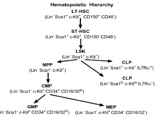
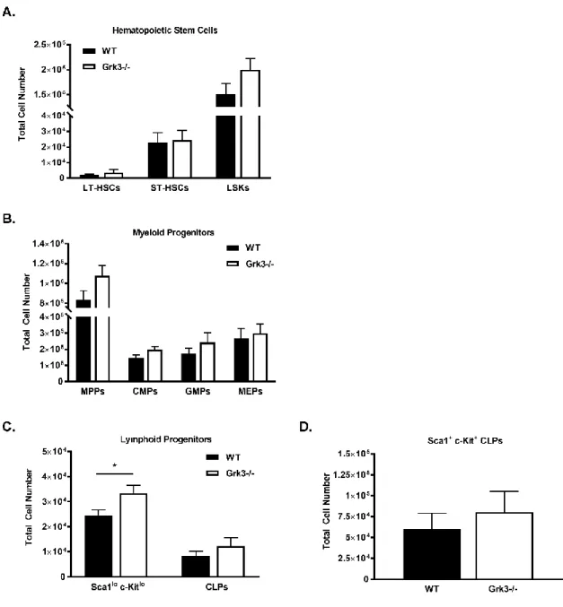
alkaline phosphatase expression………...……27 Figure 2.2- BmMSC CXCL12 ELISA and CCK-8 proliferation………...….29 Figure 2.3- Micro-computed tomography………30 Figure 2.4- SKI treatment: Osteogenic differentiation and CCK-8 proliferation………...….31 Figure 2.5- S1P-stimulated ERK1/2 signaling……….…33 Figure 2.6- β-arrestin recruitment and S1PR1 internalization……….……34 Figure 2.7- BmMSC proposed model………..……40 Figure 3.1- Bone marrow and peripheral blood analyses of WT and Grk3-/- mice…………49 Figure 3.2- Hematopoietic hierarchy of differentiation………...……50 Figure 3.3- Extended bone marrow subset analyses of WT and Grk3-/- mice………51 Figure 3.4- Colony forming unit-spleen (CFU-S): Lin- Sca1+ c-Kit+ (LSK) HSPCs……..…52 Figure 3.5- Colony forming unit-spleen (CFU-S): Whole bone marrow………...….53 Figure 3.6- Colony forming unit-granulocytic and monocytic progenitors (CFU-GM)….…54 Figure 3.7- Colony forming unit-pre-B cell progenitors (CFU-Pre-B)………...…55
xvi
LIST OF ABBREVIATIONS AND SYMBOLS -/- Deficient
± Plus/minus
≤ Less than or equal to
µ Micro
µCT Micro-computed tomography, micro-CT
α Alpha
AAALAC Association for assessment and accreditation of laboratory animal care ALP Alkaline phosphatase
AMD3100 CXCR4 antagonist ANOVA Analysis of variance APC Allophycocyanin
β Beta
BCA Bicinchoninic acid
BmMSC(s) Bone marrow(-derived) mesenchymal stem cell(s) BV/TV Bone volume/total volume (bone volume fraction) C57BL/6 C57 Black 6 or “B6” (mice)
CAR CXCL12-abundant reticular CCK-8 Cell counting kit-8
CD Cluster of differentiation CD11b Mac-1, Integrin α M (ITGAM) CD16/CD32 FcγRIII/FcγRII
CD29 Integrin β1
xvii
CD31 PECAM-1
CD34 Hematopoietic progenitor cell antigen CD34 CD44 Hermes, H-CAM
CD45 Leukocyte common antigen
CD45R B220
CD48 Slamf2
CD73 5’-nucleotidase CD105 Endoglin CD106 VCAM-1 CD150 Slamf1
cDNA Complementary deoxyribonucleic acid CEM Complete expansion media
CFU Colony forming unit CIM Complete isolation media
c-Kit Stem cell factor receptor (CD117) CLP Common lymphoid progenitor CMP Common myeloid progenitor CO2 Carbon dioxide
CXCL12 C-X-C motif chemokine ligand 12/ stromal cell-derived factor-1 CXCR4 C-X-C motif chemokine receptor 4 (CD184)
DMSO Dimethyl sulfoxide DNA Deoxyribonucleic acid
DPBS Dulbecco’s phosphate-buffered saline
xviii ECL Enhanced chemiluminescence
ELISA Enzyme-linked immunosorbent assay ERK Extracellular signal-regulated kinase FACS Fluorescence-activated cell sorting FBS Fetal bovine serum
FTY720 S1PR agonist
g Gram
GAPDH Glyceraldehyde 3-phosphate dehydrogenase, G3PDH G-CSF Granulocyte-colony stimulating factor
GMP Granulocytic-monocytic progenitor GPCR(s) G protein-coupled receptor(s) GRK G protein-coupled receptor kinase Gy Gray, unit of radiation
HBSS Hank’s balanced salt solution HCT Hematopoietic cell transplantation HRP Horseradish peroxidase
HS Horse serum
HSC(s) Hematopoietic stem cell(s)
HSPC(s) Hematopoietic stem-progenitor cell(s) IACUC Institutional animal care and use committee IDUA Iduronidase, alpha-L-
IMDM Iscove’s modified dulbecco’s medium IU International unit
xix KD, kDa Kilodalton
LIC Ligation independent cloning
Lin Lineage (Lin-, lack of specific lineage commitment) LSK Lin- Sca1+ c-Kit+
LT- Long-term
Ly-6G Neutrophil marker
m Milli
M Molar
MAPK Mitogen-activated protein kinase MEP Myeloid erythroid progenitor mL Milliliter
MPP Myeloid progenitor population mRNA Messenger ribonucleic acid
η Nano
NaF Sodium fluoride NaVO4 Sodium orthovanadate
NT Non-target
ρ Pico
PDGFR Platelet-derived growth factor receptor PE R-phycoerythrin
PMSF Phenylmethylsulfonyl fluoride P/S Penicillin/Streptomycin PTH Parathyroid hormone
xx PTHR Parathyroid hormone receptor
qRT-PCR Quantitative reverse transcription polymerase chain reaction (real-time) RNA Ribonucleic acid
RPMI Roswell park memorial institute medium S1P Sphingosine-1-phosphate
S1PR Sphingosine-1-phosphate receptor Sca1 Stem cell antigen 1, Ly-6A/E SEM Standard error of the mean SEW2871 S1PR1 agonist
SFM Serum free media
shRNA Short hairpin ribonucleic acid SKI Sphingosine kinase inhibitor SphK Sphingosine kinase
ST- Short-term
TBS(/T) Tris-buffered saline (/Tween 20) Ter119 Erythroid cell marker, Ly-76
tTA Tetracycline-controlled transactivator
V Volt(s)
VPC23019 S1PR1 and S1PR3 antagonist
WT Wildtype
YFP Yellow fluorescent protein
1
CHAPTER 1: INTRODUCTION— THE BONE MARROW NICHE1
Overview
The bone marrow microenvironment, termed niche, is an active area of ongoing research, since the niche is critical in maintaining and directing hematopoietic stem cells (HSCs)—the most primitive cells that undergo self-renewal activity and differentiate into all the hematopoietic lineages that comprise the immune system. Mesenchymal stem cells, osteoblasts, and CXCL12-abundant reticular (CAR) cells are key cellular components of the bone marrow niche that surround HSCs and direct cell development and functionality, which is mediated, at least in part, through G protein-coupled receptors (GPCRs). Here, we discuss such complexity of the bone marrow, including historical and modern-day revelations, different regions of the bone marrow and key niche cells, mechanistic GPCR interactions within the niche, and targeting such GPCRs for therapeutic applications.
Healing Properties of Hematopoietic Cell Transplantation
In the 1950s, Dr. E. Donnall Thomas discovered infusion of human bone marrow cells could establish new blood cells and repopulate the bone marrow. In 1959, Dr. Thomas performed the first successful hematopoietic cell transplantation (HCT) among monozygotic twins to treat one twin that was diagnosed with leukemia [1]. In 1968, Dr. Robert Good and
1Adapted from my first-author review article: Brozowski JM, Billard MJ, Tarrant TK. Targeting the Molecular and Cellular Interactions of the Bone Marrow Niche in Immunologic Disease. Curr Allergy Asthma Rep. 2014 February; 14(2):402, of which JM Brozowski researched the review article topics, compiled the resources, wrote the journal article, and made all the figures/tables. For this dissertation, JM Brozowski also made Figure 1.1.
2
colleagues performed the first successful HCT among individuals whom were not identical twins to treat one sibling diagnosed with an immune deficiency [2]. Further, in 1969, Dr.
Thomas and colleagues performed the first HCT using a HLA-matched sibling donor [1].
Due to these historical revelations, HCT has become the standard of care to treat several forms of malignant and non-malignant diseases in modern-day [3].
Historical Relevance of the Bone Marrow Niche
While physicians were discovering the healing properties of the bone marrow during HCT in the 1950s-1960s, research scientists were beginning to understand the cellular rationale and importance of the bone marrow. Research scientists Till and McCulloch first observed proliferative bone marrow cells in vivo in the early 1960s [4]. After irradiation and injection of donor bone marrow cells, nodules were visualized on a mouse spleen, and nodules were comprised of cells that visually resembled hematopoietic cells upon microscopy evaluation. Till and McCulloch proposed each nodule stems from one self- renewing cell, which was experimentally tested in 1963 and established the foundation for the current colony forming unit-spleen (CFU-S) assay [5]. Today, we understand that due to murine extramedullary hematopoiesis in the spleen, transplanted bone marrow cells with self- renewal capabilities home to the spleen and form clonal nodules, termed colonies, which can be easily visible and enumerated by eye [6]. This CFU-S assay is commonly used today as a short-term transplantation assay to evaluate in vivo HSC function. Furthermore, in 1968, Friedenstein observed that a single bone marrow cell was capable of forming fibroblastic–
like cells, which uncovered the identification of bone marrow stromal cells [7]. The research of Till, McCulloch, and Friedenstein through the 1960s and 1970s demonstrated the bone
3
marrow is comprised of a heterogenous population composed of hematopoietic [4, 5, 8, 9]
and stromal cells [7, 10-15], and later in 1978, Schofield proposed the concept of a niche, where these cells associate with one another to elicit cellular behaviors [16, 17]. Thus, in 1978 the bone marrow niche was born.
Cellular Components of the Bone Marrow Niche Hematopoietic stem cells.
The hematopoietic cells Till and McCulloch identified in vivo that had created a clonal nodule were, in fact, hematopoietic stem cells (HSCs). HSCs are the most primitive cells within the bone marrow that have the abilities to duplicate themselves over and over, termed self-renewing, and differentiate into several (hematopoietic) lineages that constitute the immune system, termed multipotent.
Murine hematopoietic stem-progenitor cells (HSPCs) include all multipotent hematopoietic cells, identified by having a lack of markers indicative of specific lineage commitment, termed lineage marker negative (Lin-), and two positive markers: Sca1 and c- Kit (CD117), termed LSK HSPCs. Within the LSK HSPC population reside the most primitive murine HSCs—long-term HSCs (LT-HSCs) identified as LSK Slamf1(CD150)+ and Slamf2 (CD48)- [18-20] and short-term HSCs (ST-HSCs) identified as LSK
Slamf1(CD150)- and CD48- [18, 21]. ST-HSCs possess less self-renewal capabilities in comparison to LT-HSCs.
Importantly, these multipotent HSCs differentiate into progenitors with more limited differentiation potential, termed oligopotent progenitors. The mature myeloid and erythroid lineage blood cells differentiate through an oligopotent progenitor, termed common myeloid
4
progenitor (CMP), which terminally differentiates into neutrophils, monocytes, basophils, eosinophils, erythrocytes and megakaryocytes/thrombocytes. Alternatively, multipotent HSCs can also differentiate into the lymphoid lineage blood cells through an oligopotent progenitor, termed common lymphoid progenitor (CLP), which terminally differentiates into T lymphocytes, B lymphocytes, and natural killer cells [22].
Interestingly, while the self-renewing, multipotent HSCs are most commonly located within the bone marrow, they may also mobilize into the peripheral blood upon stress
responses, such as during inflammation or bleeding [23, 24], or upon regimen-treatments used to mobilize stem cells as a source for transplantation. Thus, the self-renewal activity (proliferation), multipotency (differentiation), and mobilization (migration/homing) are key functions of HSCs, which will be discussed in Chapter 3.
Mesenchymal stem cells.
Bone marrow-derived MSCs (BmMSCs) are stromal cells that originate within the bone marrow and possess self-renewing and multipotent characteristics, similar to HSCs, though differentiate into mesodermal lineages, such as osteocytes (bone), chondrocytes (cartilage), and adipocytes (fat)[25]. BmMSCs are a heterogenous population that must adhere to three criteria to be deemed a true stromal mesenchymal stem cell, (1) adhere to plastic during culture (with stromal-like morphology), (2) possess multipotency, and (3) express certain surface marker proteins[26]. While the International Society for Cellular Therapy (ISCT) has strict definitions for human BmMSCs marker proteins, those for murine are less well-defined, though may include Sca1, CD44, CD29, CD73, CD105 or CD106 [27-30].
5
BmMSCs are a key component of the bone marrow niche, as they secrete soluble factors that are essential for HSPC development and function, such as chemokine CXCL12 (ligand for CXCR4) [31-33]. Depletion of either CXCL12 or total numbers of BmMSCs decrease the HSC pool and repopulating activity, as well as affect homing abilities of HSPCs after transplantation [32-34], suggesting BmMSCs impact hematopoiesis. However, further characterization of these BmMSCs is warranted to aid a better understanding of their
biological activity, role within the bone marrow, and therapeutic potential. As such, the work within this dissertation further characterizes the regulation and functionality of BmMSCs through GPCR signaling, which will be discussed in Chapter 2.
Osteoblasts.
Osteoblasts are located within the bone marrow alongside of the endosteum and medullary cavity interface and form bone mediated through synthesis, deposition and mineralization of extracellular matrix. Earlier studies demonstrated osteoblasts support in vitro HSC expansion and secrete factors, including CXCL12 similar to BmMSCs, to regulate HSC function [35, 36]. Further studies have shown an increase in osteoblast numbers
enhances the number of HSPCs in vivo [37, 38], and conditional ablation of osteoblasts reduced bone marrow cellularity and thus, the number of HSCs [39], suggesting osteoblasts are key cells within the bone marrow affecting HSPC activity; however, direct interactions between HSCs and osteoblasts are an ongoing area of scientific investigations. More recent findings now suggest osteoblasts may play a more prominent role in supporting lymphoid progenitors within the bone marrow niche rather than a direct role with HSCs[32, 33].
6 CXCL12-abundant reticular (CAR) cells.
The Nagasawa laboratory identified a distinct population of reticular-like cells with long cellular processes that highly secrete chemokine CXCL12 and were equally distributed throughout the bone marrow. These newly identified reticular cells were termed, CXCL12- abundant reticular (CAR) cells, and are located surrounding sinusoidal endothelial cells or near the endosteum [40]. CAR cells have positive cell marker expression of VCAM-1, CD44, PDGFRα/β and are negative for Sca1, CD45, or CD31 cell marker expression[41].
HSCs are found in contact with CAR cells; thus, disruption of the CAR cells impacts the bone marrow niche with decreased size and number of HSCs with specific reduction in lymphoid and erythroid progenitors, as well as reduced secretion of both CXCL12 and stem cell factor and adipogenic and osteogenic differentiation potential of marrow cells. Depletion of CAR cells, however, did not affect osteoblasts or endothelial cells [42]. Thus, CAR cells are believed to be required for the development and maintenance of HSCs and
lymphoid/erythroid progenitors [41, 42].
In summary, BmMSCs, osteoblasts, and CAR cells are all key inhabitants of the bone marrow niche that aid hematopoietic cell development and function. As previously described, BmMSCs, osteoblasts, and CAR cells each secrete a key bone marrow niche chemokine, CXCL12. However, conditional-knockout of CXCL12 in each of these cells contributes to a very specific bone marrow phenotype. Depletion of CXCL12 from BmMSCs reduces HSCs, long-term repopulating activity, quiescence, and CLPs; however, depletion of CXCL12 from CAR cells and osteoblasts did not affect HSCs, but reduces lymphoid progenitors.
Interestingly, depletion of CXCL12 from mineralizing osteoblasts did not affect HSCs or
7
lymphoid progenitors [32, 33]. Therefore, such data suggest BmMSCs, osteoblasts, and CAR cells and the soluble factor CXCL12 participate in an intricate complexity within the bone marrow niche during immune cell development.
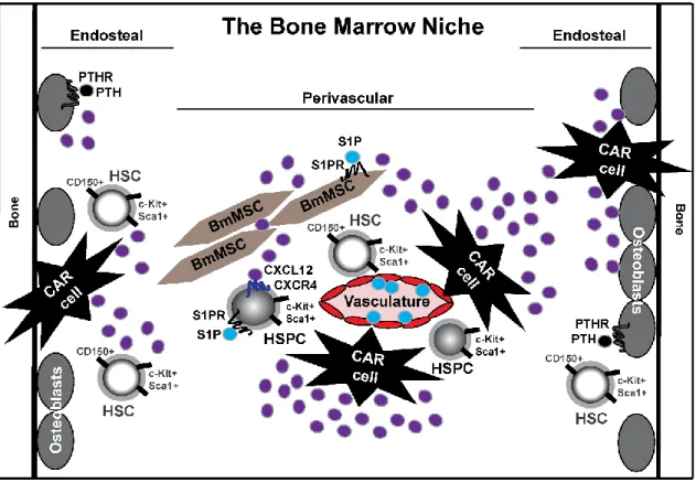
Regions of the Bone Marrow Niche
The bone marrow niche can be described into two functional regions: the endosteal niche and the perivascular niche. The endosteal niche is at the endosteum and medullary cavity interface with osteoblasts as a key cellular inhabitant, whereas the perivascular niche resides along the sinusoidal vessels surrounded by endothelial cells and stromal BmMSCs. It has been a long-standing belief that dormant, quiescent HSCs are located near the endosteal niche; however, recent studies now suggest HSCs are distributed throughout the bone
marrow with less than 20% within 10 µm of the endosteum in the endosteal niche and instead preferential associate near sinusoid vessels[19, 43-45]. In addition, conditional deletion studies showing endosteal niche osteoblast CXCL12 secretions have little to no effect on primitive HSCs [32, 33]. Interestingly, after irradiation, HSCs preferentially locate to the endosteum, which is believed to be because irradiation causes destruction to the sinusoids leaving only arteriolar vessels near the endosteum[45]. Teasing out the niche location and developmental stages of HSCs within the bone marrow is an active, ongoing area of investigation.
8
G Protein-Coupled Receptor (GPCR) Signaling within the Bone Marrow Niche Background on GPCRs.
G protein-coupled receptors (GPCRs) comprise 3-5% of the human genome and are necessary for several physiological functions. As such, GPCRs serve as therapeutic targets in 20-50% of marketed pharmaceuticals[46]. GPCRs have seven transmembrane domains with an intracellular carboxyl terminus tail, which is essential for receptor regulation. Intracellular signaling is induced upon extracellular ligand stimulation, eliciting a conformational change and dissociation of guanine nucleotide binding proteins, termed G proteins (α and β/γ) that elicit downstream cellular signaling for effector function [47].
GPCR regulation: G protein-coupled receptor kinases (GRKs) and β-arrestin.
After ligand-stimulation, G protein-coupled receptor kinases (GRKs) can terminate intracellular signaling by phosphorylating the carboxyl terminus of GPCRs, a process termed desensitization, which can recruit β-arrestin—protein necessary for GPCR internalization and another pathway of GPCR regulation in addition to receptor desensitization[47].
There are seven GRKs that are classified into three subfamilies: GRK1 (GRK1 and 7), GRK2 (GRK2 and 3), and GRK4 (GRK4-6); however, GRK1/7 and GRK4 are tissue- selective with expression in ocular tissues and testes, respectively. Therefore, only GRK2, GRK3, GRK5 and GRK6 are ubiquitously expressed[48-52]. While there is sequence homology amongst these GRKs, there are clear functional differences with selective phosphorylation sites and receptor-specific GRK regulation. Studies utilizing conventional murine models of GRK3, GRK5, and GRK6 deficiency have aided our initial understanding of the differential role of these GRKs; however, GRK2-deficient mice are embryonically
9
lethal with cardiac hypoplasia, and thus require either heterozygotic or conditional knockout models for any research purposes[53]. By utilizing GRK3-deficient (Grk3-/-) mice[54, 55], the content of this dissertation reveals a newly identified regulation process by GRK3 on sphingosine-1-phosphate (S1P)/S1P receptor (S1PR) signaling to direct the functionality of BmMSCs (Chapter 2) and discusses how GRK3 regulation on HSPCs impacts hematopoiesis mediated, at least in part, through CXCL12/CXCR4 (Chapter 3).
Ligand-induced GPCR signaling within the bone marrow niche.
GPCR signaling in the bone marrow is essential for fundamental processes for immune cell development and function [46]. In particular, cellular survival, proliferation, differentiation, and trafficking may be hindered if GPCR signaling is impaired. Three GPCRs: parathyroid hormone receptor (PTHR), chemokine receptor CXCR4, and S1PR, have been described as essential GPCRs in the bone marrow niche and will be discussed.
PTH/PTHR signaling. Parathyroid hormone (PTH) is produced by the parathyroid glands and binds PTHR located on bone marrow osteoblasts [56], cells derived from mesenchymal stem cells that are responsible for building bone. The Calvi laboratory described that mice injected with PTH had increased numbers of HSCs and enhanced survival post-transplantation, which is believed to be induced by increased levels of ligand Jagged-1 secreted by osteoblasts that binds Notch receptor on HSCs [37, 38]. However, when PTH is constitutively activated in terminally differentiated osteoblastic cells, termed
osteocytes, HSC function and numbers were not altered, suggesting HSC expansion within the bone marrow niche mediated through PTH stimulation is cell-specific [57]. Interestingly,
10
despite such promising pre-clinical studies and a phase I study [58], a phase II clinical trial that tested whether PTH treatment after transplantation enhanced HSC growth and ability to produce new blood cells, termed immune reconstitution, was terminated prematurely due to early mortality and lack of efficacy [59]. As previously mentioned, more recent findings now suggest osteoblasts may play a more prominent role in supporting lymphoid progenitors within the bone marrow niche rather than a direct role with HSCs[32, 33], which may partially explain the phase II negative outcome.
CXCL12/CXCR4 signaling. GPCR chemokine receptor CXCR4 binds its cognate ligand CXCL12 (stromal cell-derived factor-1, SDF-1)[60], and such CXCL12/CXCR4 signaling is a key signaling mechanism within the bone marrow niche for steady state immune cell development and function. CXCR4 is expressed on HSCs, CMPs, granulocytic- monocytic progenitors (GMPs), neutrophils, monocytes, and T/B lymphocytes, with low expression on myeloid erythroid progenitors (MEPs)[61, 62]. Thus, several leukocyte
subsets within bone marrow utilize CXCL12/CXCR4 signaling to modulate development and function [31].
Interestingly, others have demonstrated GRK3 has selective regulation of CXCR4 with phosphorylation at serine residues 346/347 (S346/347)[63, 64]. Our lab has further demonstrated how GRK3 regulation of CXCL12/CXCR4 signaling is particularly vital for mature hematopoietic cell function [55], and the work within this dissertation further
suggests GRK3 deficiency impacts the function of immature hematopoietic cells, mediated at least in part through CXCL12/CXCR4 (Chapter 3). GRK2[65-67] and GRK6 [68, 69] have also been described to regulate CXCR4 in cell lines and mature hematopoietic cells,
11
respectively; however, recent findings suggest the most prevalent GRK in the LSK HSPC population is GRK3, and data suggest GRK6 phosphorylation does not play a critical role in CXCL12/CXCR4 signaling that is essential during immature hematopoiesis [70].
S1P/S1PR signaling. The precursor sphingosine is phosphorylated by sphingosine kinases (Sphk1 and Sphk2) to produce active ligand S1P that can bind to GPCR S1PRs1-5.
Most cells contain sphingosine kinase activity in the cytosol and thus, secrete S1P to act in an autocrine or paracrine manner[71]. S1P levels are controlled by S1P lyase, which is an enzyme that is ubiquitously expressed and irreversibly degrades S1P[72]. S1P has vast physiological roles, including serving as a chemoattractant for HSCs. This is achieved with lower S1P expression within the bone marrow and high concentrations within the blood, thus, creating a chemoattractant gradient for the trafficking of HSCs [73]. S1P also has been shown to affect mesenchymal stem cell osteogenic differentiation [74-76] and proliferation [77, 78], of which is more clearly defined mechanistically within this dissertation (Chapter 2). While physiological roles of S1PRs in the bone marrow niche are becoming more defined, the regulation of signaling mechanisms are less well understood. Though, Arnon et al. elucidated the importance of GRK2 desensitization on S1PR1 to facilitate lymphocyte egress from circulation into the tissues [79], and the work described within this dissertation suggests the importance of GRK3 regulation on S1PR signaling to modulate BmMSC function. Taken together, both studies suggest the importance of S1P/S1PR signaling regulation on immune cells and bone marrow niche cells.
12
Targeting GPCRs as Therapeutic Strategies in the Bone Marrow
Hematopoietic cells are retained within the bone marrow by retention signals through GPCR signaling. Manipulation of retention signals, such as CXCL12/CXCR4 and S1P/S1PR among hematopoietic cells, has been utilized as therapeutic strategies for the treatment of various diseases and disorders and will be discussed.
Granulocyte-colony stimulating factor (G-CSF).
G-CSF has been commonly utilized to enhance egress of HSCs from the bone marrow into the peripheral blood[80], for the collection of HSCs to be used in HCT. Data have demonstrated G-CSF acts as a HSC mobilizer through disrupting CXCL12/CXCR4 signaling, as G-CSF treatment induces protease activity in the niche that cleaves both CXCL12 and CXCR4 [81]; however, further studies have demonstrated protease-dependent and protease-independent mechanisms decrease CXCL12 in the bone marrow [82]. G-CSF treatment has also increased S1P levels in the blood[83, 84], which may play a large role in facilitating HSC mobilization due to the S1P/S1PR chemotactic gradient.
AMD3100.
CXCL12/CXCR4 signaling has been discussed as a critical GPCR signaling mechanism in the bone marrow niche to aid HSPC development and function. As such, CXCL12/CXCR4 serves as a retention signal that is targeted therapeutically by AMD3100, a CXCR4 antagonist, to also induce HSC egress from the bone marrow into the peripheral blood [85]. Furthermore, combination treatment of G-CSF with AMD3100 enhanced HSC egress into the peripheral blood in comparison to G-CSF treatment alone [86].
13 FTY720 and SEW2871.
FTY720 binds to the S1PR family as an agonist eliciting receptor internalization and degradation, thus, suppressing S1P/S1PR signaling due to the reduction of S1PR on the cell surface. FTY720 treatment has been shown to suppress the mobilization of HSCs, as well as other cells such as T/B lymphocytes [87-90]. Therefore, in contrast to AMD3100, which antagonizes CXCL12/CXCR4 signaling and initiates release of hematopoietic cells into the periphery, FTY720 suppresses S1P/S1PR signaling through degradation of the receptor and inhibits mobilization. As such, FTY720 was FDA approved in 2010 for the treatment of relapsing multiple sclerosis by blocking immune cell infiltration into the central nervous system[91]. SEW2871 is a S1PR1-specific agonist that recycles the receptor and does not undergo degradation. As such, combination therapy of SEW2871 with AMD3100
substantially enhanced the mobilization of HSCs and progenitors [92].
Conclusion and Research Goals
The bone marrow microenvironment, termed niche, supports hematopoietic cell development and thus, is vital for establishment of the immune system. Within the niche reside complex molecular and cellular interactions, evident by the previously discussed regions of the bone marrow (endosteal vs. perivascular), key inhabitants (HSCs, BmMSCs, osteoblasts, CAR cells), and mechanistic GPCR interactions (PTH/PTHR, CXCL12/CXCR4, S1P/S1PR) (Figure 1.1). Novel therapies have been developed by beginning to understand the bone marrow niche; however, there is still much to reveal in regards to the intracellular signaling and regulation of bone marrow niche cells.
14
Our previous work revealed mice deficient in GRK3 (Grk3-/-), an intracellular protein kinase that regulates GPCR signaling, have a hypercellular bone marrow with increased LSK HSPCs, oligopotent progenitors, and evident leukocytosis—increased white blood cells (termed leukocytes) in the peripheral blood[55]. This observation suggests hematopoiesis is enhanced within Grk3-/- mice, though without a clear mechanistic understanding. As such, we proposed (1) GRK3 may be directly impacting HSPC GPCR
Figure 1.1. The bone marrow niche. The bone marrow is categorized into two regions:
endosteal niche and perivascular niche. Hematopoietic stem cells (HSCs, Lin- Sca1+ c- Kit+ CD150+ CD48-)/hematopoietic stem-progenitor cells (HSPCs, Lin- Sca1+ c-Kit+), bone marrow mesenchymal stem cells (BmMSCs), osteoblasts, and CXCL12-abundant reticular (CAR) cells are key inhabitants of the bone marrow niche. GPCR signaling is prevalent within the bone marrow niche: CXCL12/CXCR4 and sphingosine-1-phosphate (S1P)/S1P receptor (S1PR) signaling is evident on HSC/HSPCs, and the work within this dissertation further elucidates the importance of S1P/S1PR signaling on BmMSCs (Chapter 2). Parathyroid hormone (PTH)/PTH receptor (PTHR) signaling is evident on osteoblasts, cells responsible for bone formation.
15
signaling to elicit an enhanced phenotype and/or (2) GRK3 may be affecting other
surrounding bone marrow niche cells via GPCR signaling that may be contributing indirectly to the enhanced hematopoietic phenotype.
This dissertation initiates the investigations to address these possibilities by exploring regulation of cellular signaling and function of (1) BmMSCs—cells that surround
hematopoietic cells to support their development, maintenance, and function, and (2) HSPCs— cells that are essential for the establishment of hematopoietic lineages of the immune system. Specifically, the research goals of this dissertation were to investigate how GPCR regulation by GRK3 may impact these two bone marrow niche stem cell populations with crucial roles in hematopoiesis. As such, I hypothesized regulation of GPCR signaling by GRK3 controls cellular functions of both BmMSCs and HSPCs.
In chapter 2, my work demonstrates the importance of GRK3 regulation of S1P/S1PR signaling in BmMSCs, leading to enhanced BmMSC proliferation and osteogenic
differentiation when GRK3 regulation is absent. In chapter 3, my work implicates the
importance of GRK3 regulation of CXCL12/CXCR4 on HSPCs, leading to increased colony forming unit (CFU) numbers when GRK3 regulation is absent, evidenced by in vivo short- term transplantation and ex vivo CFU culture studies.
Given the important role of BmMSCs within the bone marrow niche, it may be possible that such an altered functionality of Grk3-/- BmMSCs revealed herein (Chapter 2) has the potential to impact surrounding environments in the bone marrow and affect hematopoiesis, though future investigations are warranted and proposed in Chapter 4. In addition, since GRK3 deficiency enhances CFU numbers (Chapter 3), and given the positive
16
correlation between the CFU content of a stem cell graft and the success rate of immune reconstitution after HCT, it may be possible that GRK3 deficiency could positively impact HCT, though future investigations are warranted and proposed in Chapter 4.
17
CHAPTER 2: G PROTEIN-COUPLED RECEPTOR KINASE 3 MODULATES MESENCHYMAL STEM CELL PROLIFERATION AND DIFFERENTIATION
THROUGH SPHINGOSINE-1-PHOSPHATE RECEPTOR REGULATION2
Overview
The bone marrow niche supports hematopoietic cell development through intimate contact with multipotent stromal mesenchymal stem cells; however, the intracellular signaling, function, and regulation of such supportive niche cells are still being defined.
Thus, our study was designed to better define the regulation of cellular signaling and functional mechanisms of bone marrow-derived mesenchymal stem cells (BmMSCs). Our data suggest G protein-coupled receptor kinase 3 (GRK3) functions as a negative regulator of G protein-coupled receptor signaling of sphingosine-1-phosphate receptor (S1PR) on
BmMSCs. BmMSCs isolated from GRK3-deficient (Grk3-/-) mice have enhanced
proliferation and osteogenic differentiation ex vivo compared to wildtype (WT) BmMSCs in identical culture conditions and passages. Grk3-/- BmMSC cultures also have higher levels of CXCL12, an essential chemokine for hematopoietic stem and progenitor cell development, and Grk3-/- mice have increased hematopoietic cell numbers isolated from the bone marrow.
2Brozowski JM, Timoshchenko RG, Serafin DS, Allyn B, Koontz J, Ren Y, Eudy AM, Harris TF, Abraham D, Mattox D, Rubin CT, Hilton MJ, Rubin J, Allbritton NL, Billard MJ, Tarrant TK. G protein-coupled receptor kinase 3 modulates mesenchymal stem cell proliferation and differentiation through sphingosine-1-phosphate receptor regulation. Stem Cells. Submitted September 28, 2017. In revisions for resubmission.
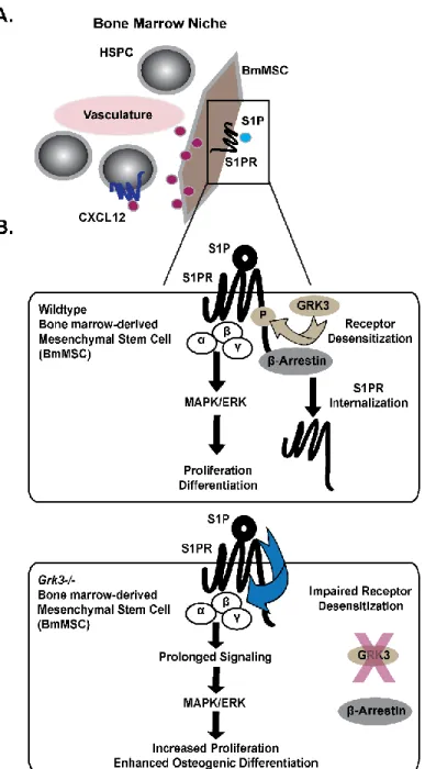
JM Brozowski performed experiments for Figure 2.1, Figure 2.2, Figure 2.4, Figure 2.5, Figure 2.6B, as well as appendices 1, 2 (performed replicated run to confirm previous data by RGT), 3, 4, 6, 7, 8 and 13. JM Brozowski wrote the journal article, as well as made the figures, including illustration for Figure 2.7. Note: Appendices 3, 7, 8, and 13 were not originally included within the submitted manuscript on September 28, 2017.
18
However, Grk3-/- mice do not have an enhanced trabecular bone volume fraction, suggesting GRK3 deficiency in niche BmMSCs may have more pronounced effects on hematopoiesis as opposed to mature bone development in vivo. Both Grk3-/- BmMSC proliferation and
osteogenic differentiation were reduced to WT level upon reduction of sphingosine-1- phosphate (S1P), and Grk3-/- BmMSCs have sustained ERK1/2 signaling upon stimulation of S1PR with S1P in comparison to WT BmMSCs. In addition, we report GRK3 recruits β- arrestin, a protein necessary for receptor internalization, to the C-terminus of S1PR1, and we demonstrate BmMSCs lacking GRK3 regulation have impaired S1PR1 internalization. Our work suggests GRK3 regulates S1PR on BmMSCs, and lack of such regulation affects BmMSC functionality.
Significance Statement
Mesenchymal stem cells are self-renewing cells that have the potential to differentiate into multiple different tissue types. This has prompted pre-clinical and clinical investigations studying mesenchymal stem cells for the treatment of various diseases; however, rigorous studies are ongoing to learn more about tissue-specific signaling, function, and regulation.
Our work demonstrates the importance of G protein-coupled receptor kinase 3 (GRK3) in bone marrow-derived mesenchymal stem cell (BmMSC) signaling and during development into pre-osteoblasts through its regulation of sphingosine-1-phosphate receptor. Lack of GRK3 receptor regulation enhances BmMSC proliferation and function toward becoming osteoblast precursors, which can then impact surrounding environments in the bone marrow and affect the production of blood cells.
19 Introduction
Mesenchymal stem cells are multipotent stromal cells that possess the ability to differentiate into mesodermal tissues, such as chondrocytes, adipocytes, and osteocytes [25].
Mesenchymal stem cells that reside within the bone marrow microenvironment, often
referred to as bone marrow niche mesenchymal stem cells (BmMSCs), secrete high levels of chemokine CXCL12 that binds to G protein-coupled receptor (GPCR) CXCR4 on
hematopoietic stem and progenitor cells (HSPCs) to affect their growth and development [31, 93]. BmMSCs impact hematopoiesis as depletion of either CXCL12 or total numbers of mesenchymal stem cells from the bone marrow decrease the hematopoietic stem cell pool and repopulating activity, as well as affect homing abilities of HSPC after transplantation [32-34].
We previously observed mice deficient in G protein-coupled receptor kinase 3 (GRK3), an intracellular kinase that negatively regulates GPCRs, have (1) a hypercellular bone marrow comprised of significantly increased Lin- Sca1+ c-Kit+ (LSK) HSPCs and selective downstream committed progenitors, and (2) an increase of total white blood cells in the peripheral blood (leukocytosis) compared to WT mice [55]. Our data showed GRK3 deficiency affects CXCR4 regulation on hematopoietic cells [55]; however, due to the
importance of stromal cells within the niche that impact HSPC development and function, we were also interested in understanding the regulatory role of GRK3 on BmMSCs, in which GRK3 is expressed [54].
We observed Grk3-/- BmMSCs have enhanced osteogenic differentiation, higher levels of CXCL12, and increased proliferation. Accumulating evidence supports that
phospholipid sphingosine-1-phosphate (S1P) affects mesenchymal stem cell function [46] in
20
osteogenic differentiation [74-76] and proliferation [77, 78]. Since the S1P receptors (S1PRs) are GPCRs, our study aimed to elucidate whether GRK3 regulated S1PR on BmMSCs to affect their functionality.
Materials and Methods Animals.
Wildtype (WT) C57BL/6 and GRK3-deficient (Grk3-/-) age-matched (8-12 week- old) mice were used under standard IACUC-approved protocols in the AAALAC-accredited vivarium of UNC, and care of animals was in accordance with institutional guidelines. The Grk3-/- mouse strain was provided by Dr. Robert J. Lefkowitz (Duke University) and backcrossed >12 generations on the C57BL/6 background.
Bone marrow-derived mesenchymal stem cell (BmMSC) isolations.
BmMSC were isolated from flushed femurs and tibias of two male mice and cultured in complete isolation media (CIM) containing RPMI (Corning, 10040CV) with 10% fetal bovine serum (FBS, Atlanta Biologicals, S12450), 10% horse serum (HS, HyClone, SH30074.03), 1% 100 IU/mL penicillin G/100 µg/mL streptomycin (P/S, Corning, 30-002- Cl), and 12 µM final concentration of L-Glutamine (Corning, 25-005-Cl), as similar methodologies have been previously described [29, 94, 95]. BmMSC expansion was in complete expansion media (CEM) containing IMDM (Gibco, 12440-053), 10% FBS, 10%
HS, 1% P/S, and 12 µM final concentration of L-Glutamine [29, 95], which was followed by hematopoietic CD45 (Stem Cell Technologies, 19771) and CD11b (Miltenyi, 130-049-601 or 130-093-634) depletion, as recommended by [94], via magnetic negative selection at early
21
passages 1-3. BmMSCs were passaged at 70-80% confluency and used for experiments at passages 4-15.
Chondrogenic, adipogenic, and osteogenic differentiation.
Chondrogenic differentiation. BmMSCs were suspended in CEM at 1.6x10⁷ viable cells/mL. Micromasses were made by adding 5 µL droplets of the cell suspension onto a 6- well plate and given 3-4 hours to attach. Chondrogenic media (Gibco StemPro®
Chondrogenesis Differentiation Kit, A10071-01) supplemented with penicillin streptomycin was added to each well and incubated for 21 days. Cells were fixed with 10% formalin and stained using Alcian Blue. Adipogenic differentiation. BmMSCs were plated at 1x10⁵ cells/well of a 6-well plate in CEM supplemented with 50 µM indomethacin, 5 µg/mL insulin, and 0.1µM dexamethasone. Cells were fixed with 10% formalin and stained using Oil Red O. Osteogenic differentiation. BmMSCs were plated at 1x10⁵ cells/well of a 6-well plate in CEM supplemented with 50 µg/mL ascorbic acid and 20 mM ß-glycerophosphate.
Cells were fixed with 10% formalin and stained using Alizarin Red. For SKI-treated osteogenic differentiation, BmMSCs were plated at 2x10⁴ cells/well in a 24-well plate in CEM, and osteogenic differentiation was induced after an overnight rest. Cells were treated with sphingosine kinase inhibitor 2 (SKI, Cayman Chemical, 10009222) at a final
concentration of 5 µM or vehicle (DMSO). Fresh media changes occurred every third day using CEM plus SKI or vehicle. Cells were fixed with 10% formalin and stained using Alizarin Red stain for analysis. Images were captured using the Olympus 1X-81 microscope and MetaMorph software.
22 Bone marker mRNA expression (qRT-PCR).
Total RNA from BmMSCs undergoing osteogenic differentiation was prepared using the RNeasy Mini/ Plus kit (Qiagen) according to manufacturer’s instructions. Reverse transcriptase cDNA synthesis was performed using iScript cDNA synthesis kit (Bio-Rad, 170-8891). qRT-PCR was performed in duplicate (SYBR® Green, Bio-Rad, 172-5271) and normalized to housekeeping gene IDUA. Mean fold change of alkaline phosphatase was determined by 2-ΔΔCt with WT day 0 as control. Primers utilized for qRT-PCR were Alkaline Phosphatase forward: AAG GCT TCT TCT TGC TGG TG, Alkaline Phosphatase reverse:
GCC TTA CCC TCA TGA TGT CC; IDUA forward: GCA TCC AAG TGG GTG AAG TT and IDUA reverse: CAT TGA GCA GGT CCG GAT AC.
ELISA.
BmMSCs were plated at 1x10⁵ cells/well of a 6-well plate in CEM and rested overnight for attachment before supernatant collections began at baseline (day 1) and each subsequent collection. Supernatants of undifferentiated BmMSC monolayers were collected and analyzed for CXCL12 protein using the CXCL12 DuoSet ELISA kit (R&D Systems, DY460), as per instructions.
Micro-computed tomography (µCT).
For 8-12 week-old mice, µCT imaging was used to analyze the trabecular bone morphology at the distal femur at 12 micron resolution. The metaphyseal region of the distal femur was scanned beginning 720 microns proximal to the growth plate and extending 1500 microns towards the diaphysis of the femur. An automatic script was used to analyze the
23
region of interest to separate the trabecular and cortical regions of the bone and quantify bone morphology. Trabecular analysis includes quantification of BV/TV (bone volume/total volume). For 17-20 month-old aged mice, µCT imaging morphology (VivaCT80, Scanco Medical, Basserdorf, Switzerland) was used to analyze the trabecular bone. Briefly, metaphysis region was selected for 100 slices under the femur growth plate. Trabecular analysis includes quantification of BV/TV. Analyses were conducted at 12 μm slice
increment with an integration time of 300 ms, a current of 145 mA, and an energy setting of 55 kV. The threshold was chosen using 2D evaluation of several slices in the transverse anatomic plane so that mineralized bone was identified but surrounding soft tissue was excluded.
Cellular proliferation.
Proliferation of 5x10⁴ BmMSCs/well of a 6-well plate was analyzed after exogenous CCK-8 (Dojindo Molecular Technologies) was added to each well at the indicated
timepoints. Absorbance was measured after 3-hour incubation using the Promega Glomax®
Multi + Detection System. Data were analyzed by deducting background (media and CCK-8) absorbance from raw absorbance reads. For S1P studies, BmMSCs were plated at 1x10⁴ cells/well in a 24-well plate and treated with SKI at a final concentration of 5 µM.
Immunoblotting.
BmMSCs were plated in CEM at 2.25x10⁵ cell density in a 6-well plate and incubated overnight. DPBS rinsed cells were rinsed three times with IMDM containing 20% charcoal- stripped FBS (to remove serum S1P) and 1% P/S, and incubated at 37ºC for 15 minutes.
24
Fresh media was added, and BmMSCs were stimulated with 1 µM S1P for indicated timepoints. Unstimulated BmMSCs served as 0 minute timepoint control. Following stimulation with S1P, BmMSC cells were rinsed with DPBS and lysed in cold HBSS + 1%
TritonX100 lysis buffer containing protease inhibitors (1 mM PMSF, 1 µg/mL aprotinin, 1 µg/mL pepstatin, and 1 µg/mL leupeptin) and phosphatase inhibitors (5 mM NaF and 2 mM NaVO4). All WT and Grk3-/- BmMSC lysates were normalized via Bicinchoninic acid (BCA) assay, and 6 µg of total protein in laemmli sample buffer (non-reducing) was freshly loaded onto AnyKD Mini-PROTEAN®TGX precast protein gel (Bio-Rad, 4569036). Gels were run at 100V for 1.5 - 2 hours in 1XTris/Glycine SDS buffer. Proteins were transferred overnight at 4ºC onto nitrocellulose membrane in Tris base (25 mM)/Glycine (192 mM) transfer buffer containing 20% methanol. The membrane was blocked in 3% fatty-acid free BSA in TBS plus 0.1% Tween-20 (TBS/T) for 2 hours at 25ºC and incubated with primary antibody 1:2,000 phospho-p44/42 MAPK or 1:2,000-3,000 p44/42 MAPK (Cell Signaling Technologies, 4370/4695) overnight at 4ºC, or 1:10,000 GAPDH (Trevigen, 2275-PC-100) for 2 hours at 4ºC. The membrane was washed three times for 10 minutes in TBS/T,
incubated with secondary antibody 1:5,000 anti-rabbit IgG HRP (Cell Signaling
Technologies, 7074) for 1 hour at 25ºC, and washed twice for 10 minutes in TBS/T and once in TBS. Detection was performed via ECL Prime or ECL Select (GE Healthcare, RPN2232/
RPN2235) and imaged on GeneSys image acquisition software. Densitometry was obtained by measuring ratio of phospho-ERK (pERK) over total ERK using Image J software.
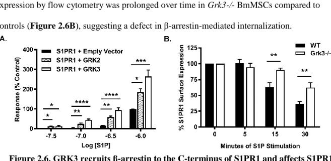
25 β-arrestin recruitment assay.
GRK recruitment of β-arrestin to the S1PR1 carboxy-terminus was measured using agonist-stimulation in a modified-TANGO assay, as previously reported [96]. HTLA cells were transfected with a S1PR1-TCS-tTA receptor construct after removing the V2
vasopressin sequence to prevent nonspecific β-arrestin recruitment to the wildtype S1PR1.
GRK over-expression was achieved via plasmids: GRK2 pcMyc_LIC and GRK3
pcMyc_LIC, and utilized a separate expression vector encoding yellow-fluorescent protein (YFP) that was simultaneously transfected to serve as a transfection control. HTLA cells were transiently transfected with 6.5 μg of total plasmid DNA (3 μg of S1P-Tango, 0.5 μg of YFP, and either 3 μg of empty-vector control, GRK2, or GRK3) via calcium-phosphate precipitation. Transfection efficiency was determined by YFP epifluorescence to be consistently >70% at 24 hours post-transfection. Cells were serum starved and then
stimulated with S1P ligand at varied concentrations up to 1 µM. BriteGlo reagent (Promega, Madison, WI, USA) was added for luminescence detection via Promega Glomax® Multi + Detection System (0.5 sec/ well). Raw data were normalized by subtracting background for each independent run and setting the lowest concentration of the control condition at 0% and highest concentration at 100%.
S1PR1 internalization assay.
BmMSCs cultured in CEM were rinsed with DPBS and incubated in serum-free CEM (SFM) for 2.5 hours. BmMSCs were detached using Accutase® (Sigma, A6964), rinsed once with cold SFM containing 5% charcoal-stripped FBS (to remove serum S1P), and
resuspended in cold DPBS. 1x10⁵ BmMSCs were stimulated with 1 µM S1P ligand in
26
100 µL FACS buffer (DPBS1X + 0.2% fatty acid free BSA + 0.1% sodium azide) at specific timepoints. Unstimulated BmMSCs served as 0 minute timepoint control. S1PR1
internalization was halted with 2 mL of ice cold FACS buffer and sample tubes were placed on ice. BmMSCs were stained for Sca1 (eBioscience clone: D7, APC-conjugated) and
S1PR1 (R&D Systems clone: 713412, PE-conjugated) for 30 minutes on ice in 100 µL FACS buffer, rinsed, and analyzed by flow cytometry.
Statistical analyses.
All data were graphed utilizing GraphPad Prism v.7 and statistically evaluated using GraphPad Prism v.7 or Microsoft Office Excel program. Taking into consideration time and strain (WT and Grk3-/-), the bone marker qRT-PCR, CCK-8 proliferation, and S1PR1 internalization was statistically analyzed using a RM two-way ANOVA with Sidak’s multiple comparison test, a method preferred over Bonferroni due to increased power [97, 98]. Similarly, taking into consideration time and strain (WT and Grk3-/-) but with multiple treatment groups, the SKI-treated CCK-8 proliferation was analyzed using a RM two-way ANOVA with Tukey’s multiple comparison test for pairwise comparisons [97, 99]. Student’s t-test compared two independent groups (WT and Grk3-/-) for the CXCL12-detection
ELISA, micro-computed tomography data, and western blot densitometry. Taking into consideration three independent groups (empty vector, GRK2, and GRK3), the β-arrestin recruitment assay was analyzed by one-way ANOVA with Dunnett’s multiple comparison test, which compared each group to the control (empty vector) [97, 100].
27 Results
Grk3-/- BmMSCs have enhanced osteogenic differentiation.
In our previous studies, we observed Grk3-/- mice had increased HSPC numbers compared to WT mice [55]. Since it is well established that bone marrow stromal cells enhance hematopoiesis [32-34, 101-103], we aimed to test whether there were differences between Grk3-/- and WT murine stromal BmMSCs that could contribute to this phenotype.
BmMSCs isolated from WT and Grk3-/- mice adhered to culture plastic, underwent
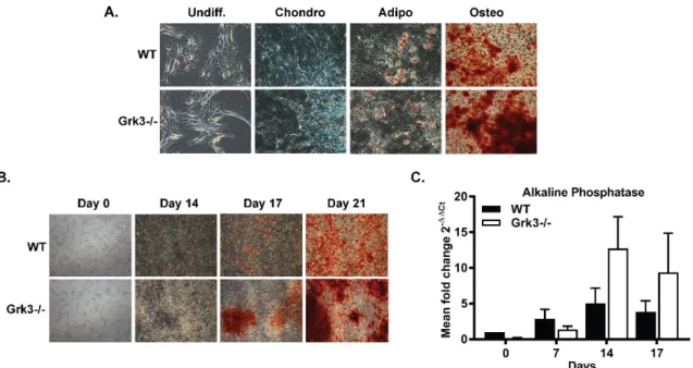
differentiation into tissue-specific lineages to ensure multipotency (Figure 2.1A), and lacked
Figure 2.1. Bone marrow-derived mesenchymal stem cells (BmMSCs) deficient in G protein-coupled receptor kinase 3 (Grk3-/-) have enhanced osteogenic differentiation in comparison to wildtype (WT) BmMSCs. (A) Multipotent differentiation of BmMSCs isolated ex vivo from Grk3-/- and WT mice into chondrocytes (Alcian Blue stain), adipocytes (Oil Red O stain), and osteoblasts (Alizarin Red stain) at day 21; images acquired at 10X magnification. Differentiation analyses n=3 chondrogenic/adipogenic and n=10 osteogenic.
(B) Time-course Grk3-/- and WT BmMSC osteogenic differentiation (Alizarin Red stain) at 2X magnification. Representative images, n=4. (C) Mineralization of bone marker alkaline phosphatase expression during osteogenic differentiation relative to housekeeping gene IDUA. Data represent mean ± SEM, n=7.
28
expression of CD45 and CD11b (hematopoietic cell markers) and consistently expressed mouse mesenchymal stem cell markers Sca1, CD106, CD73, CD44, CD29 (Appendix 1).
During differentiation analysis, we observed no substantial differences between WT and Grk3-/- BmMSC chondrogenic or adipogenic differentiation cultures. However, we observed enhanced and earlier osteogenic differentiation in Grk3-/- BmMSC cultures in comparison to WT cultures, as demonstrated by positive alizarin red stain (Figure 2.1B) and higher mRNA expression levels of alkaline phosphatase (ALP), a marker of osteoblast differentiation that peaks near day 14 (Figure 2.1C) [104, 105]. We noticed the enhanced osteogenic
differentiation phenotype was repeatedly consistent and was reproduced in four separate isolations. To further ensure there were no isolation differences between WT and Grk3-/- BmMSCs that may induce such a phenotype, we utilized shRNA to knockdown GRK3 (GRK3-KD) from WT BmMSCs and induced multipotent differentiation, which again showed the identical phenotype of enhanced osteogenic differentiation in GRK3-KD BmMSCs in comparison to non-target (NT) control BmMSCs (Appendix 2).
Grk3-/- BmMSC cultures have higher levels of CXCL12.
Niche BmMSCs, CXCL12-abundant reticular (CAR) cells in the bone marrow, and pre-osteoblasts secrete CXCL12 [32, 33], an essential chemokine in the niche for HSPC development and/or function. Since we observed our Grk3-/- mice had increased HSPCs [55], we next tested whether there may be differences in CXCL12 levels between WT and Grk3-/- BmMSCs ex vivo. Our data revealed Grk3-/- BmMSC cultures had higher levels of CXCL12 detected in comparison to WT BmMSC cultures at baseline, as well as each following time point evaluated (Figure 2.2A).
29 GRK3 deficiency increases proliferation ex vivo.
Due to the enhanced detection of CXCL12 in Grk3-/- BmMSC culture, we wanted to further investigate whether this was the result of enhanced secretory function or a
proliferation response, since we also observed increased cell counts when passaging Grk3-/- BmMSCs. Cell counting kit-8 (CCK-8) quantitates proliferation by absorbance detection of increased formazan dye production from viable cells. Using this assay of cellular
quantification, Grk3-/- BmMSCs have enhanced proliferation over time in comparison to WT BmMSCs (Figure 2.2B).
GRK3 deficiency does not affect mature bone formation in vivo.
As a result of our observed enhancement of Grk3-/- BmMSC osteogenic differentiation and proliferation ex vivo, we proposed to investigate whether GRK3
Figure 2.2. Grk3-/- BmMSCs have higher levels of CXCL12 detected and proliferate more in comparison to WT BmMSCs. (A) Quantification of CXCL12 protein
concentration from supernatant of BmMSC cultures. Lower limit of detection at 46.9 ρg/mL. Data represent mean ± SEM, n=4. (B) BmMSC proliferation determined by increased formazan dye production from viable cells over time using CCK-8 proliferation assay. Data represent mean ± SEM, n=3, except day 0 n=2.
* P<0.05 ** P<0.01 *** P<0.001 **** P<0.0001