Methyl 3-[(
E
)-furfurylidene]dithio-carbazate
Shang Shan,* Shan-Heng Wang, Ying-Li Xu, Pei-Jin Xie and Yu-Liang Tian
College of Chemical Engineering and Materials Science, Zhejiang University of Technology, People’s Republic of China
Correspondence e-mail: [email protected] Received 31 March 2008; accepted 29 April 2008
Key indicators: single-crystal X-ray study;T= 294 K; mean(C–C) = 0.003 A˚;
Rfactor = 0.032;wRfactor = 0.097; data-to-parameter ratio = 14.6.
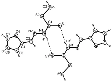
The molecule of the title Schiff base compound, C7H8N2OS2, prepared by the reaction of methyl dithiocarbazate and furfural in an ethanol solution under reflux, adopts an E configuration; the dithiocarbazate and furan units are located on opposite sides of the C N double bond. The planar dithiocarbazate group is twisted slightly with respect to the furan ring, making a dihedral angle of 5.2 (1). Adjacent
molecules are linked by N—H S hydrogen bonding to form a supramolecular dimer across an inversion center.
Related literature
For general background, see: Okabeet al.(1993); Shanet al. (2002, 2003). For a related structure, see: Chen et al. (2007). For the synthesis, see: Huet al.(2001).
Experimental
Crystal data
C7H8N2OS2
Mr= 200.27 Triclinic,P1 a= 4.0866 (8) A˚ b= 8.8698 (12) A˚ c= 12.8453 (15) A˚
= 93.970 (14) = 91.856 (12)
= 98.293 (12)
V= 459.21 (12) A˚3 Z= 2
MoKradiation
= 0.53 mm1
T= 294 (2) K 0.340.280.20 mm
Data collection
Rigaku R-AXIS RAPID IP diffractometer
Absorption correction: multi-scan (ABSCOR; Higashi, 1995) Tmin= 0.850,Tmax= 0.950
(expected range = 0.804–0.899)
4733 measured reflections 1608 independent reflections 1349 reflections withI> 2(I) Rint= 0.030
Refinement
R[F2> 2(F2)] = 0.031 wR(F2) = 0.096
S= 1.09 1608 reflections
110 parameters
H-atom parameters constrained max= 0.19 e A˚
3
min=0.28 e A˚
3
Table 1
Hydrogen-bond geometry (A˚ ,).
D—H A D—H H A D A D—H A
N1—H1 S1i
0.86 2.65 3.4892 (17) 165
Symmetry code: (i)x;yþ1;zþ1.
Data collection: PROCESS-AUTO (Rigaku, 1998); cell refine-ment:PROCESS-AUTO; data reduction:CrystalStructure(Rigaku/ MSC, 2002); program(s) used to solve structure:SIR92(Altomareet al., 1993); program(s) used to refine structure: SHELXL97 (Shel-drick, 2008); molecular graphics:ORTEP-3 for Windows(Farrugia, 1997); software used to prepare material for publication: WinGX
(Farrugia, 1999).
The project was supported by the Natural Science Foun-dation of Zhejiang Province, China (grant No. M203027).
Supplementary data and figures for this paper are available from the IUCr electronic archives (Reference: XU2417).
References
Altomare, A., Cascarano, G., Giacovazzo, C. & Guagliardi, A. (1993).J. Appl. Cryst.26, 343–350.
Chen, Z.-Y., Wu, G.-Q., Jiang, F.-X., Tian, Y.-L. & Shan, S. (2007).Acta Cryst. E63, o1919–o1920.
Farrugia, L. J. (1997).J. Appl. Cryst.30, 565. Farrugia, L. J. (1999).J. Appl. Cryst.32, 837–838.
Higashi, T. (1995).ABSCOR. Rigaku Corporation, Tokyo, Japan. Hu, W., Sun, N. & Yang, Z. (2001).Chem. J. Chin. Univ.22, 2014–2017. Okabe, N., Nakamura, T. & Fukuda, H. (1993).Acta Cryst.C49, 1678–1680. Rigaku (1998).PROCESS-AUTO. Rigaku Corporation, Tokyo, Japan. Rigaku/MSC (2002).CrystalStructure. Rigaku/MSC, The Woodlands, Texas,
USA.
Shan, S., Xu, D.-J., Hung, C.-H., Wu, J.-Y. & Chiang, M. Y. (2003).Acta Cryst. C59, o135–o136.
Shan, S., Xu, D.-J., Wu, J.-Y. & Chiang, M. Y. (2002).Acta Cryst.E58, o1333– o1335.
Sheldrick, G. M. (2008).Acta Cryst.A64, 112–122. Acta Crystallographica Section E
Structure Reports
Online
supporting information
Acta Cryst. (2008). E64, o970 [doi:10.1107/S1600536808012506]
Methyl 3-[(
E
)-furfurylidene]dithiocarbazate
Shang Shan, Shan-Heng Wang, Ying-Li Xu, Pei-Jin Xie and Yu-Liang Tian
S1. Comment
Since some hydrazone derivatives have shown the potential bioactivity as DNA-damaging or mutagenic agents (Okabe et
al., 1993), a lots of new hydrazone compounds has been synthesized in our laboratory (Shan et al., 2002, 2003). As part
of the ongoing investigation on hydrazone, we present here the crystal structure of the title compound.
The molecular structure is shown in Fig. 1. The N2═C3 bond distance of 1.284 (2) Å clearly indicates the double bond
character for the Schiff base compound. The molecule adopts an E configuration, the carbazate and furan moieties
located on the opposite positions of the N2═C3 bond; similar to that found in a related structure (Chen et al., 2007). The
dithiocarbazate moiety is well co-planar, the maximum atomic deviation being 0.037 (1) Å (S2), and the dithiocarbazate
mean plane is slightly twisted with respect to the furan plane by a smaller dihedral angle of 5.2 (1)°. This shows the
whole molecule is nearly co-planar.
Inter-molecular N—H···S hydrogen bonding links adjacent molecules to form the centro-symmetric supra-molecular
dimmer (Fig. 1 and Table 1).
S2. Experimental
Methyl dithiocarbazate was synthesized in the manner reported previously (Hu et al., 2001). Methyl dithiocarbazate (1.24
g, 10 mmol) and furfural (0.96 g, 10 mmol) were dissolved in ethanol (10 ml) and refluxed for 4 h. Yellow crystalline
product appeared after cooling to room temperature. They were separated and washed with cold water. Single crystals of
the title compound were obtained by recrystallization from an ethanol solution.
S3. Refinement
Methyl H atoms were placed in calculated positions with C—H = 0.96 Å and torsion angle was refined to fit electron
density, Uiso(H) = 1.5Ueq(C). Other H atoms were placed in calculated positions with C—H = 0.93 and N—H = 0.86 Å,
Figure 1
The molecular structure of the title compound with 40% probability displacement ellipsoids (arbitrary spheres for H
atoms) [symmetry code: (i) -x, 1 - y, 1 - z].
Methyl 3-[(E)-furfurylidene]dithiocarbazate
Crystal data
C7H8N2OS2
Mr = 200.27
Triclinic, P1 Hall symbol: -P 1
a = 4.0866 (8) Å
b = 8.8698 (12) Å
c = 12.8453 (15) Å
α = 93.970 (14)°
β = 91.856 (12)°
γ = 98.293 (12)°
V = 459.21 (12) Å3
Z = 2
F(000) = 208
Dx = 1.448 Mg m−3 Melting point = 414–416 K Mo Kα radiation, λ = 0.71073 Å Cell parameters from 2276 reflections
θ = 2.0–25.0°
µ = 0.53 mm−1
T = 294 K Prism, yellow
0.34 × 0.28 × 0.20 mm
Data collection
Rigaku R-AXIS RAPID IP diffractometer
Radiation source: fine-focus sealed tube Graphite monochromator
Detector resolution: 10.0 pixels mm-1
ω scans
Absorption correction: multi-scan (ABSCOR; Higashi, 1995)
Tmin = 0.850, Tmax = 0.950
4733 measured reflections 1608 independent reflections 1349 reflections with I > 2σ(I)
Rint = 0.030
θmax = 25.2°, θmin = 1.6°
h = −4→4
k = −10→10
Refinement
Refinement on F2 Least-squares matrix: full
R[F2 > 2σ(F2)] = 0.031
wR(F2) = 0.096
S = 1.09 1608 reflections 110 parameters 0 restraints
Primary atom site location: structure-invariant direct methods
Secondary atom site location: difference Fourier map
Hydrogen site location: inferred from neighbouring sites
H-atom parameters constrained
w = 1/[σ2(F
o2) + (0.0598P)2] where P = (Fo2 + 2Fc2)/3 (Δ/σ)max = 0.001
Δρmax = 0.19 e Å−3 Δρmin = −0.28 e Å−3
Special details
Geometry. All e.s.d.'s (except the e.s.d. in the dihedral angle between two l.s. planes) are estimated using the full covariance matrix. The cell e.s.d.'s are taken into account individually in the estimation of e.s.d.'s in distances, angles and torsion angles; correlations between e.s.d.'s in cell parameters are only used when they are defined by crystal symmetry. An approximate (isotropic) treatment of cell e.s.d.'s is used for estimating e.s.d.'s involving l.s. planes.
Refinement. Refinement of F2 against ALL reflections. The weighted R-factor wR and goodness of fit S are based on F2, conventional R-factors R are based on F, with F set to zero for negative F2. The threshold expression of F2 > σ(F2) is used only for calculating R-factors(gt) etc. and is not relevant to the choice of reflections for refinement. R-factors based on F2 are statistically about twice as large as those based on F, and R- factors based on ALL data will be even larger.
Fractional atomic coordinates and isotropic or equivalent isotropic displacement parameters (Å2)
x y z Uiso*/Ueq
S1 0.20815 (13) 0.27619 (5) 0.50863 (4) 0.0516 (2)
S2 0.38692 (13) 0.20142 (5) 0.28541 (4) 0.0506 (2)
N1 0.1859 (4) 0.45265 (16) 0.35473 (12) 0.0464 (4)
H1 0.1098 0.5149 0.3988 0.056*
N2 0.2353 (4) 0.49265 (16) 0.25394 (11) 0.0449 (4)
O1 0.3206 (4) 0.60245 (14) 0.05809 (10) 0.0553 (4)
C1 0.2537 (4) 0.3197 (2) 0.38518 (14) 0.0407 (4)
C2 0.4665 (5) 0.0393 (2) 0.35393 (17) 0.0557 (5)
H2A 0.6311 0.0720 0.4090 0.084*
H2B 0.5448 −0.0343 0.3062 0.084*
H2C 0.2659 −0.0063 0.3831 0.084*
C3 0.1483 (5) 0.6216 (2) 0.23454 (16) 0.0502 (5)
H3 0.0577 0.6772 0.2875 0.060*
C4 0.1847 (5) 0.6835 (2) 0.13501 (16) 0.0481 (5)
C5 0.1076 (7) 0.8131 (3) 0.09881 (19) 0.0685 (6)
H5 0.0139 0.8886 0.1360 0.082*
C6 0.1961 (6) 0.8126 (3) −0.00661 (18) 0.0673 (6)
H6 0.1705 0.8873 −0.0523 0.081*
C7 0.3227 (6) 0.6849 (3) −0.02770 (17) 0.0608 (6)
H7 0.4018 0.6557 −0.0920 0.073*
Atomic displacement parameters (Å2)
U11 U22 U33 U12 U13 U23
S2 0.0694 (4) 0.0482 (3) 0.0378 (3) 0.0171 (2) 0.0114 (2) 0.0072 (2)
N1 0.0681 (10) 0.0401 (8) 0.0326 (9) 0.0104 (7) 0.0108 (7) 0.0056 (7)
N2 0.0614 (10) 0.0393 (8) 0.0352 (9) 0.0078 (7) 0.0060 (7) 0.0077 (7)
O1 0.0837 (10) 0.0443 (7) 0.0418 (8) 0.0168 (7) 0.0145 (7) 0.0112 (6)
C1 0.0456 (10) 0.0405 (9) 0.0345 (10) 0.0013 (8) 0.0024 (8) 0.0026 (8)
C2 0.0701 (14) 0.0467 (11) 0.0534 (13) 0.0161 (9) 0.0056 (10) 0.0087 (9)
C3 0.0655 (13) 0.0478 (11) 0.0390 (11) 0.0114 (9) 0.0094 (9) 0.0051 (9)
C4 0.0631 (12) 0.0419 (10) 0.0419 (11) 0.0129 (9) 0.0072 (9) 0.0076 (8)
C5 0.0988 (17) 0.0596 (13) 0.0577 (14) 0.0391 (12) 0.0168 (12) 0.0165 (11)
C6 0.0947 (17) 0.0596 (13) 0.0544 (15) 0.0224 (12) 0.0054 (12) 0.0278 (11)
C7 0.0875 (16) 0.0583 (12) 0.0393 (12) 0.0115 (11) 0.0109 (10) 0.0173 (9)
Geometric parameters (Å, º)
S1—C1 1.6675 (18) C2—H2B 0.9600
S2—C1 1.7500 (19) C2—H2C 0.9600
S2—C2 1.800 (2) C3—C4 1.430 (3)
N1—N2 1.379 (2) C3—H3 0.9300
N1—C1 1.331 (2) C4—C5 1.344 (3)
N1—H1 0.8600 C5—C6 1.413 (3)
N2—C3 1.284 (2) C5—H5 0.9300
O1—C4 1.361 (2) C6—C7 1.327 (4)
O1—C7 1.364 (2) C6—H6 0.9300
C2—H2A 0.9600 C7—H7 0.9300
C1—S2—C2 101.98 (9) N2—C3—C4 122.79 (19)
C1—N1—N2 121.34 (16) N2—C3—H3 118.6
C1—N1—H1 119.3 C4—C3—H3 118.6
N2—N1—H1 119.3 C5—C4—O1 109.57 (17)
C3—N2—N1 114.59 (16) C5—C4—C3 132.1 (2)
C4—O1—C7 106.45 (15) O1—C4—C3 118.37 (16)
N1—C1—S1 120.76 (14) C4—C5—C6 106.8 (2)
N1—C1—S2 114.05 (13) C4—C5—H5 126.6
S1—C1—S2 125.19 (11) C6—C5—H5 126.6
S2—C2—H2A 109.5 C7—C6—C5 106.67 (19)
S2—C2—H2B 109.5 C7—C6—H6 126.7
H2A—C2—H2B 109.5 C5—C6—H6 126.7
S2—C2—H2C 109.5 C6—C7—O1 110.5 (2)
H2A—C2—H2C 109.5 C6—C7—H7 124.8
H2B—C2—H2C 109.5 O1—C7—H7 124.8
C1—N1—N2—C3 177.62 (17) N2—C3—C4—C5 179.2 (2)
N2—N1—C1—S1 177.32 (12) N2—C3—C4—O1 −0.7 (3)
N2—N1—C1—S2 −3.0 (2) O1—C4—C5—C6 0.7 (3)
C2—S2—C1—N1 178.23 (14) C3—C4—C5—C6 −179.2 (2)
C2—S2—C1—S1 −2.13 (15) C4—C5—C6—C7 −0.5 (3)
N1—N2—C3—C4 179.09 (17) C5—C6—C7—O1 0.1 (3)
C7—O1—C4—C3 179.25 (18)
Hydrogen-bond geometry (Å, º)
D—H···A D—H H···A D···A D—H···A
N1—H1···S1i 0.86 2.65 3.4892 (17) 165