N
,4-Dimethylbenzamide
Jia-Ying Xua* and Wei-Hua Chengb
aCollege of Chemical and Biological Engineering, Yancheng Institute of Technology, Yinbing Road No. 9 Yancheng, Yancheng 224051, People’s Republic of China, and bDepartment of Chemical Engineering, Yancheng College of Textile Technology, Yancheng 224051, People’s Republic of China
Correspondence e-mail: [email protected]
Received 20 January 2011; accepted 27 January 2011
Key indicators: single-crystal X-ray study;T= 293 K; mean(C–C) = 0.003 A˚; Rfactor = 0.050;wRfactor = 0.166; data-to-parameter ratio = 14.7.
In the crystal of the title compound, C9H11NO, molecules are
connected via intermolecular N—H O hydrogen bonds, forming a one-dimensional network in the b-axis direction. The dihedral angle between the amide group and the benzyl ring is 13.8 (2).
Related literature
For the synthetic procedure, see: Leeet al.(2009). For bond-length data, see: Allenet al.(1987). ?show [softreturn]>
Experimental
Crystal data
C9H11NO Mr= 149.19
Monoclinic,P21=n a= 6.7670 (14) A˚
b= 9.946 (2) A˚
c= 12.229 (2) A˚
= 92.63 (3)
V= 822.2 (3) A˚3
Z= 4
MoKradiation
= 0.08 mm1 T= 293 K
0.300.200.10 mm
Data collection
Enraf–Nonius CAD-4 diffractometer
Absorption correction: scan (Northet al., 1968)
Tmin= 0.977,Tmax= 0.992
3362 measured reflections
1510 independent reflections 1062 reflections withI> 2(I)
Rint= 0.033
3 standard reflections every 200 reflections
intensity decay: 1%
Refinement
R[F2> 2(F2)] = 0.050
wR(F2) = 0.166 S= 1.01 1510 reflections
103 parameters
H-atom parameters constrained max= 0.20 e A˚
3
min=0.15 e A˚
3
Table 1
Hydrogen-bond geometry (A˚ ,).
D—H A D—H H A D A D—H A
N—H0A Oi
0.86 2.10 2.912 (2) 158
Symmetry code: (i)xþ1 2;y
1 2;zþ
1 2.
Data collection: CAD-4 Software (Enraf–Nonius, 1985); cell refinement: CAD-4 Software; data reduction: XCAD4 (Harms & Wocadlo,1995); program(s) used to solve structure: SHELXS97 (Sheldrick, 2008); program(s) used to refine structure:SHELXL97 (Sheldrick, 2008); molecular graphics:SHELXTL(Sheldrick, 2008); software used to prepare material for publication:SHELXTL.
The authors thank the Center of Testing and Analysis, Nanjing University, for the data collection.
Supplementary data and figures for this paper are available from the IUCr electronic archives (Reference: VM2076).
References
Allen, F. H., Kennard, O., Watson, D. G., Brammer, L., Orpen, A. G. & Taylor, R. (1987).J. Chem. Soc. Perkin Trans. 2, pp. S1–19.
Enraf–Nonius (1985).CAD-4 Software. Enraf–Nonius, Delft, The Nether-lands.
Harms, K. & Wocadlo, S. (1995).XCAD4. University of Marburg, Germany. Lee, S., Song, K. H., Choe, J., Ju, J. & Jo, Y. (2009).J. Org. Chem.74, 6358–
6361.
North, A. C. T., Phillips, D. C. & Mathews, F. S. (1968).Acta Cryst.A24, 351– 359.
Sheldrick, G. M. (2008).Acta Cryst.A64, 112–122.
Acta Crystallographica Section E
Structure Reports Online
supporting information
Acta Cryst. (2011). E67, o557 [doi:10.1107/S1600536811003527]
N
,4-Dimethylbenzamide
Jia-Ying Xu and Wei-Hua Cheng
S1. Comment
Benzamide derivatives exhibit interesting biological activities such as antibacterial and antifungal effects (Lee et al.,
2009) We report here the crystal structure of the title compound N,4-dimethylbenzamide (I), an important organic
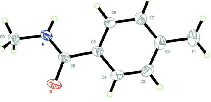
intermediate (Fig. 1). Bond lengths and angles are within normal ranges (Allen et al., 1987).
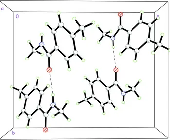
In the crystal packing of (I) the molecules are connected together via N—H···O intermolecular hydrogen bonds to form
a one-dimensional network in the b direction (Table 1, graph set C1,1(4)), which seems to be very effective in the
stabilization of the crystal structure.
S2. Experimental
The title compound, (I) was prepared by a method reported in literature (Lee et al. (2009)). Crystals were obtained by
dissolving (I) (0.2 g, 1.34 mmol) in ethanol (25 ml) and evaporating the solvent slowly at room temperature for about 7 d.
S3. Refinement
All H atoms were positioned geometrically and constrained to ride on their parent atoms, with C—H = 0.93 Å for
aromatic H, 0.96 Å for methyl H and 0.86 Å for N—H, respectively. The Uiso(H) = xUeq(C), where x = 1.2 for aromatic H
[image:2.610.132.483.455.626.2]and N—H, and x = 1.5 for methyl H.
Figure 1
The molecular structure of (I), with the atom-numbering scheme. Displacement ellipsoids are drawn at the 50%
Figure 2
Packing diagram for (I) showing the N—H···O hydrogen bonds as dashed lines.
N,4-Dimethylbenzamide
Crystal data
C9H11NO
Mr = 149.19
Monoclinic, P21/n
Hall symbol: -P 2yn
a = 6.7670 (14) Å
b = 9.946 (2) Å
c = 12.229 (2) Å
β = 92.63 (3)°
V = 822.2 (3) Å3
Z = 4
F(000) = 320
Dx = 1.205 Mg m−3
Mo Kα radiation, λ = 0.71073 Å Cell parameters from 25 reflections
θ = 9–13°
µ = 0.08 mm−1
T = 293 K Block, colourless 0.30 × 0.20 × 0.10 mm
Data collection
Enraf–Nonius CAD-4 diffractometer
Radiation source: fine-focus sealed tube Graphite monochromator
ω/2θ scans
Absorption correction: ψ scan (North et al., 1968)
Tmin = 0.977, Tmax = 0.992
3362 measured reflections
1510 independent reflections 1062 reflections with I > 2σ(I)
Rint = 0.033
θmax = 25.4°, θmin = 2.6°
h = 0→8
k = −11→11
l = −14→14
Refinement
Refinement on F2
Least-squares matrix: full
R[F2 > 2σ(F2)] = 0.050
wR(F2) = 0.166
S = 1.01 1510 reflections 103 parameters 0 restraints
Primary atom site location: structure-invariant direct methods
Secondary atom site location: difference Fourier map
Hydrogen site location: inferred from neighbouring sites
H-atom parameters constrained
w = 1/[σ2(F
o2) + (0.1P)2 + 0.080P]
where P = (Fo2 + 2Fc2)/3
(Δ/σ)max < 0.001
Δρmax = 0.20 e Å−3
Δρmin = −0.15 e Å−3
Extinction correction: SHELXL97 (Sheldrick, 2008), Fc*=kFc[1+0.001xFc2λ3/sin(2θ)]-1/4
Extinction coefficient: 0.028 (8)
Special details
Geometry. All e.s.d.'s (except the e.s.d. in the dihedral angle between two l.s. planes) are estimated using the full covariance matrix. The cell e.s.d.'s are taken into account individually in the estimation of e.s.d.'s in distances, angles and torsion angles; correlations between e.s.d.'s in cell parameters are only used when they are defined by crystal symmetry. An approximate (isotropic) treatment of cell e.s.d.'s is used for estimating e.s.d.'s involving l.s. planes.
Refinement. Refinement of F2 against ALL reflections. The weighted R-factor wR and goodness of fit S are based on F2,
conventional R-factors R are based on F, with F set to zero for negative F2. The threshold expression of F2 > σ(F2) is used
only for calculating R-factors(gt) etc. and is not relevant to the choice of reflections for refinement. R-factors based on F2
are statistically about twice as large as those based on F, and R- factors based on ALL data will be even larger.
Fractional atomic coordinates and isotropic or equivalent isotropic displacement parameters (Å2)
x y z Uiso*/Ueq
O 0.2041 (3) 0.99892 (14) 0.23558 (16) 0.0773 (6)
N 0.2849 (2) 0.78998 (15) 0.28900 (14) 0.0514 (5)
H0A 0.2554 0.7059 0.2868 0.062*
C1 −0.5644 (4) 0.6995 (3) 0.0146 (2) 0.0766 (8)
H1A −0.6295 0.6353 0.0592 0.115*
H1B −0.5367 0.6591 −0.0543 0.115*
H1C −0.6485 0.7763 0.0023 0.115*
C2 −0.3742 (3) 0.7431 (2) 0.07191 (17) 0.0550 (6)
C3 −0.2949 (4) 0.8698 (2) 0.05499 (19) 0.0652 (7)
H3A −0.3599 0.9283 0.0062 0.078*
C4 −0.1226 (3) 0.9106 (2) 0.10867 (18) 0.0591 (6)
H4A −0.0750 0.9968 0.0969 0.071*
C5 −0.0184 (3) 0.82556 (17) 0.18015 (15) 0.0440 (5)
C6 −0.0961 (3) 0.69834 (18) 0.19627 (17) 0.0530 (6)
H6A −0.0291 0.6387 0.2434 0.064*
C7 −0.2701 (3) 0.6593 (2) 0.14374 (18) 0.0581 (6)
H7A −0.3195 0.5738 0.1569 0.070*
C8 0.1657 (3) 0.87739 (18) 0.23661 (16) 0.0479 (5)
C9 0.4623 (3) 0.8321 (2) 0.3494 (2) 0.0613 (6)
H9A 0.5168 0.7575 0.3904 0.092*
H9B 0.4309 0.9034 0.3987 0.092*
Atomic displacement parameters (Å2)
U11 U22 U33 U12 U13 U23
O 0.0747 (11) 0.0273 (8) 0.1272 (15) −0.0054 (7) −0.0245 (10) 0.0023 (8)
N 0.0498 (10) 0.0296 (8) 0.0737 (12) −0.0025 (7) −0.0084 (8) −0.0006 (7)
C1 0.0590 (15) 0.0951 (19) 0.0743 (16) −0.0004 (13) −0.0132 (12) −0.0086 (14)
C2 0.0481 (12) 0.0610 (13) 0.0555 (12) 0.0047 (10) −0.0017 (10) −0.0086 (10)
C3 0.0654 (15) 0.0563 (13) 0.0721 (15) 0.0125 (11) −0.0159 (12) 0.0065 (11)
C4 0.0631 (14) 0.0396 (11) 0.0739 (14) 0.0040 (10) −0.0038 (12) 0.0083 (10)
C5 0.0446 (11) 0.0321 (9) 0.0552 (11) 0.0050 (8) 0.0007 (9) −0.0030 (8)
C6 0.0531 (12) 0.0351 (10) 0.0695 (13) 0.0000 (9) −0.0108 (10) 0.0047 (9)
C7 0.0547 (13) 0.0463 (12) 0.0724 (14) −0.0055 (9) −0.0068 (11) −0.0010 (10)
C8 0.0503 (12) 0.0288 (9) 0.0647 (12) 0.0016 (8) 0.0017 (9) −0.0024 (8)
C9 0.0537 (13) 0.0506 (12) 0.0783 (15) −0.0037 (10) −0.0122 (11) −0.0019 (11)
Geometric parameters (Å, º)
O—C8 1.237 (2) C3—H3A 0.9300
N—C8 1.330 (2) C4—C5 1.386 (3)
N—C9 1.442 (3) C4—H4A 0.9300
N—H0A 0.8600 C5—C6 1.388 (3)
C1—C2 1.501 (3) C5—C8 1.489 (3)
C1—H1A 0.9600 C6—C7 1.372 (3)
C1—H1B 0.9600 C6—H6A 0.9300
C1—H1C 0.9600 C7—H7A 0.9300
C2—C7 1.381 (3) C9—H9A 0.9600
C2—C3 1.389 (3) C9—H9B 0.9600
C3—C4 1.373 (3) C9—H9C 0.9600
C8—N—C9 121.90 (17) C4—C5—C6 117.46 (19)
C8—N—H0A 119.0 C4—C5—C8 118.17 (17)
C9—N—H0A 119.0 C6—C5—C8 124.35 (17)
C2—C1—H1A 109.5 C7—C6—C5 120.96 (19)
C2—C1—H1B 109.5 C7—C6—H6A 119.5
H1A—C1—H1B 109.5 C5—C6—H6A 119.5
C2—C1—H1C 109.5 C6—C7—C2 121.9 (2)
H1A—C1—H1C 109.5 C6—C7—H7A 119.0
H1B—C1—H1C 109.5 C2—C7—H7A 119.0
C7—C2—C3 117.0 (2) O—C8—N 121.39 (19)
C7—C2—C1 121.6 (2) O—C8—C5 120.36 (18)
C3—C2—C1 121.4 (2) N—C8—C5 118.24 (16)
C4—C3—C2 121.5 (2) N—C9—H9A 109.5
C4—C3—H3A 119.2 N—C9—H9B 109.5
C2—C3—H3A 119.2 H9A—C9—H9B 109.5
C3—C4—C5 121.2 (2) N—C9—H9C 109.5
C3—C4—H4A 119.4 H9A—C9—H9C 109.5
C7—C2—C3—C4 −1.1 (3) C3—C2—C7—C6 −0.1 (3)
C1—C2—C3—C4 178.9 (2) C1—C2—C7—C6 179.9 (2)
C2—C3—C4—C5 1.5 (4) C9—N—C8—O 1.5 (3)
C3—C4—C5—C6 −0.7 (3) C9—N—C8—C5 −177.53 (18)
C3—C4—C5—C8 −179.17 (19) C4—C5—C8—O 13.1 (3)
C4—C5—C6—C7 −0.4 (3) C6—C5—C8—O −165.2 (2)
C8—C5—C6—C7 177.91 (18) C4—C5—C8—N −167.81 (18)
C5—C6—C7—C2 0.8 (3) C6—C5—C8—N 13.8 (3)
Hydrogen-bond geometry (Å, º)
D—H···A D—H H···A D···A D—H···A
N—H0A···Oi 0.86 2.10 2.912 (2) 158
![Crystal structure of 2 methyl N [(4 methylpyridin 2 yl)carbamothioyl]benzamide](data:image/gif;base64,R0lGODlhAQABAIAAAP///wAAACH5BAEAAAAALAAAAAABAAEAAAICRAEAOw==)