(1
E
,2
E
)-1,2-Bis(2,2-diphenylhydrazin-1-ylidene)ethane
Angel Mendoza,a* Blanca M. Cabrera-Vivas,bRuth Mele´ndrez-Luevano,bJuan C. Ramı´rezband Marcos Flores-Alamoc
aCentro de Quı´mica, ICUAP, Beneme´rita Universidad Auto´noma de Puebla, Puebla, Pue., Mexico,bFacultad de Ciencias Quı´micas, Beneme´rita Universidad Auto´noma de Puebla, Puebla, Pue., Mexico, andcFacultad de Quı´mica, Universidad Nacional Auto´noma de Me´xico, 04510 Me´xico DF, Mexico
Correspondence e-mail: [email protected]
Received 3 August 2010; accepted 10 August 2010
Key indicators: single-crystal X-ray study;T= 298 K; mean(C–C) = 0.003 A˚; Rfactor = 0.044;wRfactor = 0.113; data-to-parameter ratio = 13.8.
In the crystal structure of the title compound, C26H22N4, the molecule is located on an inversion centre and shows an E configuration with respect to each C N bond. The dihedral angle between the phenyl rings in the diphenylhydrazone group is 83.69 (11). These two rings make dihedral angles of
30.53 (15) and 84.53 (16) with the central N—N C—
C N—N dihydrazonoethane plane. Intermolecular C— H interactions are observed.
Related literature
For applications of hydrazones, see: Angell et al. (2006); Iban˜ez et al.(2002). For related structures, see: Clulowet al. (2008); Mendozaet al.(2010). For bond-length data, see: Allen et al.(1987).
Experimental
Crystal data
= 103.924 (16)
V= 1047.2 (3) A˚3
Z= 2
CuKradiation
= 0.58 mm1
T= 298 K
0.190.110.05 mm
Data collection
Oxford Xcalibur Atlas Gemini diffractometer
Absorption correction: analytical (CrysAlis PRO; Oxford Diffraction, 2010)
Tmin= 0.978,Tmax= 0.993
3621 measured reflections 1892 independent reflections 1163 reflections withI> 2(I)
Rint= 0.038
Refinement
R[F2> 2(F2)] = 0.044
wR(F2) = 0.113
S= 1.01 1892 reflections
137 parameters
H-atom parameters constrained max= 0.13 e A˚
3
min=0.14 e A˚
[image:1.610.315.565.306.346.2]3
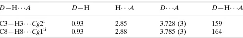
Table 1
Hydrogen-bond geometry (A˚ ,).
Cg1 andCg2 are the centroids of the C1–C6 and C7–C12 rings, respectively.
D—H A D—H H A D A D—H A
C3—H3 Cg2i 0.93 2.85 3.728 (3) 159
C8—H8 Cg1ii
0.93 2.88 3.785 (3) 164
Symmetry codes: (i)xþ1;yþ1;zþ1; (ii)x1;y;z.
Data collection: CrysAlis CCD (Oxford Diffraction, 2009); cell refinement: CrysAlis RED (Oxford Diffraction, 2009); data reduc-tion:CrysAlis RED; program(s) used to solve structure:SHELXS97 (Sheldrick, 2008); program(s) used to refine structure:SHELXL97 (Sheldrick, 2008); molecular graphics: ORTEP-3 for Windows (Farrugia, 1997); software used to prepare material for publication: WinGX(Farrugia, 1999).
The authors gratefully acknowledge financial support from the Facultad de Ciencias Quı´micas (BUAP).
Supplementary data and figures for this paper are available from the IUCr electronic archives (Reference: IS2589).
References
Allen, F. H., Kennard, O., Watson, D. G., Brammer, L., Orpen, A. G. & Taylor, R. (1987).J. Chem. Soc. Perkin Trans. 2, pp. S1–19.
Angell, S. E., Rogers, C. W., Zhang, Y., Wolf, M. O. & Jones, W. E. Jr (2006).
Coord. Chem. Rev.250, 1829–1841.
Clulow, A. J., Selby, J. D., Cushion, M. G., Schwarz, A. D. & Mountford, P. (2008).Inorg. Chem.47, 12049–12062.
Farrugia, L. J. (1997).J. Appl. Cryst.30, 565. Farrugia, L. J. (1999).J. Appl. Cryst.32, 837–838.
Iban˜ez, G. A., Escandar, G. M. & Olivieri, A. C. (2002).J. Mol. Struct.605, 17– 26.
Mendoza, A., Cabrera-Vivas, B. M., Mele´ndrez-Luevano, R., Pacheco-A´ lvarez, T. & Carranza, V. (2010).Acta Cryst.E66, o2058.
Oxford Diffraction (2009). CrysAlis CCD and CrysAlis RED. Oxford Diffraction Ltd, Yarnton, Oxfordshire, England.
Oxford Diffraction (2010).CrysAlis PRO. Oxford Diffraction Ltd, Yarnton, Acta Crystallographica Section E
Structure Reports
Online
supporting information
Acta Cryst. (2010). E66, o2349 [https://doi.org/10.1107/S1600536810032198]
(1
E
,2
E
)-1,2-Bis(2,2-diphenylhydrazin-1-ylidene)ethane
Angel Mendoza, Blanca M. Cabrera-Vivas, Ruth Mel
é
ndrez-Luevano, Juan C. Ram
í
rez and
Marcos Flores-Alamo
S1. Comment
Among the most interesting applications of hydrazones, molecular sensing is worth mentioning. They are being used
widely to detect chemical and biological species (Angell et al., 2006). Also, hydrazones are being applied as plasticizer agents, polymerization initiators and antioxidants (Ibañez et al., 2002). There are pigments, as 1-fenilazo-2-naftol, that show an azo/hydrazone tautomery in which the main tautomer exist as hydrazone form.
The asymmetric unit of the title compound I consist of C13H11N2 with a Z′ = 0.5 showing a centrosymmetrical structure.
The compound I (C26H22N4) present an E configuration for each C═N double bond (Fig. 1), with N,N-diphenyl group
opposite to second C═N group. The molecule shows a non-planar structure for phenyl rings respect to N—N group, with
a torsion angle between them C2—C1—N1—C7 = 46.6 (3)°. The torsion angle of phenyl ring C1/C2/C3/C4/C5/C6 to N
—N═C group is -173.48 (18)°, and the other ring C7/C8/C9/C10/C11/C12 shows a torsion angle of -14.9 (3)° to the
same group. The N—N distance [1.364 (2) Å] is shorter than found in free diphenylhydrazine [1.418 (2) Å] (Clulow et al., 2008). Imine bond distance, N2═C13 [1.287 (2) Å], is longer than N═C typical bond (Allen et al., 1987), but similar [1.286 (3) Å] to related structures with N,N-diphenylhidrazone group (Mendoza et al., 2010).
S2. Experimental
N,N-diphenylhydrazine (2.74 mg, 12.4 mmol) was dissolved in ethanol and acetic acid (0.5 ml) was added slowly into this solution while stirring. Glyoxal (300 mg, 5.1 mmol) was added drop by drop into the above solution with strong
stirring and the resulting mixture was kept at atmospheric temperature until it became yellow solution. After three hours,
the amber solution turns to be precipitated. The mixture was separated with filtration in vacuum system and the
precipitate was washed three times with cold methanol. Recrystallization was performed several times with acetonitrile,
to obtain needle crystals suitable for X-ray analysis. Yield: 1.79 g (90%) at 25 °C, mp. 185–189 °C. FT–IR (film): (cm-1):
3062 ν(C—H), 1750–2000 ν(Ph), 1591, 1544, 1490 ν(C═N). EI–MS: m/z 390 M+.
S3. Refinement
H atoms were placed in geometrical idealized positions (C—H = 0.93 Å) and refined as riding on their parent atoms, with
Figure 1
The molecular structure of compound I, with atom labels and 50% probability displacement ellipsoids for non-H atoms.
(1E,2E)-1,2-Bis(2,2-diphenylhydrazin-1-ylidene)ethane
Crystal data C26H22N4
Mr = 390.48
Monoclinic, P21/n
a = 12.2210 (19) Å b = 5.612 (1) Å c = 15.731 (3) Å β = 103.924 (16)° V = 1047.2 (3) Å3
Z = 2
F(000) = 412 Dx = 1.238 Mg m−3
Cu Kα radiation, λ = 1.5418 Å Cell parameters from 864 reflections θ = 3.7–68.0°
µ = 0.58 mm−1
T = 298 K Prism, colourless 0.19 × 0.11 × 0.05 mm
Data collection
Oxford Xcalibur Atlas Gemini diffractometer
Graphite monochromator
Detector resolution: 10.4685 pixels mm-1
ω scans
Absorption correction: analytical
(CrysAlis PRO; Oxford Diffraction, 2010) Tmin = 0.978, Tmax = 0.993
3621 measured reflections 1892 independent reflections 1163 reflections with I > 2σ(I) Rint = 0.038
θmax = 68.2°, θmin = 4.1°
h = −14→14 k = −4→6 l = −18→10
Refinement
Hydrogen site location: inferred from neighbouring sites
H-atom parameters constrained w = 1/[σ2(F
o2) + (0.0496P)2 + 0.0674P]
where P = (Fo2 + 2Fc2)/3
(Δ/σ)max < 0.001
Δρmax = 0.13 e Å−3
Δρmin = −0.14 e Å−3
Extinction correction: SHELXL97 (Sheldrick, 2008), Fc*=kFc[1+0.001xFc2λ3/sin(2θ)]-1/4
Extinction coefficient: 0.0134 (9)
Special details
Geometry. All s.u.'s (except the s.u. in the dihedral angle between two l.s. planes) are estimated using the full covariance matrix. The cell s.u.'s are taken into account individually in the estimation of s.u.'s in distances, angles and torsion angles; correlations between s.u.'s in cell parameters are only used when they are defined by crystal symmetry. An approximate (isotropic) treatment of cell s.u.'s is used for estimating s.u.'s involving l.s. planes.
Refinement. Refinement of F2 against ALL reflections. The weighted R-factor wR and goodness of fit S are based on F2,
conventional R-factors R are based on F, with F set to zero for negative F2. The threshold expression of F2 > 2σ(F2) is
used only for calculating R-factors(gt) etc. and is not relevant to the choice of reflections for refinement. R-factors based on F2 are statistically about twice as large as those based on F, and R- factors based on ALL data will be even larger.
Fractional atomic coordinates and isotropic or equivalent isotropic displacement parameters (Å2)
x y z Uiso*/Ueq
N2 0.09341 (13) 0.1892 (3) 0.46419 (10) 0.0451 (5)
N1 0.20416 (13) 0.2544 (3) 0.48657 (10) 0.0481 (5)
C1 0.23555 (16) 0.4384 (4) 0.43534 (12) 0.0421 (5)
C13 0.05715 (15) 0.0396 (4) 0.51307 (13) 0.0451 (5)
H13 0.1046 −0.0161 0.5646 0.054*
C12 0.27665 (18) 0.3696 (4) 0.63743 (14) 0.0541 (6)
H12 0.2307 0.5037 0.6265 0.065*
C7 0.27471 (15) 0.2080 (4) 0.57158 (12) 0.0415 (5)
C6 0.16000 (18) 0.6077 (4) 0.39339 (13) 0.0494 (5)
H6 0.0859 0.6044 0.3988 0.059*
C2 0.34652 (17) 0.4494 (4) 0.42837 (13) 0.0532 (6)
H2 0.3988 0.3382 0.4574 0.064*
C5 0.1938 (2) 0.7827 (4) 0.34321 (14) 0.0587 (6)
H5 0.1422 0.8956 0.3147 0.07*
C8 0.33966 (18) 0.0069 (4) 0.58755 (15) 0.0572 (6)
H8 0.3374 −0.1049 0.5435 0.069*
C3 0.3793 (2) 0.6255 (4) 0.37837 (15) 0.0623 (7)
H3 0.4538 0.6319 0.3739 0.075*
C4 0.3036 (2) 0.7904 (4) 0.33537 (15) 0.0640 (7)
H4 0.3261 0.9069 0.3011 0.077*
C10 0.4116 (2) 0.1382 (6) 0.73586 (17) 0.0726 (8)
H10 0.4585 0.115 0.7913 0.087*
C9 0.40936 (19) −0.0277 (5) 0.67093 (19) 0.0722 (8)
H9 0.4544 −0.163 0.6827 0.087*
C11 0.3458 (2) 0.3350 (5) 0.71930 (15) 0.0708 (8)
Atomic displacement parameters (Å2)
U11 U22 U33 U12 U13 U23
N2 0.0382 (9) 0.0534 (11) 0.0436 (10) −0.0137 (8) 0.0099 (7) −0.0026 (8) N1 0.0372 (9) 0.0609 (11) 0.0446 (10) −0.0163 (8) 0.0066 (7) 0.0080 (9) C1 0.0439 (11) 0.0468 (12) 0.0357 (10) −0.0134 (10) 0.0096 (8) −0.0006 (9) C13 0.0391 (10) 0.0537 (13) 0.0428 (11) −0.0106 (10) 0.0101 (9) 0.0036 (11) C12 0.0544 (13) 0.0597 (14) 0.0500 (13) −0.0014 (12) 0.0161 (10) 0.0011 (12) C7 0.0352 (10) 0.0470 (12) 0.0438 (11) −0.0101 (10) 0.0125 (8) 0.0036 (10) C6 0.0480 (12) 0.0532 (13) 0.0470 (12) −0.0083 (11) 0.0113 (10) −0.0035 (11) C2 0.0461 (12) 0.0587 (14) 0.0564 (13) −0.0081 (11) 0.0157 (10) 0.0069 (11) C5 0.0705 (16) 0.0501 (14) 0.0523 (13) −0.0041 (12) 0.0087 (11) 0.0069 (11) C8 0.0512 (13) 0.0509 (13) 0.0713 (16) −0.0074 (12) 0.0184 (12) 0.0017 (13) C3 0.0574 (14) 0.0731 (16) 0.0640 (15) −0.0158 (13) 0.0297 (12) 0.0068 (13) C4 0.0806 (17) 0.0612 (16) 0.0540 (14) −0.0189 (14) 0.0234 (12) 0.0076 (12) C10 0.0544 (14) 0.102 (2) 0.0554 (16) −0.0156 (16) 0.0024 (12) 0.0226 (16) C9 0.0484 (13) 0.0676 (17) 0.098 (2) 0.0028 (13) 0.0116 (14) 0.0304 (16) C11 0.0731 (16) 0.092 (2) 0.0463 (14) −0.0098 (16) 0.0125 (12) −0.0014 (14)
Geometric parameters (Å, º)
N2—C13 1.287 (2) C2—C3 1.381 (3)
N2—N1 1.364 (2) C2—H2 0.93
N1—C1 1.418 (2) C5—C4 1.377 (3)
N1—C7 1.430 (2) C5—H5 0.93
C1—C6 1.377 (3) C8—C9 1.395 (3)
C1—C2 1.388 (3) C8—H8 0.93
C13—C13i 1.429 (4) C3—C4 1.367 (3)
C13—H13 0.93 C3—H3 0.93
C12—C7 1.373 (3) C4—H4 0.93
C12—C11 1.373 (3) C10—C11 1.354 (3)
C12—H12 0.93 C10—C9 1.377 (4)
C7—C8 1.368 (3) C10—H10 0.93
C6—C5 1.384 (3) C9—H9 0.93
C6—H6 0.93 C11—H11 0.93
C13—N2—N1 118.93 (16) C4—C5—C6 120.3 (2)
N2—N1—C1 115.85 (16) C4—C5—H5 119.9
N2—N1—C7 122.01 (14) C6—C5—H5 119.9
C1—N1—C7 118.63 (15) C7—C8—C9 118.9 (2)
C6—C1—C2 119.11 (19) C7—C8—H8 120.5
C6—C1—N1 122.18 (18) C9—C8—H8 120.5
C2—C1—N1 118.71 (19) C4—C3—C2 120.8 (2)
N2—C13—C13i 119.0 (2) C4—C3—H3 119.6
C11—C12—H12 119.7 C11—C10—C9 120.2 (2)
C8—C7—C12 120.2 (2) C11—C10—H10 119.9
C8—C7—N1 120.98 (19) C9—C10—H10 119.9
C12—C7—N1 118.86 (19) C10—C9—C8 120.1 (2)
C1—C6—C5 120.3 (2) C10—C9—H9 119.9
C1—C6—H6 119.8 C8—C9—H9 119.9
C5—C6—H6 119.8 C10—C11—C12 119.9 (2)
C3—C2—C1 120.0 (2) C10—C11—H11 120.1
C3—C2—H2 120 C12—C11—H11 120.1
C1—C2—H2 120
C13—N2—N1—C1 −173.48 (18) N1—C1—C6—C5 −179.26 (18)
C13—N2—N1—C7 −14.9 (3) C6—C1—C2—C3 −1.3 (3)
N2—N1—C1—C6 26.8 (3) N1—C1—C2—C3 179.48 (18)
C7—N1—C1—C6 −132.5 (2) C1—C6—C5—C4 −0.6 (3)
N2—N1—C1—C2 −154.06 (18) C12—C7—C8—C9 −1.5 (3)
C7—N1—C1—C2 46.6 (3) N1—C7—C8—C9 178.36 (18)
N1—N2—C13—C13i −176.3 (2) C1—C2—C3—C4 0.0 (3)
C11—C12—C7—C8 1.7 (3) C2—C3—C4—C5 1.0 (4)
C11—C12—C7—N1 −178.09 (18) C6—C5—C4—C3 −0.8 (3)
N2—N1—C7—C8 93.9 (2) C11—C10—C9—C8 0.4 (4)
C1—N1—C7—C8 −108.1 (2) C7—C8—C9—C10 0.4 (3)
N2—N1—C7—C12 −86.3 (2) C9—C10—C11—C12 −0.1 (4)
C1—N1—C7—C12 71.7 (2) C7—C12—C11—C10 −0.9 (3)
C2—C1—C6—C5 1.6 (3)
Symmetry code: (i) −x, −y, −z+1.
Hydrogen-bond geometry (Å, º)
D—H···A D—H H···A D···A D—H···A
C3—H3···Cg2ii 0.93 2.85 3.728 (3) 159
C8—H8···Cg1iii 0.93 2.88 3.785 (3) 164