2-Chloro-3-nitropyridine
Seik Weng Ng
Department of Chemistry, University of Malaya, 50603 Kuala Lumpur, Malaysia Correspondence e-mail: [email protected]
Received 22 February 2010; accepted 30 March 2010
Key indicators: single-crystal X-ray study;T= 293 K; mean(C–C) = 0.003 A˚;
Rfactor = 0.037;wRfactor = 0.108; data-to-parameter ratio = 14.0.
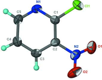
In the title compound, C5H3ClN2O2, the nitro group is twisted by 38.5 (2) with respect to the pyridine ring. In the crystal,
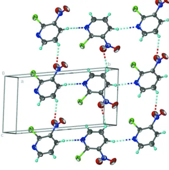
adjacent molecules are linked by non-classical C—H N and C—H O hydrogen bonds, forming a layer motif.
Related literature
For the crystal structure of isostructural 2-iodo-3-nitro-pyridine, see: Mao & Chen (2009). For the crystal structure of 2-chloro-5-nitropyridine, see: Ng (2010).
Experimental
Crystal data
C5H3ClN2O2 Mr= 158.54 Monoclinic,P21=n a= 7.613 (1) A˚ b= 12.232 (2) A˚ c= 7.716 (1) A˚ = 118.485 (2)
V= 631.5 (2) A˚3 Z= 4
MoKradiation = 0.53 mm1 T= 293 K
0.300.200.05 mm
Bruker SMART APEX diffractometer
Absorption correction: multi-scan (SADABS; Sheldrick, 1996) Tmin= 0.771,Tmax= 0.862
5889 measured reflections 1445 independent reflections 1061 reflections withI> 2(I) Rint= 0.040
Refinement
R[F2> 2(F2)] = 0.037 wR(F2) = 0.108 S= 1.02 1445 reflections 103 parameters
3 restraints
All H-atom parameters refined max= 0.22 e A˚
3
min=0.29 e A˚
3
Table 1
Hydrogen-bond geometry (A˚ ,).
D—H A D—H H A D A D—H A
C3—H3 N1i
0.93 (1) 2.53 (1) 3.430 (3) 166 (2) C4—H4 O1ii 0.93 (1) 2.64 (2) 3.327 (3) 132 (2)
Symmetry codes: (i)xþ3 2;yþ
1 2;zþ
1
2; (ii)x;y;z1.
Data collection:APEX2(Bruker, 2009); cell refinement:SAINT
(Bruker, 2009); data reduction:SAINT; program(s) used to solve structure:SHELXS97(Sheldrick, 2008); program(s) used to refine structure: SHELXL97 (Sheldrick, 2008); molecular graphics: X-SEED (Barbour, 2001); software used to prepare material for publication:publCIF(Westrip, 2010).
I thank the University of Malaya for supporting this study.
Supplementary data and figures for this paper are available from the IUCr electronic archives (Reference: IM2183).
References
Barbour, L. J. (2001).J. Supramol. Chem.1, 189–191.
Bruker (2009).APEX2andSAINT. Bruker AXS Inc., Madison, Wisconsin, USA.
Mao, L.-H. & Chen, Y. (2009).Acta Cryst.E65, o1428. Ng, S. W. (2010).Acta Cryst.E66, o848.
Sheldrick, G. M. (1996).SADABS. University of Go¨ttingen, Germany. Sheldrick, G. M. (2008).Acta Cryst.A64, 112–122.
Westrip, S. P. (2010).publCIF. In preparation. Structure Reports
Online
supporting information
Acta Cryst. (2010). E66, o1020 [https://doi.org/10.1107/S1600536810011955]
2-Chloro-3-nitropyridine
Seik Weng Ng
S1. Comment
According to a recent report on the crystal structure of 2-chloro-5-nitropyridine the respective molecule is planar
(maximum r.m.s. deviation of non-hydrogen atoms is 0.090 Å). This molecule has the electron withdrawing substituents
para to each other. The substituents interact through a short Cl···O contact of 3.068 (4) Å to generate a chain motif (Ng,
2010).
In the title compound 2-chloro-3-nitropyridine with the nitro group ortho to the chlorine substituent (Scheme I, Fig. 1),
a similar Cl···O contact is also observed but the nitro group is twisted to avoid repulsion. Adjacent molecules are linked
by non-classical C–H···N and C–H···O hydrogen bonds to form a layer motif (Fig. 2, Table 1). The C–H···N interaction is
almost linear (Table 1).
2-Chloro-3-nitropyridine is isostructural with the iodo analog. In the iodo compound, the I···O contact is necessarily
longer (Mao & Chen, 2009).
S2. Experimental
2-Chloro-3-nitropyridine was obtained from the Aldrich Chemical Company, and was recrystallized from ethyl acetate.
S3. Refinement
Carbon bound H-atoms were located in a difference Fourier map. They were refined with a distance restraint of C–H
Figure 1
Thermal ellipsoid plot (Barbour, 2001) of C5H3ClNO2 at the 50% probability level; hydrogen atoms are drawn as spheres
Figure 2
Non-classical hydrogen-bonded layer motif.
2-chloro-3-nitropyridine
Crystal data
C5H3ClN2O2 Mr = 158.54
Monoclinic, P21/n Hall symbol: -P 2yn a = 7.613 (1) Å b = 12.232 (2) Å c = 7.716 (1) Å β = 118.485 (2)° V = 631.5 (2) Å3 Z = 4
F(000) = 320 Dx = 1.668 Mg m−3
Mo Kα radiation, λ = 0.71073 Å Cell parameters from 1393 reflections θ = 3.3–24.8°
µ = 0.53 mm−1 T = 293 K
Block, faint yellow 0.30 × 0.20 × 0.05 mm
Data collection
Bruker SMART APEX diffractometer
θmax = 27.5°, θmin = 3.1° h = −9→9
l = −9→10
Refinement
Refinement on F2 Least-squares matrix: full R[F2 > 2σ(F2)] = 0.037 wR(F2) = 0.108 S = 1.02 1445 reflections 103 parameters 3 restraints
Primary atom site location: structure-invariant direct methods
Secondary atom site location: difference Fourier map
Hydrogen site location: inferred from neighbouring sites
All H-atom parameters refined
w = 1/[σ2(Fo2) + (0.0539P)2 + 0.1169P] where P = (Fo2 + 2Fc2)/3
(Δ/σ)max = 0.001 Δρmax = 0.22 e Å−3 Δρmin = −0.29 e Å−3
Fractional atomic coordinates and isotropic or equivalent isotropic displacement parameters (Å2)
x y z Uiso*/Ueq
Cl1 0.67770 (9) 0.67094 (4) 0.54558 (9) 0.0583 (2)
O1 0.5971 (3) 0.89254 (15) 0.6322 (3) 0.0793 (6)
O2 0.8242 (3) 1.00241 (15) 0.6464 (3) 0.0804 (6)
N1 0.6852 (3) 0.71189 (13) 0.2214 (3) 0.0471 (4)
N2 0.7123 (3) 0.92525 (15) 0.5760 (3) 0.0514 (5)
C1 0.6891 (3) 0.75880 (14) 0.3767 (3) 0.0381 (4)
C2 0.7109 (3) 0.87120 (14) 0.4062 (3) 0.0375 (4)
C3 0.7322 (3) 0.93592 (16) 0.2719 (3) 0.0468 (5)
C4 0.7252 (4) 0.88674 (18) 0.1090 (3) 0.0522 (5)
C5 0.7011 (3) 0.77541 (19) 0.0896 (3) 0.0516 (5)
H3 0.751 (3) 1.0104 (9) 0.294 (3) 0.060 (7)*
H4 0.740 (3) 0.9261 (17) 0.013 (3) 0.061 (7)*
H5 0.693 (3) 0.7407 (18) −0.021 (2) 0.060 (7)*
Atomic displacement parameters (Å2)
U11 U22 U33 U12 U13 U23
Geometric parameters (Å, º)
Cl1—C1 1.7226 (18) C2—C3 1.374 (3)
O1—N2 1.217 (2) C3—C4 1.371 (3)
O2—N2 1.213 (2) C3—H3 0.925 (9)
N1—C1 1.317 (2) C4—C5 1.373 (3)
N1—C5 1.330 (3) C4—H4 0.930 (10)
N2—C2 1.462 (2) C5—H5 0.929 (10)
C1—C2 1.391 (3)
C1—N1—C5 118.07 (17) C4—C3—C2 118.15 (18)
O2—N2—O1 124.60 (19) C4—C3—H3 122.2 (15)
O2—N2—C2 117.20 (19) C2—C3—H3 119.7 (15)
O1—N2—C2 118.15 (18) C3—C4—C5 118.67 (19)
N1—C1—C2 121.95 (16) C3—C4—H4 122.2 (15)
N1—C1—Cl1 115.43 (14) C5—C4—H4 119.2 (15)
C2—C1—Cl1 122.55 (14) N1—C5—C4 123.54 (18)
C3—C2—C1 119.58 (17) N1—C5—H5 116.5 (15)
C3—C2—N2 117.52 (16) C4—C5—H5 120.0 (15)
C1—C2—N2 122.90 (17)
C5—N1—C1—C2 −0.5 (3) O2—N2—C2—C1 143.2 (2)
C5—N1—C1—Cl1 −177.56 (15) O1—N2—C2—C1 −39.3 (3)
N1—C1—C2—C3 −1.1 (3) C1—C2—C3—C4 1.9 (3)
Cl1—C1—C2—C3 175.72 (15) N2—C2—C3—C4 −178.09 (19)
N1—C1—C2—N2 178.96 (18) C2—C3—C4—C5 −1.2 (3)
Cl1—C1—C2—N2 −4.2 (3) C1—N1—C5—C4 1.3 (3)
O2—N2—C2—C3 −36.8 (3) C3—C4—C5—N1 −0.4 (4)
O1—N2—C2—C3 140.7 (2)
Hydrogen-bond geometry (Å, º)
D—H···A D—H H···A D···A D—H···A
C3—H3···N1i 0.93 (1) 2.53 (1) 3.430 (3) 166 (2)
C4—H4···O1ii 0.93 (1) 2.64 (2) 3.327 (3) 132 (2)