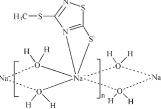
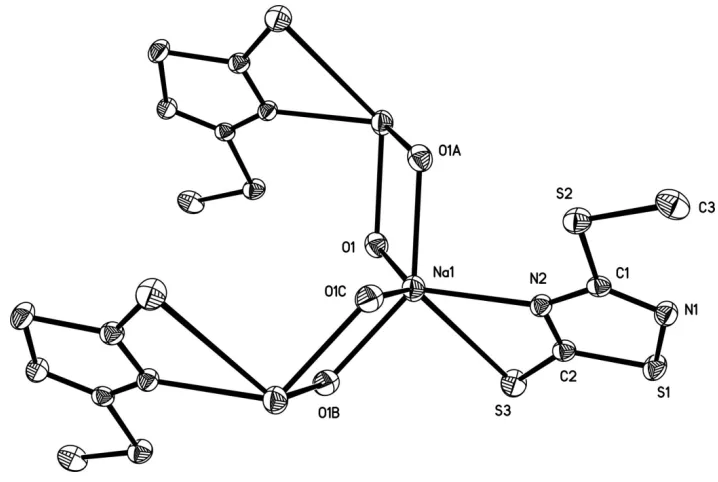
catena
-Poly[[(3-methylsulfanyl-1,2,4-thiadiazole-5-thiolato)sodium]di-
l
-aqua-j
4O
:
O
]
Jun-Hong Zhang,a,bChun-Lin Ma,bRu-Fen Zhang,b Hai-Zeng Wanga* and Guo-Jia Fub
aCollege of Chemistry and Chemical Engineering, Ocean University of China,
Qingdao 266100, Shandong, People’s Republic of China, andbCollege of Chemistry
and Chemical Engineering, Liaocheng University, Shandong 252059, People’s Republic of China
Correspondence e-mail: zhangjunhong@lcu.edu.cn
Received 17 November 2009; accepted 1 December 2009
Key indicators: single-crystal X-ray study;T= 298 K; mean(S–N) = 0.003 A˚;
Rfactor = 0.024;wRfactor = 0.064; data-to-parameter ratio = 13.5.
The crystal structure of the title compound, [Na(C3H3N2S3
)-(H2O)2]n, features polymeric chains made up of O O
edge-shared NaSN(H2O)4units running along thebaxis. The Na +
ion and all non-H atoms of the thiadiazole ligand lie on a mirror plane.
Related literature
For related structures, see: Guo (2004); Wanget al.(2007).
Experimental
Crystal data [Na(C3H3N2S3)(H2O)2] Mr= 222.28
Monoclinic,P21=m a= 7.5794 (8) A˚
b= 6.9736 (6) A˚
c= 8.6879 (12) A˚
= 102.027 (1)
V= 449.13 (9) A˚3 Z= 2
MoKradiation
= 0.83 mm1 T= 298 K
0.390.270.15 mm
Data collection
Siemens SMART CCD area-detector diffractometer Absorption correction: multi-scan
(SADABS; Sheldrick, 1996)
Tmin= 0.739,Tmax= 0.886
2256 measured reflections 862 independent reflections 728 reflections withI> 2(I)
Rint= 0.017
Refinement
R[F2> 2(F2)] = 0.024 wR(F2) = 0.064 S= 1.07 862 reflections
64 parameters
H-atom parameters constrained
max= 0.28 e A˚
3
min=0.21 e A˚
[image:1.610.90.251.474.583.2]3
Table 1
Selected bond lengths (A˚ ).
Na1—O1 2.4493 (16) Na1—N2 2.467 (2)
Na1—O1i
2.4736 (16) Na1—S3 3.1271 (14)
Symmetry code: (i)x;y;zþ2.
Data collection:SMART(Siemens, 1996); cell refinement:SAINT
(Siemens, 1996); data reduction:SAINT; program(s) used to solve structure:SHELXS97(Sheldrick, 2008); program(s) used to refine structure: SHELXL97 (Sheldrick, 2008); molecular graphics:
SHELXTL(Sheldrick, 2008); software used to prepare material for publication:SHELXTL.
The authors thank the National Natural Science Foundation of China (grant No. 20741008) and the State Key Laboratory of Crystalline Materials, Liaocheng University, People’s Republic of China.
Supplementary data and figures for this paper are available from the IUCr electronic archives (Reference: CI2968).
References
Guo, M.-L. (2004).Acta Cryst.E60, m1684–m1685.
Sheldrick, G. M. (1996).SADABS. University of Go¨ttingen, Germany. Sheldrick, G. M. (2008).Acta Cryst.A64, 112–122.
Siemens (1996).SMARTandSAINT. Siemens Analytical X-ray Instruments Inc., Madison, Wisconsin, USA.
Wang, X.-H., Hu, M.-C. & Yang, Z.-Q. (2007).Acta Cryst.E63, m1527–m1529.
Acta Crystallographica Section E Structure Reports
Online
supporting information
Acta Cryst. (2010). E66, m15 [doi:10.1107/S1600536809051721]
catena
-Poly[[(3-methylsulfanyl-1,2,4-thiadiazole-5-thiolato)sodium]di-
µ
-aqua-κ
4O
:
O
]
Jun-Hong Zhang, Chun-Lin Ma, Ru-Fen Zhang, Hai-Zeng Wang and Guo-Jia Fu
S1. Comment
In the title compound (Fig.1), each Na+ ion has a six-coordinate environment formed by a S atom and a N atom of the
3-methylsulfanyl-1,2,4-thiadiazole-5-thiolate ligand, and four bridging water O atoms O1, O1A, O1B and O1C. The adjacent NaSNO4 units share O1···O1A and O1B···O1C edges, producing chains running along the b axis (Fig. 2). Similar
chains were found in the crystal structure of sodium carboxynitrobenzoate tetrahydrate (Guo, 2004). The Na—O [2.4493 (16) and 2.4736 (16) Å], Na—S [3.1271 (14) Å] and Na—N [2.467 (2) Å] distances are comparable to those observed in a related structure (Wang et al., 2007).
S2. Experimental
To a solution of 3-methylmercapto-5-mercapto-1,2,4-thiadiazole (10 mmol) in 60 ml of doubly-distilled water, a solution of an equimolar amount (10 mmol) of sodium hydroxide in 40 ml of doubly-distilled water was added dropwise at room temperature. After vigorous stirring for 6 h, the resulting mixture was evaporated in vacuo to a volume of about 20 ml and filtered hot. The filtrate was then set aside for crystallization at room temperature. Two weeks later, colourless single crystals suitable for X-ray diffraction were obtained.
S3. Refinement
H atoms of the water molecules were initially located a difference Fourier map and later refined using a riding model with O-H = 0.85Å and Uiso(H) = 0.05 Å2. C-bound H atoms were positioned geometrically and treated as riding on their
Figure 1
The coordination environment around the Na+ ion. Displacement ellipsoids are drawn at the 30% probability level.
[Symmetry codes: (A) -x, -y, 2-z; (B) x, 1/2-y, z; (C) -x, 1/2+y, 2-z].
Figure 2
Part of the polymeric chain parallel to the b axis. H atoms have been omitted for clarity.
catena-Poly[[(3-methylsulfanyl-1,2,4-thiadiazole-5-thiolato)sodium]di- µ-aqua-κ4O:O]
Crystal data
[Na(C3H3N2S3)(H2O)2] Mr = 222.28
Monoclinic, P21/m
Hall symbol: -P 2yb
a = 7.5794 (8) Å
b = 6.9736 (6) Å
c = 8.6879 (12) Å
β = 102.027 (1)°
V = 449.13 (9) Å3 Z = 2
F(000) = 228
Dx = 1.644 Mg m−3
Mo Kα radiation, λ = 0.71073 Å Cell parameters from 1437 reflections
θ = 2.4–27.9°
[image:3.610.124.483.363.537.2]T = 298 K Block, colourless
0.39 × 0.27 × 0.15 mm
Data collection
Siemens SMART CCD area-detector diffractometer
Radiation source: fine-focus sealed tube Graphite monochromator
φ and ω scans
Absorption correction: multi-scan (SADABS; Sheldrick, 1996)
Tmin = 0.739, Tmax = 0.886
2256 measured reflections 862 independent reflections 728 reflections with I > 2σ(I)
Rint = 0.017
θmax = 25.0°, θmin = 2.4°
h = −7→9
k = −8→8
l = −10→10
Refinement
Refinement on F2
Least-squares matrix: full
R[F2 > 2σ(F2)] = 0.024 wR(F2) = 0.064 S = 1.07 862 reflections 64 parameters 0 restraints
Primary atom site location: structure-invariant direct methods
Secondary atom site location: difference Fourier map
Hydrogen site location: inferred from neighbouring sites
H-atom parameters constrained
w = 1/[σ2(F
o2) + (0.027P)2 + 0.2249P]
where P = (Fo2 + 2Fc2)/3
(Δ/σ)max = 0.001
Δρmax = 0.28 e Å−3
Δρmin = −0.21 e Å−3
Special details
Geometry. All e.s.d.'s (except the e.s.d. in the dihedral angle between two l.s. planes) are estimated using the full covariance matrix. The cell e.s.d.'s are taken into account individually in the estimation of e.s.d.'s in distances, angles and torsion angles; correlations between e.s.d.'s in cell parameters are only used when they are defined by crystal symmetry. An approximate (isotropic) treatment of cell e.s.d.'s is used for estimating e.s.d.'s involving l.s. planes.
Refinement. Refinement of F2 against ALL reflections. The weighted R-factor wR and goodness of fit S are based on F2,
conventional R-factors R are based on F, with F set to zero for negative F2. The threshold expression of F2 > σ(F2) is used
only for calculating R-factors(gt) etc. and is not relevant to the choice of reflections for refinement. R-factors based on F2
are statistically about twice as large as those based on F, and R- factors based on ALL data will be even larger.
Fractional atomic coordinates and isotropic or equivalent isotropic displacement parameters (Å2)
x y z Uiso*/Ueq Occ. (<1)
Na1 0.07064 (15) 0.2500 0.93636 (13) 0.0400 (3) N1 0.1651 (3) 0.2500 0.4084 (3) 0.0339 (5) N2 0.1364 (3) 0.2500 0.6700 (2) 0.0291 (5) O1 0.16585 (17) −0.0019 (2) 1.13259 (15) 0.0413 (4)
H1A 0.1666 0.0502 1.2212 0.050*
H1B 0.2701 −0.0493 1.1374 0.050* S1 0.37894 (9) 0.2500 0.51077 (9) 0.0386 (2) S2 −0.17326 (9) 0.2500 0.46612 (9) 0.0378 (2) S3 0.46031 (10) 0.2500 0.86933 (9) 0.0448 (2) C1 0.0626 (3) 0.2500 0.5131 (3) 0.0283 (6) C2 0.3165 (3) 0.2500 0.6910 (3) 0.0310 (6) C3 −0.2220 (4) 0.2500 0.2548 (3) 0.0470 (8) H3A −0.3503 0.2500 0.2163 0.071*
H3C −0.1709 0.3624 0.2175 0.071* 0.50
Atomic displacement parameters (Å2)
U11 U22 U33 U12 U13 U23
Na1 0.0488 (7) 0.0382 (7) 0.0350 (6) 0.000 0.0134 (5) 0.000 N1 0.0315 (12) 0.0391 (14) 0.0321 (13) 0.000 0.0085 (10) 0.000 N2 0.0261 (11) 0.0318 (12) 0.0302 (12) 0.000 0.0074 (9) 0.000 O1 0.0393 (8) 0.0461 (9) 0.0374 (8) 0.0029 (6) 0.0054 (6) −0.0041 (7) S1 0.0274 (4) 0.0514 (5) 0.0394 (4) 0.000 0.0122 (3) 0.000 S2 0.0256 (4) 0.0444 (5) 0.0423 (4) 0.000 0.0043 (3) 0.000 S3 0.0319 (4) 0.0626 (6) 0.0364 (4) 0.000 −0.0009 (3) 0.000 C1 0.0286 (13) 0.0214 (13) 0.0350 (15) 0.000 0.0067 (11) 0.000 C2 0.0280 (13) 0.0295 (15) 0.0358 (15) 0.000 0.0070 (11) 0.000 C3 0.0439 (17) 0.0468 (19) 0.0426 (18) 0.000 −0.0088 (14) 0.000
Geometric parameters (Å, º)
Na1—O1i 2.4493 (16) N2—C1 1.361 (3)
Na1—O1 2.4493 (16) O1—Na1ii 2.4736 (16)
Na1—N2 2.467 (2) O1—H1A 0.85
Na1—O1ii 2.4736 (16) O1—H1B 0.85
Na1—O1iii 2.4736 (16) S1—C2 1.727 (3)
Na1—S3 3.1271 (14) S2—C1 1.749 (3)
Na1—Na1ii 3.8756 (10) S2—C3 1.796 (3)
Na1—Na1iv 3.8756 (10) S3—C2 1.698 (3)
N1—C1 1.314 (3) C3—H3A 0.96
N1—S1 1.679 (2) C3—H3B 0.96
N2—C2 1.340 (3) C3—H3C 0.96
O1i—Na1—O1 91.62 (8) Na1ii—Na1—Na1iv 128.23 (6)
O1i—Na1—N2 124.42 (5) C1—N1—S1 106.14 (18)
O1—Na1—N2 124.42 (5) C2—N2—C1 109.3 (2) O1i—Na1—O1ii 139.79 (5) C2—N2—Na1 105.76 (16)
O1—Na1—O1ii 76.14 (5) C1—N2—Na1 144.92 (16)
N2—Na1—O1ii 92.90 (6) Na1—O1—Na1ii 103.86 (5)
O1i—Na1—O1iii 76.14 (5) Na1—O1—H1A 105.7
O1—Na1—O1iii 139.79 (5) Na1ii—O1—H1A 112.7
N2—Na1—O1iii 92.90 (6) Na1—O1—H1B 116.7
O1ii—Na1—O1iii 88.79 (7) Na1ii—O1—H1B 111.0
O1i—Na1—S3 88.68 (4) H1A—O1—H1B 106.9
O1—Na1—S3 88.68 (4) N1—S1—C2 93.66 (12) N2—Na1—S3 56.09 (5) C1—S2—C3 102.79 (14) O1ii—Na1—S3 128.30 (4) C2—S3—Na1 73.65 (9)
O1iii—Na1—S3 128.30 (4) N1—C1—N2 121.0 (2)
O1i—Na1—Na1ii 120.36 (6) N1—C1—S2 124.2 (2)
O1—Na1—Na1ii 38.29 (3) N2—C1—S2 114.86 (18)
O1ii—Na1—Na1ii 37.85 (3) N2—C2—S1 109.90 (19)
O1iii—Na1—Na1ii 117.98 (6) S3—C2—S1 125.60 (16)
S3—Na1—Na1ii 112.39 (3) S2—C3—H3A 109.5
O1i—Na1—Na1iv 38.29 (3) S2—C3—H3B 109.5
O1—Na1—Na1iv 120.36 (6) H3A—C3—H3B 109.5
N2—Na1—Na1iv 112.92 (3) S2—C3—H3C 109.5
O1ii—Na1—Na1iv 117.98 (6) H3A—C3—H3C 109.5
O1iii—Na1—Na1iv 37.85 (4) H3B—C3—H3C 109.5
S3—Na1—Na1iv 112.39 (3)