www.nature.com/scientificreports
Preclinical validation of the small
molecule drug quininib as a novel
therapeutic for colorectal cancer
Adrian G. Murphy
1,2, Rory Casey
3, Aoife Maguire
4, Miriam Tosetto
1, Clare T. Butler
2,
Emer Conroy
2, Alison L. Reynolds
2, Kieran Sheahan
1, Diarmuid O’Donoghue
1,
William M. Gallagher
2, David Fennelly
1, Breandán N. Kennedy
2& Jacintha O’Sullivan
3Colorectal cancer (CRC) is a leading cause of cancer deaths. Molecularly targeted therapies (e.g. bevacizumab) have improved survival rates but drug resistance ultimately develops and newer
therapies are required. We identified quininib as a small molecule drug with anti-angiogenic activity
using in vitro, ex vivo and in vivo screening models. Quininib (2-[(E)-2-(Quinolin-2-yl) vinyl] phenol), is a small molecule drug (molecular weight 283.75 g/mol), which significantly inhibited blood vessel development in zebrafish embryos (p < 0.001). In vitro, quininib reduced endothelial tubule formation (p < 0.001), cell migration was unaffected by quininib and cell survival was reduced by quininib
(p < 0.001). Using ex vivo human CRC explants, quininib significantly reduced the secretions of IL-6, IL-8, VEGF, ENA-78, GRO-α, TNF, IL-1β and MCP-1 ex vivo (all values p < 0.01). Quininib is well tolerated in mice when administered at 50 mg/kg intraperitoneally every 3 days and significantly reduced tumour growth of HT-29-luc2 CRC tumour xenografts compared to vehicle control. In addition, quininib reduced
the signal from a αvβ3 integrin fluorescence probe in tumours 10 days after treatment initiation,
indicative of angiogenic inhibition. Furthermore, quininib reduced the expression of angiogenic genes
in xenografted tumours. Collectively, these findings support further development of quininib as a novel
therapeutic agent for CRC.
Colorectal cancer (CRC) is the leading cause of cancer deaths in the Western world with over 300,000 new cases of the disease diagnosed per year and an annual mortality of approximately 130,0001. The development of
molecularly targeted agents, specifically anti-angiogenic drugs, has improved survival in CRC2,3. Bevacizumab
(Avastin) improves overall survival in CRC by 23% when combined with cytotoxic chemotherapy although ~55% of patients do not respond to bevacizumab4. In addition, regorafenib was the first small molecule anti-angiogenic
drug to be licensed for use in CRC5. Despite these advances, tumours ultimately become resistant to these agents
therefore the development of novel anti-angiogenic agents is required.
In vitro drug discovery screening models lack any resemblance of the tumour microenvironment which is important for modelling tumour behaviour. In contrast, in vivo models retain the physiological complexity between a tumour and the whole-organism system. Zebrafish (Danio rerio) are increasingly being used as a vertebrate model organism for cancer research6. Advantages include their small size, low costs of maintenance,
high rates of fecundity and the availability of transgenic strains. Tg[fli1:EGFP] zebrafish drive EGFP (enhanced green fluorescent protein) expression using the fli1 promoter and have been used extensively in anti-angiogenic drug discovery7,8. The fli1 promoter is the earliest endothelial cell marker in zebrafish which persists in blood
vessels until 2 days post fertilisation (dpf) and EGFP-tagged blood vessels are easily visualised using fluores-cence microscopy9. Chemical screens in zebrafish larvae have previously detected promising novel compounds
with anti-cancer activity10–12. White et al. identified leflonamide, a dihydrorotate dehydrogenase inhibitor which
affected cell cycle activity in zebrafish and showed that it reduced melanoma development when combined with BRAF inhibition in a preclinical murine model13. Unbiased zebrafish drug screens allow exploration of the
“chemical space” to detect compounds with previously unknown anti-angiogenic activity14.
1Centre for Colorectal Disease, St Vincent’s University Hospital, Elm Park, Dublin 4, Ireland. 2UCD School of
Biomolecular and Biomedical Science Conway Institute, University College Dublin, Dublin 4, Ireland. 3Trinity
Translational Medicine Institute, Department of Surgery, Trinity College Dublin, St James’s Hospital, Dublin 8, Ireland. 4Department of Histopathology, St. James’s Hospital, Dublin 8, Ireland. Correspondence and requests for
materials should be addressed to J.O’S. (email: osullij4@tcd.ie) received: 08 February 2016
accepted: 15 September 2016 Published: 14 October 2016
www.nature.com/scientificreports/
In this study, we demonstrate that a small molecule inhibitor quininib has robust anti-angiogenic and anti-tumour efficacy using in vitro, ex vivo and in vivo models of colorectal cancer.
Results
Unbiased chemical screening with zebrafish larvae detects novel anti-angiogenic agent,
quininib.
An unbiased phenotype-based screen of 1000 chemicals from a subset of the Chembridge DIVERSet library revealed quininib as the lead hit in intersegmental developmental angiogenesis assays (Fig. 1A). Quininib (or 2-[(E)-2-(Quinolin-2-yl)vinyl]phenol ) is a small molecule compound which contains a benzene ring fused to a quinoline ring12.In our studies, quininib was shown to elicit the greatest effect at 10 μ M, reducing intersegmental vessel (ISV) development by 67% (p < 0.001, Fig. 1D). Larvae treated with 10 μ M quininib were noted to have pericardial oedema (defined as excess fluid in the region of the developing heart) and found to be 23% shorter at 48 hpf (Fig. 1E) whereas no larvae treated with vehicle control displayed these phenotypes. Larvae treated with quininib did not exhibit abnormal locomotor behaviours and no additional morphological abnormalities were observed.
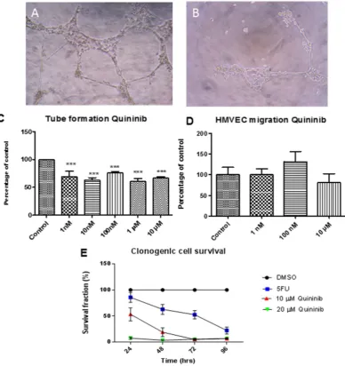
[image:2.595.159.544.48.446.2]Quininib reduces endothelial tubule formation and cancer cell survival.
In dermal-derived human microvascular endothelial cells HMVEC-D, concentrations of 1 nM to 10 μ M quininib reduced tubule formation,www.nature.com/scientificreports/
(p < 0.001) (Fig. 2C). The migration rate of HMVEC-Ds was not affected by 10 μ M quininib (Fig. 2D). Using clonogenic assays, 10 μ M and 20 μ M quininib significantly reduced cell survival (p < 0.0001) compared to vehicle control (Fig. 2E).
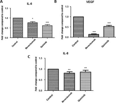
Quininib reduces VEGF, IL-6 and IL-8 secretion in a human CRC explant model.
We measured the effects of quininib on the protein secretions of angiogenic growth factors and inflammatory cytokines in humanex vivo colorectal cancer explants from 40 patients (10 per stage) with CRC. Treatment with 10 μ M quininib resulted in significant reduction in secretions of important angiogenic mediators IL-6 (37.8%), VEGF (47.3%) and IL-8 (13.2%), (Fig. 3A–C respectively). Treatment with 10 μ g/ml bevacizumab also resulted in the reduced secretion of IL-8, IL-6 and VEGF. The secretion of other mediators including ENA-78, GROα , MCP-1, TNF and IL-1β were also significantly reduced following quininib treatment (all p < 0.01, Supplementary Fig. S1). Quininib elicited similar effects in decreasing the above analytes across all stages of CRC (stages I–IV).
Quininib reduces tumour growth and bioluminescence in a colorectal cancer mouse
model.
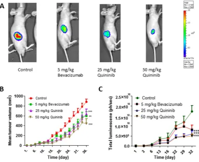
We created a xenograft model of colorectal cancer by injecting 2.5 × 10⁶ HT-29-luc2 cells into Balb/Cnu/nu mice. When tumours reached 100 mm3, we randomised the animals into treatment groups comprising
[image:3.595.160.549.46.459.2]5 mg/kg bevacizumab, 25 mg/kg quininib or 50 mg/kg quininib. We measured tumour growth using calliper
www.nature.com/scientificreports/
measurements and optical imaging (bioluminescence, BLI). Treatment with 50 mg/kg quininib resulted in signif-icant decrease in tumour volume, as measured using callipers, compared to control (average final measurements: 442.1 mm3 vs. 881.6 mm3, p < 0.001, Fig. 4B). Treatment with 5 mg/kg bevacizumab and 25 mg/kg quininib
also significantly reduced tumour volume compared to control (average final measurements: 559.3 mm3 and
639.8 mm3 respectively, p < 0.001). Treatment with 50 mg/kg quininib did not significantly reduce tumour
vol-ume compared to 5 mg/kg bevacizumab or 25 mg/kg quininib. Similarly, BLI signals were lowest in the 50 mg/kg quininib group compared to control (p < 0.001, Fig. 4C). BLI was also lower in the 25 mg/kg quininib and 5 mg/kg bevacizumab groups (all p < 0.001, Fig. 4B) and was not significantly different from 50 mg/kg quininib.
Quininib reduces angiogenic signalling in a xenograft model.
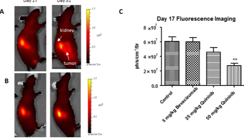
We used a fluorescent probe which binds to αvβ3 integrin in vivo and acts as a surrogate of tumour-associated angiogenesis. This probe was injected on days 17 and 31 of the study (10 and 24 days after initiation of treatments) and fluorescence was measured 4 hours after injection. We found that fluorescence was significantly reduced in the 50 mg/kg quininib (p < 0.01) com-pared to control on day 17 (Fig. 5A–C). Fluorescence was not significantly reduced in the 5 mg/kg bevacizumab or 25 mg/kg quininib groups on day 17 (Fig. 5C). When we repeated fluorescent imaging on day 31 (24 days after the initiation of treatment), there were no significant differences in any of the groups compared to control (Supplementary Fig. S2).Quininib reduces angiogenic gene expression in xenografts.
Gene expression changes in HT-29-luc2 cells treated with bevacizumab or quininib were screened using the RT2 profiler human angiogenesisarray. Of the 84 genes analysed, quininib altered the expression of 31 genes (37%) and bevacizumab altered 17 genes (20%) (Supplementary Fig. S3).
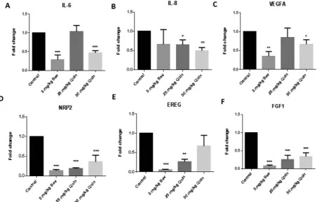
[image:4.595.156.549.47.393.2]We examined the expression of genes closely associated with angiogenesis in CRC xenografts by quantitative PCR. Quininib treatment (50 mg/kg) resulted in the reduced expression of interleukin-6 (IL6), interleukin-8 (IL8), vascular endothelial growth factor A (VEGFA), neuropilin-2 (NRP2) and fibroblast growth factor 1(FGF1)
www.nature.com/scientificreports/
(Fig. 6A–F). Bevacizumab treatment (5 mg/kg) resulted in the reduced expression of IL6, IL8, VEGFA, NRP2,
EREG (epiregulin) and FGF1.
Discussion
We conducted a chemical screen which detected quininib, a compound with anti-angiogenic activity in zebrafish embryos. In vitro, quininib reduced both survival of HT-29-luc2 CRC cells and endothelial cell tubule formation but did not reduce HMVEC-D migration rate. We cultured human colorectal tumours ex vivo and found that the secretion of different angiogenic and inflammatory mediators were reduced by quininib. Quininib reduced tumour growth and bioluminescent activity compared to vehicle control in a CRC xenograft model. In addition,
αvβ3 integrin fluorescent probe signal was reduced by quininib ten days after initiation of treatment. We further demonstrated the anti-angiogenic capacity of quininib by examining gene expression of angiogenic factors in xenograft tumours and found that the expression of IL6, IL8, NRP2, VEGFA and FGF1 was reduced by quininib treatment.
Quininib, a quinoline, detected in an unbiased small molecule chemical screen, reduced intersegmental vessel formation in Tg [fli1: EGFP] zebrafish embryos, a transgenic model allowing easy visualization of developing vasculature7,15–17. We observed reduced larval lengths when embryos were treated with 10 μ M quininib at 48 hpf.
This effect has been observed in other reported screens of anti-angiogenic agents in zebrafish where compounds were added between 6–10 hpf, and is regarded as an expected effect of inhibiting angiogenesis, rather than a toxic effect18,19. Zebrafish embryos undergo considerably rapid morphogenetic processes up to 48 hpf and small
molecule screens have to capacity to detect system-specific toxicities as early as 24 hpf8,20. Aside from pericardial
edema, another recognised anti-angiogenic effect, larvae treated with quininib did not demonstrate any addi-tional behavioural or morphological deficits, indicating that quininib is non-toxic. Previously, using a zebrafish screening approach, Ewan et al. identified rosuvastatin, a small molecule inhibitor of Wnt signalling exhibiting anti-angiogenic and anti-tumour properties in prostate cancer21,22. This study validates the use of zebrafish in
[image:5.595.157.550.46.366.2]screening anti-angiogenic compounds due to the high functional conservation between zebrafish and human vascular development23,24.
Figure 4. Quininib reduces tumour growth in a xenograft model of colorectal cancer. (A) Representative images of bioluminescent images taken from animals treated with vehicle control, 5 mg/kg bevacizumab, 25 mg/kg quininib and 50 mg/kg quininib. (B) Graph of tumour volume (mm3) shows reduced tumour development
www.nature.com/scientificreports/
Quininib did not affect endothelial cell migration rate but did reduce tubule formation in human endothe-lial cells although it possible that higher concentrations may exert a dose dependent effect. However, 10 μ M quininib was sufficient to produce anti-angiogenic effects in multiple models including zebrafish and human
ex vivo explants. In preclinical studies, bevacizumab reduces human endothelial cell VEGF165 – induced
growth and migration when stimulated with the specific ligand of this monoclonal antibody, and treat-ment with bevacizumab in the absence of VEGF165 –stimulation does not reduce endothelial cell growth or
migration25,26. The tubule formation assay, as a complex multi-step process, represents the reorganization
stage of physiological angiogenesis27,28. It is, therefore, a superior in vitro functional assay to test novel
anti-angiogenic compounds.
Regorafenib, a multi-tyrosine kinase inhibitor known to prolong survival in metastatic colorectal cancer, reduces the cell proliferation rate of a variety of cancer cell lines in vitro including a potent effect on a GIST (gas-trointestinal stromal tumour) cell line with only a modest effect on a CRC cell line5,29. This may be explained by
the relative sensitivities of selected cell lines to certain therapeutic agents or it may highlight the discrepancies that exist between in vitro assays and clinical findings.
We used human CRC explants to assess quininib’s effect on the secretion of angiogenic and inflammatory factors as this model has previously detected abundant secretion of these factors in the colorectal tumour microenvironment29–32. In this study, the secretion of important angiogenic and inflammatory mediators were
significantly reduced to different levels when ex vivo explants were treated with quininib. In addition, all data were normalized to individual explant protein content thereby accounting for any differences in cell content in the explant tissues. Overall, this indicates that these inhibitory effects were not simply due to reduced tissue size or reduced cellular content.
Explants contain the key components of the tumour microenvironment including endothelial and inflam-matory cells which engage in complex crosstalk resulting in a balance favouring/opposing angiogenic activ-ity. Bevacizumab affects serum levels of angiogenic growth factors/cytokines and serum levels of IL-6, IL-8, IL-10, epidermal growth factor and macrophage-derived chemokine may predict clinical responses in patients with metastatic colorectal cancer33. Given the economic costs of administering anti-angiogenic agents, much
interest lies in identifying biomarkers which could detect those who might benefit from anti-angiogenic therapies34,35.
We used a xenograft CRC model to demonstrate that quininib decreased tumour growth and biolumines-cent activity in vivo. Preliminary studies had shown that quininib was well tolerated and 50 mg/kg was the maximum tolerated dose (MTD, data not shown). There was no statistical difference between the effects of the MTD and 50% MTD doses on tumour growth and it is possible that lower doses may have a similar effect. We compared the MTD of quininib with the standard dose of 5 mg/kg bevacizumab used in xenograft studies as the MTD of bevacizumab has not been reported36–38. The approval of bevacizumab used in combination with
cytotoxic chemotherapy in metastatic CRC has led to prolonged overall survival4,39. It is possible that a further
[image:6.595.157.549.49.272.2]synergistic effect on tumour growth could be found if quininib was combined with a fluoropyrimidine-based chemotherapy regimen.
www.nature.com/scientificreports/
To date, regorafenib is the only small molecule anti-angiogenic drug licensed for use in treatment-refractory metastatic CRC5. However, despite the modest survival advantage of regorafenib compared to placebo, it is not
prescribed universally by oncologists due its toxicity profile, therefore there is a role for novel anti-angiogenic agents in this setting40.
In order to examine the specific effects of quininib on angiogenesis in vivo, we used a fluorescent probe specific for αvβ3 integrin. This integrin is a receptor for extracellular matrix proteins and was the first integrin dis-covered to regulate angiogenesis41. Angiogenic activity was reduced after 10 days of treatment with 50 mg/kg
quininib although this anti-angiogenic effect was not seen in those animals treated with bevacizumab. This conflicts with prior data where fluorescence from a probe bound to αvβ3 integrin was shown to be reduced at 7 days with beva-cizumab treatment, however, alternative probe/imaging techniques were used42. It is possible that quininib and
beva-cizumab may have different effects on αvβ3 integrin binding or that timing of 7 vs. 10 days is important in this process. To correlate with our in vitro and ex vivo data, we examined the effects of quininib on angiogenic gene expres-sion in xenografts. These genes (IL6, IL8, NRP2, FGF1, VEGFA) are renowned for their importance in angiogen-esis and we found that their expression was reduced by quininib43–46. Using target profiling, quininib has been
identified as a cysteinyl leukotriene receptor 1 and receptor 2 antagonist47. Although predominantly known for
their role in inflammation, cysteinyl leukotrienes have been reported to have pro-angiogenic activities48–50.
Tumours ultimately become resistant to anti-angiogenic therapies51, therefore, novel anti-angiogenic
com-pounds are needed. In summary, we have identified a novel small molecule anti-angiogenic inhibitor, quininib, which shows activity in vitro, ex vivo and in vivo in CRC models. Future work should focus on identifying addi-tional anti-tumoral effects in combination with chemotherapeutic agents and radiation.
Methods
Chemical library.
Chemicals for screening were purchased from ChemBridge DIVERSet library (California). All compounds were screened at 10 μ M concentration diluted in embryo medium to a final concentration of 0.1% DMSO. [image:7.595.101.554.46.335.2]Zebrafish husbandry.
Zebrafish (Danio rerio) experiments were performed in accordance with local guide-lines (Dept. Health and Children, Ireland). All experimental protocols were approved by the Animal Research Ethics Committee in University College Dublin. Zebrafish were maintained according to standard procedures at 28.5 °C on a 14 hr light/10 hr dark cycle. Embryos were obtained by natural spawning and developmental stages established by time and morphological criteria. Transgenic zebrafish Tg [fli1: EGFP] were used for all experiments.www.nature.com/scientificreports/
Intersegmental vessel assay.
At 6 hours post fertilisation (hpf) prior to vasculogenesis, 5 embryos (chori-onated) were incubated with an individual DIVERSet compound at 10 μ M concentration in each well of a 48 well plate with 0.1% DMSO. At 48 hpf, treated larvae were dechorionated, fixed in 4% paraformaldehyde overnight at 4 °C, washed with PBS and assessed for morphological defects including pericardial oedema using brightfield microscopy. Dose response relationship experiments were performed with 0.1, 1, 2.5, 5, 7.5 and 10 μ M of relevant compound with at least 30 larvae tested per concentration.Imaging.
Brightfield and fluorescent images were taken using an Olympus SZX16 stereo zoom microscope with an Olympus DP71 camera. The larval lengths were calculated from brightfield whole larval images using Cell^F software (Olympus) and intersegmental vessels were manually counted at high magnification when Tg [fli1: EGFP] larvae were imaged using EGFP excitation/emission filters.Cell culture.
HMVEC-D (dermal derived human microvascular endothelial cells, authenticated with alpha actin staining by Clonetics, San Diego, USA) were maintained in complete endothelial cell medium (Clonetics®
EGM®
- 2-MV Bulletkit®
) containing 500 ml of Endothelial Cell Basal Medium-2 supplemented with 0.5 ml hEGF, 0.2 ml hydrocortisone, 0.5 ml GA-1000, 25 ml FBS, 0.5 ml VEGF, 2 ml hFGF-B, 0.5 ml R3-IGF-1, 0.5 ml ascorbic acid in a 37 °C humidified atmosphere of 5% CO2. HMVEC-D cells were used for experiments between passages 4–8.Clonogenic assay.
The HT-29-luc2 Bioware®
Ultra cell line was purchased from PerkinElmer (UK) and maintained in McCoy’s 5A media with L-glutamine (Fisher) supplemented with 10% fetal calf serum. Cells were washed in DPBS and trypsinised using 2 ml TrypLE Express (1X) (Invitrogen). 1.5 × 10³ cells were seeded per wellof a 6 well plate and incubated for 24 hours. Cells were then treated with 10 or 20 μ M of quininib, 5-fluorouracil (5FU) or 0.1% DMSO vehicle for 24, 48, 72 and 96 hours. The drugs were removed and cells were allowed to grow in fresh media for 10 days in total. Clones were then fixed using 4% paraformaldehyde and stained with 0.5% crystal violet solution (Pro-Lab diagnostics PL.7000) at RT for 2 hours. Clone counting was performed using the ColCount system (Oxford Optronix).
Tubule formation assay.
Matrigel (BD sciences) basement membrane matrix was plated in 96 well plates after thawing on ice and allowed to polymerise at 37 °C in 5% CO2 humidified atmosphere for 1 hour. HMVEC-Dcells were trypsinised and resuspended in EGM growth medium. Cells were seeded at a density of 5 × 10⁵ cells/cm2
overnight and then incubated in the presence of quininib for 30 hours at 37 °C in 5% CO2 humidified atmosphere.
Endothelial cell tubule formation was assessed using Olympus CKX41 inverted phase contrast microscopy and photographed (ImageJ software). The tubule analysis was determined from 3 sequential fields (20X magnifica-tion) with a connecting branch between two discrete endothelial cells defined as one tube.
Explant processing and culture.
Explants were obtained from the Department of Histopathology, St Vincent’s University Hospital, Dublin. All experimental protocols were approved by the St Vincent’s University Hospital, Dublin ethics committee and experimental protocols conducted in accordance with approved guide-lines (Irish Council for Bioethics). A piece of colorectal tumour was taken following surgery and stored in a tissue wash buffer which contained PBS supplemented with 4 μ g/ml amphotericin B (Invitrogen, Paisley, UK), 100 U/ml penicillin, 100 μ g/ml streptomycin and 30 μ g/ml gentamicin. Explant tissue pieces from each of the tumours were cut into smaller pieces and placed in pre-warmed RPMI media (supplemented with 20% FCS, 4 μ g/ml ampho-tericin B, 100 U/ml penicillin, 100 μ g/ml streptomycin, 30 μ g/ml gentamicin) and incubated at 37 °C and 5% CO2for 24 hours. The media was replaced with fresh media containing 0.1% DMSO (control), 100 μ g/ml bevacizumab or 10 μ M quininib solutions and cultured for 72 hours.
ELISA (Enzyme Linked ImmunoSorbent Assay).
Tumour conditioning media (TCM) was removed from explant culture and the secretion of cytokines and angiogenic growth factors analyzed by ELISA as per the manufacturers’ instructions. To assess cytokine release, a multiplex kit was used for IL-6, TNF, IL-1β and IL-8 (MSD). Individual ELISA kits were used to assess VEGF, MCP-1, ENA-78 and GROα (R&D Duoset systems). Secretion data for all factors were normalised by explant protein content using the BCA assay (Pierce).Xenograft experiments.
Animal experiments were performed in accordance with local guidelines (Dept. Health and Children, Ireland). All experimental protocols were approved by the Animal Research Ethics Committee in University College Dublin. All animals were obtained from Harlan, UK aged 4–6 weeks and accli-matised for 7 days prior to experimental use. Animals were monitored and scored daily for physical/behavioural changes during the studies. Animals were fed autoclaved water and irradiated food ad libitum and maintained on a 12 hour light/dark cycle. Male athymic mice (Balb/C nu/nu) were subcutaneously injected into the right flank with 2.5 × 10⁶ cells in PBS/matrigel. Animals were optically imaged 1 hour post injection and twice weekly duringthe study. Tumours were measured using digital callipers three times weekly and tumour volumes were calculated according to the following formula:
= × × . .
Tumour volume(mm )3 [(Tumour width)2 (Tumour height) 0 5]
Animals were randomised to receive treatment when tumour volumes reached 100 mm3. Animals received
vehicle control, 25 mg/kg quininib or 50 mg/kg quininib intraperitoneally every 3 days for a maximum of 30 days. Quininib was dissolved in a 6% ethanol/cremaphor solution (Sigma). Animals received clinical-grade 5 mg/kg bevacizumab twice weekly intraperitoneally for a maximum of 30 days52,53. Animals were euthanised if tumours
www.nature.com/scientificreports/
Optical imaging.
Optical imaging was performed using an IVIS spectrum imaging system (PerkinElmer). Animals were anaesthetised by induction with 4–5% isoflurane (Vetflurane, Virbac, UK) and maintained with 1–2% isoflurane during imaging. For bioluminescence studies, luciferin (150 mg/kg, PerkinElmer) was admin-istered intraperitoneally to non-anaesthetised animals 10 minutes prior to imaging. For fluorescence studies, XenoLight RediJect Integrin 750 Probe (2 nmol/100 μ L, PerkinElmer) was administered via tail vein injection to anaesthetised animals 10 and 24 days post commencement of treatments. Excitation and emission wavelengths of 745 nm and 820 nm respectively, were used for the acquisition of the in vivo fluorescent images. Images and quantitative measurements of bioluminescent and fluorescent signals were obtained and analyzed using Living Image Software v3.2 (Caliper).PCR profiler array and Quantitative PCR.
RNA was extracted from tumours using TriReagent (Molecular Research Center, Ohio, USA). cDNA was synthesised using reverse transcriptase enzyme and buffer (Bioline, Kilkenny, Ireland) and random primers (Invitrogen. CA, USA). The RT2 profiler Human AngiogenesisPCR array (Super Array Bioscience Corp, USA) was used to analyse the expression of 84 genes related to angi-ogenesis in HT-29-luc2 cells treated with 100 μ g/ml bevacizumab or 10 μ M quininib. Real time PCR was per-formed in triplicate samples on ABI Prism 6500 (ABI Biosystems, CA, USA) for 40 cycles and the threshold cycle (Ct) calculated for each sample relative to the 18s ribosomal RNA control expression. Specific primers were used for VEGFA (Hs00900055_m1), EREG (Hs00914313_m1), NRP2 (Hs00187290_m1), FGF1 (Hs00265254_m1),
IL6 (Hs00985639_m1) and IL8 (Hs01567912_m1) (ABI Biosystems, CA, USA).
Statistical analysis.
Statistical analyses were performed using GraphPad Prism for Windows (version 5.01). Differences between means from multiple groups were assessed by using one way analysis of variance (ANOVA) with the posthoc Dunnett’s multiple comparison test to look for individual differences compared to the control group. For bioluminescence measurements comparing two groups, Mann-Whitney U test was used. Paired data were compared using Wilcoxon signed rank test. All error bars reflect the standard error of the mean. Statistical significance of p < 0.05 is represented as *p < 0.01 as ** and p < 0.001 as ***.References
1. Ferlay, J. et al. Cancer incidence and mortality patterns in Europe: estimates for 40 countries in 2012. Eur. J. Cancer49, 1374–1403 (2013). 2. Asghar, U., Hawkes, E. & Cunningham, D. Predictive and prognostic biomarkers for targeted therapy in metastatic colorectal cancer.
Clin. Colorectal Cancer9, 274–281 (2010).
3. Vale, C. L. et al. Does anti-EGFR therapy improve outcome in advanced colorectal cancer? A systematic review and meta-analysis. Cancer Treat. Rev.38, 618–625 (2012).
4. Hurwitz, H. et al. Bevacizumab plus irinotecan, fluorouracil, and leucovorin for metastatic colorectal cancer. N. Engl. J. Med.350, 2335–2342 (2004).
5. Grothey, A. et al. Regorafenib monotherapy for previously treated metastatic colorectal cancer (CORRECT): an international, multicentre, randomised, placebo-controlled, phase 3 trial. Lancet381, 303–312 (2013).
6. Zon, L. I. & Peterson, R. T. In vivo drug discovery in the zebrafish. Nat. Rev. Drug Discov.4, 35–44 (2005).
7. Tran, T. C. et al. Automated, quantitative screening assay for antiangiogenic compounds using transgenic zebrafish. Cancer Res. 67, 11386–11392 (2007).
8. Peterson, R. T., Link, B. A., Dowling, J. E. & Schreiber, S. L. Small molecule developmental screens reveal the logic and timing of vertebrate development. Proc. Natl. Acad. Sci.97, 12965–12969 (2000).
9. Lawson, N. D. & Weinstein, B. M. In vivo imaging of embryonic vascular development using transgenic zebrafish. Dev. Biol.248, 307–318 (2002).
10. Jacoby, E. & Mozzarelli, A. Chemogenomic strategies to expand the bioactive chemical space. Current medicinal chemistry16, 4374–4381 (2009).
11. Letamendia, A. et al. Development and validation of an automated high-throughput system for zebrafish in vivo screenings. PloS one 7, e36690 (2012).
12. Kennedy, B., Alvarez, Y. & O’Sullivan, J. Patent: Anti-angiogenic compound US8916586 B2 (2014).
13. White, R. M. et al. DHODH modulates transcriptional elongation in the neural crest and melanoma. Nature471, 518–522 (2011). 14. Sun, Q., Heilmann, J. & Konig, B. Natural phenolic metabolites with anti-angiogenic properties - a review from the chemical point
of view. Beilstein J. Org. Chem.11, 249–264 (2015).
15. Isogai, S., Horiguchi, M. & Weinstein, B. M. The vascular anatomy of the developing zebrafish: an atlas of embryonic and early larval development. Dev. Biol.230, 278–301 (2001).
16. Gore, A. V. et al. Vascular development in the zebrafish. Cold Spring Harb. Perspect Med. 2, a006684 (2012).
17. Galvin, O. et al. A sustained release formulation of novel quininib-hyaluronan microneedles inhibits angiogenesis and retinal vascular permeability in vivo. J. Control Release, 233, 198–207 (2016).
18. Chimote, G. et al. Comparison of effects of angiogenic agents in the zebrafish efficacy-toxicity model for translational anti-angiogenic drug discovery. Drug Des. Devel. Ther. 8, 1107–1123 (2014).
19. Chan, J., Bayliss, P. E., Wood, J. M. & Roberts, T. M. Dissection of angiogenic signaling in zebrafish using a chemical genetic approach. Cancer cell1, 257–267 (2002).
20. Kimmel, C. B. et al. Stages of embryonic development of the zebrafish. Dev. Dyn.203, 253–310 (1995).
21. Ewan, K. et al. A useful approach to identify novel small-molecule inhibitors of Wnt-dependent transcription. Cancer Res. 70, 5963–5973 (2010).
22. Wang, C. et al. Rosuvastatin, identified from a zebrafish chemical genetic screen for antiangiogenic compounds, suppresses the growth of prostate cancer. Eur. Urol.58, 418–426 (2010).
23. Habeck, H. et al. Analysis of a zebrafish VEGF receptor mutant reveals specific disruption of angiogenesis. Curr. Biol.12, 1405–1412 (2002).
24. van Rooijen, E. et al. von Hippel-Lindau tumor suppressor mutants faithfully model pathological hypoxia-driven angiogenesis and vascular retinopathies in zebrafish. Dis. Model. Mech. 3, 343–353 (2010).
25. Falcao, M., Soares, R., Azevdep. I., Falcao-Reis, F. & Carneiro, A. Effects of bevacizumab on HUVEC migration and capillary formation. Acta Ophthal.85, Supplement s240 (2007).
www.nature.com/scientificreports/
27. Arnaoutova, I. & Kleinman, H. K. In vitro angiogenesis: endothelial cell tube formation on gelled basement membrane extract. Nat. Protoc.5, 628–635 (2010).
28. Francescone, R. A. 3rd, Faibish, M. & Shao, R. A Matrigel-based tube formation assay to assess the vasculogenic activity of tumor cells. J. Vis. Exp. 55, 3040 (2011).
29. Wilhelm, S. M. et al. BAY 43-9006 exhibits broad spectrum oral antitumor activity and targets the RAF/MEK/ERK pathway and receptor tyrosine kinases involved in tumor progression and angiogenesis. Cancer Res. 64, 7099–7109 (2004).
30. Michielsen, A. J. et al. Tumour tissue microenvironment can inhibit dendritic cell maturation in colorectal cancer. PloS One6, e27944 (2011).
31. Michielsen, A. J. et al. Inhibition of dendritic cell maturation by the tumor microenvironment correlates with the survival of colorectal cancer patients following bevacizumab treatment. Mol. Cancer Ther.11, 1829–1837 (2012).
32. Michielsen, A. J., O’Sullivan, J. N. & Ryan, E. J. Tumor conditioned media from colorectal cancer patients inhibits dendritic cell maturation. Oncoimmunology1, 751–753 (2012).
33. Abajo, A. et al. Identification of predictive circulating biomarkers of bevacizumab-containing regimen efficacy in pre-treated metastatic colorectal cancer patients. Br. J. Cancer.107, 287–290 (2012).
34. Clarke, S. et al. An Australian translational study to evaluate the prognostic role of inflammatory markers in patients with metastatic ColorEctal caNcer Treated with bevacizumab (Avastin) [ASCENT]. BMC cancer13, 120 (2013).
35. Martin, P. et al. Predicting response to vascular endothelial growth factor inhibitor and chemotherapy in metastatic colorectal cancer. BMC cancer14, 887 (2014).
36. Federal Drug Administration. Review and Evaluation of Toxicology Data (Avastin). BLA STN # 125085, http://www.accessdata.fda. gov/drugsatfda_docs/nda/2004/STN-125085_Avastin_Toxr.pdf (2004).
37. Higgins, B. et al. Antitumor activity of capecitabine and bevacizumab combination in a human estrogen receptor-negative breast adenocarcinoma xenograft model. Anticancer Res. 27, 2279–2287 (2007).
38. Kolinsky, K. et al.In vivo activity of novel capecitabine regimens alone and with bevacizumab and oxaliplatin in colorectal cancer xenograft models. Mol. Cancer. Ther.8, 75–82 (2009).
39. Kabbinavar, F. F. et al. Addition of bevacizumab to bolus fluorouracil and leucovorin in first-line metastatic colorectal cancer: results of a randomized phase II trial. J. Clin. Oncol.23, 3697–3705 (2005).
40. Argiles, G. et al. Regorafenib plus modified FOLFOX6 as first-line treatment of metastatic colorectal cancer: A phase II trial. Eur. J. Cancer51, 942–949 (2015).
41. Brooks, P. C., Clark, R. A. & Cheresh, D. A. Requirement of vascular integrin alpha v beta 3 for angiogenesis. Science264, 569–571 (1994). 42. Kossodo, S. et al. Dual in vivo quantification of integrin-targeted and protease-activated agents in cancer using fluorescence
molecular tomography (FMT). Mol. Imaging Biol.12, 488–499 (2010).
43. Guo, Y. et al. Interleukin-6 signaling pathway in targeted therapy for cancer. Cancer Treat. Rev.38, 904–910 (2012).
44. Ning, Y. et al. Interleukin-8 is associated with proliferation, migration, angiogenesis and chemosensitivity in vitro and in vivo in colon cancer cell line models. Int. J. Cancer128, 2038–2049 (2011).
45. Pan, Q. et al. Blocking neuropilin-1 function has an additive effect with anti-VEGF to inhibit tumor growth. Cancer cell11, 53–67 (2007). 46. Compagni, A. et al. Fibroblast growth factors are required for efficient tumor angiogenesis. Cancer Res.60, 7163–7169 (2000). 47. Reynolds AL et al. Phenotype-based discovery of 2-[(E)-2-(quinolin-2-YL)vinylphenol as a novel inhibitor of developmental and
pathological ocular angiogenesis. J. Biol. Chem.291, 7242–7255 (2015).
48. Kanayasu, T. et al. Leukotriene C4 stimulates angiogenesis in bovine carotid artery endothelial cells in vitro. Biochem. Biophys. Res. Commun.159, 572–578 (1989).
49. Tsopanoglou, N. E., Pipili-Synetos, E. & Maragoudakis, M. E. Leukotrienes. C4 and D4 promote angiogenesis via a receptor-mediated interaction. Eur. J. Pharmacol.258, 151–154 (1994).
50. Burke, L., Butler, C. T., Murphy, A., Moran, B., Gallagher, W. M., O’Sullivan, J. & Kennedy, B. N. Evaluation of Cysteinyl Leukotriene Signaling as a Therapeutic Target for Colorectal Cancer. Front. Cell Dev. Biol.4(103), doi: 10.3389/fcell.2016.00103 (2016). 51. Bergers, G. & Hanahan, D. Modes of resistance to anti-angiogenic therapy. Nat. Rev. Cancer8, 592–603 (2008).
52. Gerber, H. P. & Ferrara, N. Pharmacology and pharmacodynamics of bevacizumab as monotherapy or in combination with cytotoxic therapy in preclinical studies. Cancer Res. 65, 671–680 (2005).
53. von Baumgarten, L. et al. Bevacizumab has differential and dose-dependent effects on glioma blood vessels and tumor cells. Clin. Cancer. Res.17, 6192–6205 (2011).
Acknowledgements
Adrian Murphy was funded by a Newman Fellowship from the University College Dublin Newman Foundation to conduct this work. This work was also funded by a Science Foundation Ireland TIDA grant 11/TIDA/B1966.
Author Contributions
A.G.M. carried out experimental work, wrote the main manuscript text and prepared the Figures. R.C. assisted with in vivo work and RT-PCR experiments. A.G.M., E.C. and W.M.G. assisted with in vivo work. M.T. and C.T.B. assisted with in vitro experiments. A.L.R. assisted with zebrafish experiments. K.S., D.O’D. and D.F. assisted with
ex vivo explant experiments. B.N.K. and J.O’S. co-supervised this work to completion. All authors have read and approved the manuscript.
Additional Information
Supplementary information accompanies this paper at http://www.nature.com/srep
Competing financial interests: Quininib is the subject of the patent:Anti-Angiogenic compounds. WO 2012/095836 A1, granted Dec 2014.
How to cite this article: Murphy, A. G. et al. Preclinical validation of the small molecule drug quininib as a novel therapeutic for colorectal cancer. Sci. Rep.6, 34523; doi: 10.1038/srep34523 (2016).
This work is licensed under a Creative Commons Attribution 4.0 International License. The images or other third party material in this article are included in the article’s Creative Commons license, unless indicated otherwise in the credit line; if the material is not included under the Creative Commons license, users will need to obtain permission from the license holder to reproduce the material. To view a copy of this license, visit http://creativecommons.org/licenses/by/4.0/