A thesis submitted to the University of Southampton for the degree of Doctor of Philosophy
ACKNOWLEDGEMENTS
I am greatly indebted to my supervisor Dr. C. Anthony for the help and encouragement he has given throughout this s tudy.
I also wish to acknowledge the helpful advice of Dr. W.T. Drabble and Professor M. Akhtar, and to thank Professor K. Munday for providing the excellent facilities used in this work.
CONTENTS
Page CHAPTER 1
A review
1.1 Introduction 1
PART A The oxidation and assimilation of
C^-compounds 1
1.2 Bacteria capable of growth on C^-compounds 1
a) Obligate methylotrophs 3
b) Facultative methylotrophs 4
c) Restricted facultative methylotrophs 5 1.3 The oxidation of C^-compounds 10
a) Methane oxidation 11
b) Methanol oxidation 12
c) Formaldehyde and formate oxidation 15 d) Oxidation of N-methyl compounds 16 e) The electron transport chain of methylotrophs 20 1.4 The assimilation of C^-compounds 21 a) The ribulose monophosphate cycle 21
b) The serine pathway 25
1.5 The assimilation of acetyl-CoA produced by the
serine pathway 27
a) Bacteria which form isocitrate lyase 27 b) Oxidation of acetyl-CoA to glyoxylate in
Pseudomonas AMI 29
X X
Page 1.8 The enzymes of poly-^~hydroxybutyrate
biosynthesis 35
1.9 The enzymes of poly-?-hydroxybutyrate
degradation 36
a) Extracellular PHB degradation 36 b) Intracellular PHB degradation 38 1.10 Regulation of poly-^-hydroxybutyrate metabolism 40
PART C Regulation and functions of the TCA cycle
in bacteria 44
1.11 Functions of the TCA cycle 44 1.12 Bacteria lacking a complete TCA cycle 46 a) Gram-negative facultative anaerobes 46 b) Gram-positive facultative anaerobes 48
c) Autotrophic bacteria 48
d) Methylotrophic bacteria 49
1.13 Regulation of citrate synthase activity 51 a) Bacteria which have a complete TCA cycle 51 b) Bacteria which lack a complete TCA cycle 51 1.14 The molecular weights of citrate synthase from
different bacteria 52
CHAPTER 2 Materials and methods
2.1 Materials 55
a) Chemicals 55
b) Preparation of CoA derivatives 57
Page 1 4
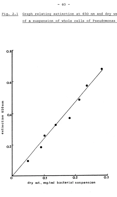
2.2 Purification of U- C-acetate 57 2.3 Media and maintenance of cultures 58 2.4 Growth and harvesting of cultures 59 2.5 Estimation of the dry weight of cells in
suspension 59
2.6 Isolation of mutants of Pseudomonas AMI 61 2.7 Characterisation of the growth response of mutant
strains 62
2.8 Isolation of revertants 63
2.9 Measurement of oxygen uptake by bacterial
suspensions 64
2.10 Preparation of sonic extracts 64
2.11 Protein assay 65
2.12 Enzyme and cytochrome assays 65 a) Enzymes of f-hydroxybutyrate metabolism 66 b) Enzymes of the TCA cycle 66
c) Enzymes of metabolism 67
d) Enzymes of metabolism 67
e) Enzymes of the glyoxylate bypass 68
f) Cytochrome assays 68
2.13 Molecular weight and kinetic studies of citrate
synthase 68
2.14 Partial purification of acetyl-CoA synthetase
from Pseudomonas AMI 70
2.15 Assay of ADP production and adenylate kinase 73 2.16 Amino acid analysis and poly-^-hydroxybutyrate
I V
Page 2.17 Bacterial incorporation of ^^C-acetate 74
2.18 Chromatographic analysis 74
2.19 Detection of radioactive compounds 75 2.20 Co-chromatography of radioactive compounds 7 6
14
2.21 Incorporation of radioactivity from C-acetate
into growing cultures of bacteria 76
CHAPTER 3
The formation of acetyl-CoA during growth of Pseudomonas AMI on ^-hydroxybutyrate, ethanol and C^-compounds
3.1 Introduction 78
3.2 The enzymology of ^-hydroxybutyrate oxidation
to acetyl-CoA 78
a) p-hydroxybutyrate dehydrogenase 79 b) Acetoacetate:succinate CoA transferase 79
c) ^-ketothiolase 81
d) Acetoacetyl-CoA reductase 83
3.3 Factors affecting the poly-p-hydroxybutyrate
content of Pseudomonas AMI 85
3.4 The enzymology of ethanol metabolism to
acetyl-CoA 88
a) Alcohol (methanol) dehydrogenase 88 b) Oxidation of acetaldehyde 88 c) Activation of acetate to acetyl-CoA 90 3.5 The significance of acetyl-CoA synthetase
induction during growth on f-hydroxybutyrate 92 3.6 Identification of the products of acetyl-CoA
Page a) Measurement of ADP production 93 b) Separation of adenylate kinase and
acetyl-CoA synthetase 96
c) Identification of acetyl-CoA 98 3.7 Characterisation of mutant ICT 54 99 3.8 The metabolism of malonate to acetyl-CoA 102 3.9 Oxidation of lactate and pyruvate to acetyl-CoA 103 3.10 Acetyl-CoA synthetase activities in extracts of
Pseudomonas AMI 103
3.11 Summary 105
CHAPTER 4
The assimilation of acetyl-CoA during growth of Pseudomonas AMI on C^-compounds, P-hydroxybutyrate, ethanol and C^-compounds
4.1 Introduction 106
4.2 The malate synthase pathways 106 4.3 Characterisation of mutant ICT 5 110 4.4 Characterisation of mutant ICT 51 114 4.5 Properties of mutant ICT SIR 116 4.6 Properties of mutant PCT 57 119 4.7 The involvement of glycollate during acetyl-CoA
assimilation by the malate synthase pathway 120 4.8 Metabolism of U-^^C-acetate by
f-hydroxybutyrate-grown wild-type Pseudomonas AMI and mutant 20 BL 123 4.9 Metabolism of U-^^C-acetate by
V I
Page 4.10 Incorporation of ^'^C-acetate into cultures of
Pseudomonas AMI and mutant ICT 54 growing on
P-hydroxybutyrate 127
4.11 Summary and discussion 129
CHAPTER 5
Properties of a mutant of Pseudomonas AMI lacking 2-oxoglutarate dehydrogenase: a biochemical basis for obligate methylotrophy
5. 1 Introduction 133
5. 2 Growth and oxidative properties of mutant ICT 41 134 5. 3 Activities of TCA cycle enzymes in mutant ICT 41 138 5. 4 Properties of revertants of mutant ICT 41 143 5. 5 Amino acid accumulation by mutant ICT 41 143
5. 6 Summary and discussion 149
CHAPTER 6
Regulation of the tricarboxylic acid cycle and malate synthase of Pseudomonas AMI
6.1 Introduction 153
6.2 Activities of TCA cycle enzymes during growth of
Pseudomonas AMI on various substrates 153 6.3 Regulation of citrate synthase activity 155 6.4 Regulation of isocitrate dehydrogenase activity 158 6.5 Regulation of malate synthase activity 160
Page CHAPTER 7
Unsolved problems and areas for further investigation
7.1 Introduction 172
7.2 Unsolved problems of metabolism of C^-compounds
by Pseudomonas AMI 172
7.3 Areas for further investigation 174 7.4 Mutants unable to oxidise methanol 176
ABSTRACT i^cu^noF
PHYSIOLOGY AND BIOCEEMISTRY
Doctor of Philosophy
TEE MICROBIAL METABOLISM OF C,,- AND Cg-COMPOUNDS by IAIN JOHN TAYLOR
In the facultative methylotroph, Pseudomonas AM1, the malate synthase pathway has been proposed as a route for the assimilation of growth substrates metabolised to acetate or acetyl-CoA; these include //-hydroxybutyrate, ethanol, malonate,lactate and pyruvate. In this study, the enzymes involved in the metabolism of these compounds to acetyl-CoA have been identified and the role of malate synthase during acetyl-CoA assimilation has been examined by the isolation of mutants lacking the enzyme. The growth properties of one mutant
(ICT 51) indicated that malate synthase activity is required during assimilation of /2-hydroxybutyrate, ethanol and malonate but not for growth on Ci-compounds, lactate or pyruvate. However, results with two further mutants lacking malate synthase activity (iCT PCT $7) suggested that an alternative to the malate synthase pathway may operate under certain conditions.
Radioactive labelling experiments with C-acetate and whole cells of mutant ICT ^4 and mutant 20 BL (lacking hydroxypyruvate reductase) showed that acetyl-CoA (not acetate) is the precursor for oxidation to glyoxylate and that the intermediate formation of glycollate is probably not involved.
Pseudomonas AM1 was shown to possess a complete tricarboxylic acid cycle during growth on all substrates. Some enzymes (isocitrate dehydrogenase, 2-oxoglutarate dehydrogenase and malate dehydrogenase) had lower specific activities during growth on Ci-compounds although a more important site of regulation of ICA cycle activity was at the level of enzyme inhibition. The NADP-specific isocitrate dehydrogenase was subject to concerted inhibition by a combination of glyoxylate and oxaloacetate and the citrate synthase (M.Wt.250,000) was inhibited by NADH. No inhibition of citrate synthase was found with 2-oxoglutarate.
Results with a mutant (ICT 41) lacking 2-oxoglutarate dehydrogenase provided evidence that the lack of this enzyme may be a sufficient biochemical basis for the obligate methylotrophy of some bacteria and demonstrated the importance of a complete TCA cycle for growth of
CHAPTER 1 1.1 Introduction
This Chapter consists of three main Parts; the first is a brief review of the literature concerning the oxida-tion and assimilaoxida-tion of C^-compounds (fully reviewed recently by Quayle, 1972 and Anthony, 1975a). This Part specifically deals with the metabolism of reduced C^-compounds and thus excludes autotrophs which are capable of utilising COg as a sole source of carbon (reviewed by Kelly, 1971). The second Part describes the metabolism and functions of the endogenous energy reserve poly-P-hydroxybutyrate found in many bacteria including those capable of growth on C^-compounds. The final Part of t^iis Chapter is a review of the functions and regulation of the tricarboxylic acid cycle with particular reference to those bacteria which lack a complete oxidative cycle.
PART A
The oxidation and assimilation of C^-compounds 1.2 Bacteria capable of growth on C^-compounds
These bacteria are known as methylotrophs and are
able to grow non-autotrophically at the expense of compounds containing one or more carbon atoms but containing no
carbon - carbon bonds (Colby and Zatitian, 1972) . Through-out this thesis the term ' C^^-compound' is used not only
for compounds containing one carbon atom, but also for compounds containing more than one carbon atom providing they contain no C - C bonds (Table 1.1).
Compounds containing one carbon atom
Compounds containing more than one carbon atom
methane CH* dimethyl ether (CHg) 2° methanol CHgOH dimethylamine (CHg) methylamine CHgNHg trimethylamine (CHg) 3N
formaldehyde HCHO tetramethylammonium (CHg) formate HCOOH trimethylamine N-oxide (CHg) ^NO formamide HCONHg trimethylsulphonium
compounds
3S-bacteria but a number of methylotrophic yeasts have now been isolated. These include species of Candida, Kloekera, Torulopsis, Pichla and Hansensula some of which have been discussed in a review by Cooney and Levine (1972) which concentrates on the use of methanol as a suitable substrate for industrial fermentations by both yeasts and bacteria.
Methylotrophs have long been divided into two major groups: obligate methylotrophs which utilise C^-compounds as their unique growth substrates upon which their growth is absolutely dependent, and facultative methylotrophs which have the added ability to grow on a variety of other
organic compounds (Colby and Zatman, 1972). A third group of methylotrophs has recently become recognised and referred to as 'restricted facultative' methylotrophs. This group of methylotrophs constitutes a group of bacteria with a very restricted range of growth substrates which differ from obligate methylotrophs in their capacity to grow on a few multicarbon (or non-C^) compounds (Colby and Zatman, 1975a).
a) Obligate methylotrophs
Until 1966, all known obligate methylotrophs could grow on methane and methanol, but not on any other carbon source (except dimethyl ether). Examples of these obligate methane-utilisers included Pseudomonas methanica (Leadbetter
methane-utilisers and divided them into five Genera on the basis of morphology, type of resting stage formed and
membrane structure (Table 1.2). This method of classifi-cation was later extended by Lawrence and Quayle (1970) to include Pseudomonas methanica (Methylomonas group), Methanomonas methanooxldans (Methylosinus group) and Methylococcus capsulatus (Methylococcus group). All the groups were shown to possess complex internal membrane structures of two different types. Type I bacteria
(Methylomonas, Methylococcus and Methylobacter) have bundles of disc—shaped vesicles while Type II bacteria (Methylosinus and Methylocystis) have a series of peripheral membranes surrounding the cytoplasm.
More recently obligate methylotrophs have ]be&n isolated which cannot utilise methane as a carbon source. Such
bacteria include Bacterium C2A1, Bacterium 4B6 (Colby and Zatman, 1973), Organism W1 (Dahl, Mehta and Hoare, 1972) and Methylomonas M-15 (Sahm and Wagner, 1975) (Table 1.2). These organisms differ morphologically from the methane-utilisers in that they do not possess internal membrane ultrastruetures.
No obligate methylotroph so far described is capable of growth on formate, and none grow anaerobically with nitrate as terminal oxidant instead of oxygen.
b) Facultative methvlotrophs
5
-malate. All the facultative methylotrophs are Gram-negative and, with the exception of Hyphomicrobium, are able to
grow on the following compounds as well as the C^-compounds shown: ethanol or acetate (or both), pyruvate and lactate, succinate and other dicarboxylic acids and at least one carbohydrate. The pink pseudomonads are the bacteria most frequently encountered and these are usually capable of growth on oxalate. Hyphomicrobium X, a stalked, budding bacterium is included in Table 1.3 although this is an atypical facultative methylotroph. It can only grow
methylotrophically at the expense of ethanol, acetate and ^-hydroxybutyrate and growth on the latter compound is extremely slow with a doubling time of 35 hours (Attwood and Harder, 1974). These growth properties suggest that Hyphomicrobium X may be classified with the restricted
I m .miiu I I I lU iii»iuiii jiiiijii im'Wuiii
facultative methylotrophs discussed below. Another unusual feature of this organism is its ability to grow anaerobically with nitrate as a terminal oxidant (Attwood and Harder,
1972).
No facultative methylotroph is capable of growth on methane and the well-defined internal membrane structures found in methane-oxidisers have not been observed in any facultative methylotroph so far described.
c) Restricted facultative methylotrophs
methylotrophs (from Anthony, 1975a)
Organism
*Methane-utilisers (Type I):
Methylomonas
(Pseudomonas methanica) Methylobacter
Methylococcus
Substrates supporting growth
methane, methanol
Reference
Quayle, 1972
*Methane-uti U s e r s (Type II):
Methylocystis Methylosinus
(Methanomonas methano-oxidans)
methane, methanol Quayle, 1972
Obligate methylotrophs unable to use methane : Bacterium 4B6
Bacterium C2A1 Organism W1
Methylomonas M-15
methylamine (not methanol or formate methylamine,
Colby and Zatman, 1973 Colby and methanol (not formate) Zatman, 1973 methylamine, methanol
(not formate)
methanol (not methyla-mine or formate)
Dahl et , 1972
Sahm and Wagner, 1975
*The generic names given are those suggested by Whittenbury et , (1970); the names in parentheses refer to previously described methane utilisers now included in these genera.
In addition to those compounds listed dimethyl ether
supports growth of all the methane-utilisers and dimethylamine and trimethylamine supports growth of Bacterium C2A1 and
ro
a\
1—!
Cfl (U •
as o 1—1 LD d fd r - CD (Ti n 4-1 1—1 CD CD
4-1 >1 CD M
d « CD
0 T)
k 4-1 rt!
d K
vo < B M w o M -P O
rH >
4-1 0) 0) > •H +J (0 -p r H 3 O (0 44 m o x : 4J 3: O M Cn Cn C -r4 4J k o g 0) w 0) 4J tC to fd d 0
Sj c
0, o A M fd o Q) C O X ! 4J •H & m t ! a 0 o r u
fO r—1
«b, G c d
CD 1—1
+3
rtS fd g r-H M CD t J VD
(0
>1 N g d d T l M
(0 fd •fc. (C <Ti d r - (C
0 "O N # (d rH (t5 cn N
o d rH 1 12 •H rH
T ) o
fO T ) f d | n3 «• N T )
D C T l Cfi d 4-1 •- d
d >1 fd 4-l| d EH a -H in OS
iQ d CDI fcj 6 rH CD
0 >1 •. +3 iy> rH >1
t—1 >1 k P O 15 T l -H CD A m
CD 4-1 lO rH r~" T ! d fO rc! CD 4J t—1
CD d Ti 0 m nS 0 CTi f ! 4J 4-1 n3 O
a< rtl r—1 U M M 1 1 CO CO c/p S U rH
0 O m d
d g g g g
g 4-1 4J 4-1 4J
4-1 d d d d d d
0 g g g g
d 4-1 T ) •P •P u
4-1 4-1
d d 0 d d d 0
g g d G fi g
4-1 rs • 3 T3
1—1 rH
O o
d d
fd fd
i H t—1 rH 1—1 X !
0 O 0 0 CD 4-> 4-1
d d d d 4J CD CD
(C fd fd fd fd g g
r d g
4-1 4J 4J 4J SH 4-1 4->
CD CD CD CD o 0 0
g g 6 g d d
g
rH ». o
CD CD (D CD CD d CD CD
d d d d d fd d CD d CD
-H -H •rH "H -H rC •H 4J •H -P
g g g g e - p e fd g g
(0 CD fd CD (d CD (0 (U fd CD fd g ro g
1—1 4-1 (—1 4-1 rH 4J rH 4J r - l g 1—1 M rH V4
> , fd >1 fd >1 fd >1 (d >1 >1 o >1 o
- d g g g ^ e r d 4-) , d 4H r d 4H
4J M 4-1 SH 4-1 U 4-1 M 4-1 0 4-1 4-1
CD O CD 0 CD 0 CD 0 CD d CD SH (D k
E w g 44 g 4H g 4-1 g g O g 0
4-1 o OJ 4-1 ro g M O 4-1 (U
C
rH E
O (0
d iH fO >1
A ^ 4J 4-)
0) CD
6 e
w (1, cn
4-1 Cfi t 3
m fd 1—1 r - CN
Xi g d s CN <
0 3 0 < S ( n
'r4 g
rQ o m CO in
0 fd fd fd
m
.
u 0 d d da o CD o 0 0
1—1 m •r-i CQ g g g
-r4 g CU o o 0
CD d 0 T l T ) t 3
I—! fd r d 0 0 0
Xi a d CD CD CD
fd U > - H (0 If) CO
o m CM A A A
in T3 fd q o e 13 0 0) W A CD > -r4 4-1 (C cn CD d I i u t—I o d (0 4J CD e CD d •H
§
1—Io <u
4-) d td fO N T5 d rtS d o - p Duro 6
r-tS CTv
m M CD 4J d B CD 4J >1 fC s g
CD O
e 4-1 4-1 o d CD d -rH
^ o
I—I d
>1 (0
^3 rd
4J 4-1 CD (U
G e
g CO < - o
13 fd S s u 13 fd rH CM
CD S (U A pq m
4-1 0} w w t s 4-1 un i n
d fd fd fd fd d CD
CD d d d d CD rH s E
g 0 o 0 0 g -H p p
Cn g g g g t j i 4-1 •H -rH
•H 0 o o o •H 0 5h iH
A n j ' O T l 13 0 , g CD CD
I 0 0 0 0 1 1 4-J 4-1
d CD CD CD 0) d d O U
0 m CO m CO 0 0 fd fd
methylotrophs (from Anthony 1975a)
Hyphomicrobium sp. grow on only those substrates listed and on ethanol, acetate and p - h y d r o x y b u t y r a t e . All the
other facultative methylotrophs are able to grow on the following: acetate and/or ethanol; pyruvate and lactate; at least one carbohydrate; succinate and other dicarboxylic acids. The pink pseudomonads are also usually able to grow on oxalate. All the bacteria listed use the serine pathway of assimilation.
Abbreviations used: dmn, dimethylamine; tmn, trimethylamine; tern, tetramethylammonium compounds; tmo, trimethylamine-N-oxide; tms, trimethylsulphonium compounds.
Table 1.4 Substrates supporting the growth of the restricted facultative methylotrophs (from Colby and Zatman,
1975a) Substrate compounds: tetramethylammonium trimethylaraine trimethylamine N-oxide dimethylamine methylamine methanol Type M W3A1 W6A
+ + + + + + Type L S2A1 PM6
+ + + + + + + + + Non-C^ compounds glucose gluconate citrate glutamate alanine betaine nutrient agar +
N.T, N.T.
Two of these isolates (W3A1 and W6A) grew only on glucose
out of 56 non-C^ compounds tested. Another two isolates
(S2A1 and Bacillus RM6) gr(^v cm betaine, D-glucose,
gluconate, citrate, L-alanine and nutrient agar (Table 1.4).
Such bacteria are therefore clearly distinguished from
the typical facultative and obligate methylotrophs previously
described. For convenience, the organisms with the
more-restricted range of growth substrates (W3A1 and W6A) were
designated type 'M' less-restricted organisms
(PM6 and S2A1) designated type 'L'. The type 'L' organisms
were positive while the type 'M' organisms
Gram-negative. These restricted facultative methylotrophs could
not utilise methane and did not possess internal membrane
structures.
1.3 The oxidation of C^-compounds
of the energy satisfaction of aerobic
methylo-trophs arises by way of the complete oxidation of the
growth substrate to carbon dioxide and water. substrates
are assimilated into cell material at the oxidation level
of formaldehyde which can exist in the free state or bound
as a tetrahydrofolate derivative. An outline of the
oxidation of C^-compounds is given below followed by a
discussion of some of Uus individual reactions:
CH^ f C H g O H t'HCHO' » HCOOH » 00%
N-methyl CELL MATERIAL
1 1
-a) Methane oxidation.
The difficulty in preparing active cell-free extracts has been a major obstacle in the study of methane oxidation. However Ferenci (1974) and Ribbons (1975) have recently
demonstrated NADH and Og-dependent methane oxidation by particulate fractions of cell-free extracts prepared from obligate methane-utilisers. With Methylococcus capsulatus
(Type I membrane structure) Ribbons was able to demonstrate the simultaneous disappearance of methane and 0^ concomitant with the oxidation of NADH. The stoichiometry of the reaction however was not established as the presumed initial product of methane oxidation (methanol) was further oxidised to formate by the preparation which also contained high NADH-oxidase activity. Using similar particulate fractions of Pseudomonas methanica (Type I) and Methylosinus trichosporium
(Type II) Ferenci showed that NADH oxidation and oxygen uptake formed a 1:1 ratio consistent with the involvement of a mono-oxygenase (methane hydroxylase) catalysed reaction. Carbon monoxide was also oxidised by this system and it was suggested that a common component of the mono-oxygenase system was involved in both methane and CO oxidation.
That molecular oxygen is involved in methane oxidation was previously demonstrated by Higgins and Quayle (1970) who detected after incubation of whole cells of
p. methanica and Methanomonas methanooxidans (Methylosinus)
18_ 18,
wi ith methane and Og. By contrast, CH^ OH was not found when these cells were incubated with methane and H^ 0.
(methane hydroxylase) reaction for methane oxidation to methanol:
CH^ + Og + NADH + H* » CH^OH + NAD* + H^O
It is of interest to note that a similar mono-oxygenase appears to catalyse methane oxidation in methane-utilisers with different membrane systems and assimilation pathways. The demonstration of methane hydroxylase activity in the particulate fractions of both types of methane utiliser, and the observation that only those methylotrophs with complex internal membrane systems can oxidise methane suggests that these membranes play a specific role during methane oxidation or associated electron transport and ATP synthesis.
b) Methanol oxidation
Two enzymes are known to be responsible for methanol oxidation to formaldehyde in micro-organisms, one exclusively in yeast and the other in bacteria.
In the methylotrophic yeasts Kloekera sp. No 2201 (Ta.ni, Miya and Ogata, 1972) and Candida boidinii (Sahm and Wagner, 1973) methanol is oxidised by an inducible FAD-dependent alcohol oxidase which has a broad substrate
.13
2H„0 CHgOH + Og » HCHO + HgOg
An inducible catalase was also present in extracts of methanol-grown C. boidinii and the overall mechanism for methanol oxidation by this organism was therefore represented as follows (Roggenkamp, Sahm and Wagner, 1974):
HCHO
CHgOH — » HgOg »HCHO
CHgOH
Electron micrographs show that methanol-grown C. boidinii contain microbodies with crystalline inclusions not found
during growth of the yeast on other substrates. The
crystalline inclusions were also absent from a mutant lack-ing methanol oxidase activity even when incubated in medium containing methanol (Sahm, Roggenkamp, Wagner and Hinklemann, 1975). That the enzyme is associated with these
micro-bodies was further demonstrated by the finding of almost
all the methanol oxidase activity in the particulate fraction of cell-free extracts.
Methanol is oxidised in bacteria by an enzyme first described by Anthony and Zatman (196 4) from Pseudomonas
4 .
M 27. The methanol dehydrogenase is independent of NAD or NADP* and extracts are absolutely dependent for activity on the artificial hydrogen acceptor phenazine methosulphate
Table 1.5 A comparison of the methanol-oxldlsinq enzymes of bacteria and yeasts
trivial name molecular weight prosthetic group
activators inhibitors specificty
Bacteria
methanol dehydrogenase 120,000 - 146,000
pteridine
NHg, CHgNHg
primary alcohols
^^l"^ll^;formaldehyde Km for methanol 0.02mM
references
Yeast
methanol oxidase
600,000
FAD
Anthony and Zatman, 1965, 1967
p-CMB, KCN
short-chain primary alcohols (C^-Cg) l-2mM
Sahm and Wagner, 1973
15
(Anthony and Zatman, 1965). This enzyme is also responsible for ethanol oxidation to acetaldehyde during growth of
several facultative methylotrophs on this compound. The
molecular weight is in the region of 120,000 and the prosthetic group appears to be a pteridine. The uniformity among bact-eria with respect to methanol oxidation has been demonstrated with the finding that the purified enzyme from the obligate methylotroph M. capsulatus is almost identical with the methanol dehydrogenase from facultative methylotrophs (e.g. Pseudomonas M27) (Patel, Bose, Mandy and Hoare, 1972).
The physiological importance of the enzyme has also been demonstrated with the isolation of mutants which lack meth-anol dehydrogenase and can neither oxidise nor grow on methanol (Dunstan, Anthony and Drabble, 1972a).
The properties of these two methanol oxidising enzymes are summarised in Table 1.5.
c) Formaldehyde and formate oxidation
Formaldehyde, either in the free state or bound, is the oxidation product of methanol and N-methyl compounds, and it is at this level of oxidation that C^-compounds are assimilated into cell-material. There is considerable variation in the enzymes capable of formaldehyde oxidation by methylotrophs:
(i) NAD^-linked aldehyde dehydrogenase (Rung and Wagner, 1970).
(iii) NAD(P)-independent aldehyde dehydrogenase (Johnson and Quayle, 1964).
(iv) AnNADP-linked methylenetetrahydrofelate dehydrogenase has been described which is present at a high level in Pseudomonas AMI and oxidises 'bound formaldehyde' to the oxidation level of formate (methenyltetro-hydrofolate).
(v) In methylotrophs capable of growth on methanol or methane, formaldehyde can also be oxidised by the PMS-dependent methanol dehydrogenase described above, suggesting that, in certain bacteria, methanol can be oxidised to formate by two consecutive steps catalysed by the same enzyme. An example of this is Methylococcus capsulatus in which no other formalde-hyde oxidising enzyme was detected (Patel and Hoare,
1971).
In all methylotrophs so far studied, formate is oxidised to carbon dioxide by an NAD-linked formate dehydrogenase
(Johnson and Quayle, 1964).
d) Oxidation of N-methyl compounds
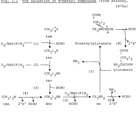
The oxidation of these compounds has recently been reviewed by Anthony (1975a) and only the major points will be discussed here. A summary of the oxidative pathways is shown in Fig. 1.1.
(i) Tetramethylammonium oxidation
17
Fig. 1.1 The oxidation of N-methyl compounds (from Anthony, 1975a) COOH
I
(4)
(CHgi+N tern
Og/NADfPlHg (1) ^ HCHO (CHgigN
tmn Og/NADfPiHg (2) ^
(CHgjgNO tmo
(3) r — H C H O
(CH^); CH^NHCHOOH
N-methylglutamate (8) Jk
HCHO
NH.
1 (7)
(CHgigN ^ » (CHgigNH tmn 2'H' HCHO dmn
Og/NADfPiHg
2'H' COOH
(CHg); NHgCHCOOH glutamate
NH.
Ts) t " HCHO
»CHgNH_ —Ar ^ HCHO (6&
mn 2'H'
Numbers in parentheses refer to the following enzymes described in the text:
(1) tetramethylammonium (tern) mono-oxygenase (2) trimethylamine (tmn) mono-oxygenase
(3) trimethylamine N-oxide (tmo) demethylase (4) trimethylamine dehydrogenase
(5) dimethylamine (dmn) mono-oxygenase (6) methylamine (mn) dehydrogenase (7) N-methylglutamate synthase
[image:28.553.67.529.80.464.2]which catalyses the incorporation of molecular oxygen into one of the methyl groups. The enzyme was induced during growth on tetramethylarranonium and the oxidation products were trimethylamine and formaldehyde (Reaction 1).
(ii) Trimethylamine oxidation
In the obligate methylotrophs Bacterium 4B6 and
Bacterium C2A1, dimethylamine and formaldehyde are produced by anaerobic oxidative demethylation of trimethylamine
catalysed by trimethylamine dehydrogenase (Reaction 4).
Artificial electron acceptors must be used for assay of this enzyme and the natural electron acceptor is unknown
(Colby and Zatman, 1973; 1974). By contrast, facultative methylotrophs which grow on N-methyl compounds use two
enzymes to effect the same overall reaction. The product of the first enzyme, trimethylamine mono-oxygenase,
(Reaction 2) is trimethylamine N-oxide which is subsequently dernethylated by the second enzyme to yield formaldehyde
and dimethylamine (Reaction 3), Examples of bacteria which oxidise trimethylamine via these two enzymes include
Bacterium 5H2, Pseudomonas 3A2 (Cox and Zatman, 1973), Hyphomicrobium vulgarae NQ and Pseudomonas aminovorans
(Large, Boulton and Crabbe, 1972). In addition to its role in oxidising trimethylamine, the trimethylamine
19
It can be seen from Fig. 1.1 that the oxidation of trimethylamine by obligate methylotrophs produces one mole-cule of reduced co-factor whereas the involvement of a mono-oxygenase during the oxidation of this compound by facult-ative methylotrophs requires a molecule of reduced co-factor (NADH or NADPH).
(iii) Dimethylamine oxidation
Dimethylamine is oxidised by a mixed-function secondary amine oxidase reported in both facultative and obligate
methylotrophs (Reaction 5). The enzyme was first demon-strated in Pseudomonas aminovorans and shown to contain a carbon monoxide-sensitive haemoprotein of the cytochrome ^420 type (Eady, Jarman and Large, 1971; Brook and Large, 1975).
(iv) Methylamine oxidation
Two routes for methylamine oxidation have been
des-cribed. The first involves the direct oxidative deamination of methylamine to formaldehyde (Reaction 6) by an inducible methylamine dehydrogenase first demonstrated in Pseudomonas AMI (Eady and Large, 19 68). The enzyme, which requires PMS as the electron acceptor, has been found in both facultative and obligate methylotrophs.
The second route for methylamine oxidation consists of two enzymes; a soluble N-methylglutamate synthase
methyl-amine oxidation in Pseudomonas MA (Bellion and Hersh, 1972), Pseudomonas MS, Pseudomonas aminovorans (Large and Carter, 1973) and possibly in the obligate methylotroph Bacterium 4B6 (Colby and Zatman, 1973).
e) The electron transport chain of methylotrophs
Little is known at the moment of the energy metabolism and respiratory pathways involved during oxidation of C^-compounds.
It has been reported that cytochromes of the a, b and c types are present in the facultative methylotrophs Pseudomonas MA (Hersh, Peterson and Thompson, 1971), Hyphomicrobium X and Pseudomonas AMI (Anthony, 1975b; Widdowson and Anthony, 1976) and also in the obligate
methylotrophs Pseudomonas methanica (Type I) and Methylosinus trichosporium (Type II) (Tonge, Knowles, Harrison and
Higgins, 1974).
In Pseudomonas MA cytochromes b and c are reduced by N-methyIglutamate in the presence of the membrane-bound N-methylglutamate dehydrogenase. Cytochrome o is the only co-binding pigment in this organism and presumably acts as the terminal oxidase catalysing the reoxidation of cyto-chrome c (Hersh et al., 1971):
2'h
N-methyglutamate cyt b —•cyt c —•cyt o —tOg glutamate HCHO
21
the cytochromes and b-type cytochromes are able to
react with carbon monoxide. In addition to this Pseudomonas AMI and Hyphomicrobium X have CO-binding c-type cytochromes. However, these CO-binding c and b-type cytochromes do not appear to have oxygenase or oxidase functions and their ability to bind CO is probably irrelevant to the normal physiology of the organisms (Widdowson and Anthony, 197 6).
The electron transport chain of Pseudomonas AMI is thought to operate as follows (from Widdowson and Anthony, 1976)
formate, formaldehyde
succinate, p-hydroxybutyrate
cytochrome b
methanol ethanol
cytochrome c
alternative route in cytochrome c-deficient mutant
cytochrome /a^ = KCN
0,
1.4 The assimilation of C^-compounds
Excluding the ribulose diphosphate cycle of autotrophic CO^ fixation, two pathways are known to effect the biosynthesis of C^- and C^-intermediates from C^-units (formaldehyde).
a) The ribulose monophosphate cycle • Ill I III I ^ *• ^
03 4j| (U U +J Ui o M G o -H +J
to X -H Q) tl CD 13 1—i E u 0 4-1 M-i o Q) )—I o o oj 4j (ts fl
cw tfi
o oj o d o qj CO o I—I 0 A •H 14 0) X! Eh cn tji •r4 k 5 w Q) G •H I—I (U -p 4-1 0 D +J •H is 1 CO (D M fo Q) -P fo rc Pu CO 0 rc fll 1 VD Q) m O 4-1 O 0 M o 0) tn (U M n3 k (U g •H O. <D I ro
0) 0)
4J CO
(0 (0 rc m
A (U
^ w
cu -h
I o
in Xi i -h Q) M
W O O Xi
M A 0 W X! O •h fl k A 04 CD CO rd CU C (U Q) D 0) 4J
its
a tfl
o 43 4J to d o o 0 I ix) I O <L)
43 tn cu o w o
O 0 4h iH OJ m Cn to o -p M to "O k >1^3 r4
43 >1 cn 43 >, 0 X T3 O 0 (U tJ 4-1 i to m (3 ! o o u 4j 0 CU A M 1 O <N 43 ! CD
A O (0 0} 43 to
0 a m 43 m o
A O 1 43 r—i Oi Cn LO Oi to 00 cTi o H <N
rH r—i rH
to CD
> 0) CO
fC CU m to 0) CO tO I—1 H n3 M 0
o
..
43 CU fO 4-1 g 1-4 CU CU C O to43 > >1 CO
4J rH CO -H CU o 0) 4-1
M > CU CU to to
O £3 4-1 CO fO ^ 4-1 -r4 to 0 G Cu
43 r4 -i4 CO CO to A 0 0
CU CU CO X o 43 CU CU 4-1 g O CD 4-1 Oi CO to
0 >1 43 43 U -H to to O N A 1 0 "T! r-4 r-4 M £3 fO M 0 0
(D CU 1 44 CU 4J t3 CU CO 0 0 CO CU M
> CU 0 43 43 O rX to
-H 43 r4 Dh A 4-1 CO 0} 4-1 4-1 0 CO CO O (3 d
to X 0 0 0 to to
C 0 CU 43 43 S4 M M 14 4J 4304 0^44 4J 4-1 CU
4J >1 rH CU
23
-obtained by Johnson and Quayle (1965) using the obligate methane utiliser Pseudomonas methanica (Type I). The key enzyme is hexulose phosphate synthase which catalyses the initial incorporation of formaldehyde (Reaction 1). The enzyme is specific for D-ribulose S-phosphate, the formalde-hyde acceptor molecule, and the product of the condensation reaction is a hexulose phosphate, D-erythro-L-glycero-3-hexulose-6-phosphate (Ferenci, Str^m and Quayle, 1974;
Kemp, 1974). The second enzyme, phospho-3-hexuloisomerase, catalyses the isomerisation of the hexulose phosphate to fructose-6-phosphate (Reaction 2). The remaining reactions of the cycle serve to regenerate the ribulose 5-phosphate acceptor and to provide as a substrate for biosynthesis of cell material. Two alternative routes for the cleavage of fructose-6-phosphate have been suggested
(Str^m, Ferenci and Quayle, 1974; Colby and Zatman, 1975b). One route involves phosphofructokinase and fructose
di-phosphate aldolase while the other involves phosphoglucose-isomerase (Reaction 9) and phospho-2-keto-3-deoxygluconate aldolase (Reaction 12) (Fig. 1.2). Another variation of the pathway occurs in some methylotrophs which lack transaldolase activity (Reaction 6). Such organisms include the two
Gram-positive, Type L restricted facultative methylotrophs (PM6 and S2A1) which use a modified cycle involving
sedoheptulose 1, 7-diphosphate and sedoheptulose diphos-phatase (Colby and Zatman, 1975b).
It has been suggested that certain enzymes of the
Fig. 1.3 Hexulose phosphate synthase-mediated cycle of formaldehyde oxidation (from Str^m et al., 1974)
Ribulose 5-P H + NADPH
NADP
+ NADPH
HCHO L
6-P-Gluconate
Glucose 6-P
3-Hexulose 6-P
Fructose 6-P
NADP
pgd, 6-phosphogluconate dehydrogenase
[image:35.553.65.489.80.530.2]25
oxidation of formaldehyde shown in Fig. 1.3. This scheme for formaldehyde oxidation has been demonstrated in P. methanica, M. capsulatus (Str0m et al., 1974) and Bacillus PM6 (Colby and Zatman, 1975b). The latter two organisms
lack alternative enzymes for formaldehyde oxidation. The cycle may also be important in all these bacteria for the generation of NADPH for biosynthetic purposes.
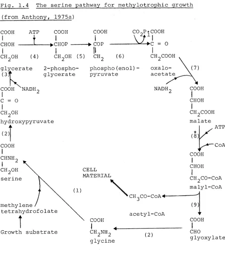
b) The serine pathway
The serine pathway was first proposed by Large, Peel and Quayle (1961) who showed that serine was the earliest
intermediate in the metabolism of ^^C-methanol and ^^C-formate by methanol and formate-grown Pseudomonas AMI. This pathway is now known to operate as a cyclic series of reactions
which effect the addition of formaldehyde and CO^ to give a molecule of acetyl-CoA (Fig. 1.4):
HCHO + COg + CoASH + 2NADH2 + 2ATP
CHgCOCoA + 2NAD + 2ADP + 2Pi + ZHgO
The enzymes which probably have an exclusive function in the serine pathway are: serine-glyoxylate aminotransferase
Fig. 1.4 The serine pathway for itiethylotrophic growth (from Anthony, 1975a)
COOH
i
»COP COOH I CHOH ATPi
chgoh (4)
COOH
I
-•chop
CH.OH (5) CH,
cogp^cooh
0
( 6 )
glycerate o f
COOH NADH, C = O
2-phospho-glycerate phospho(enol) pyruvate CHgCOOH oxalo-acetate
NADH, COOH
I
CHOH chgohhyc
i
( 2 )
COOH
I
CHNH CHgOH serine roxypyruvate methylene tetrahydrofolateGrowth substrate
CELL MATERIAL
\
CH_C0-CoA4-acetyl-CoA COOHI 4
chgnhg glycine
( 2 )
CHgCOOH malate
* ( 8 )
COOH I CHOH ATP CoA CHgCO-CoA malyl-CoA (9) COOH CHO glyoxylate
Key to enzymes:
(1) serine hydroxymethyltransferase (2) serine-glyoxylate aminotransferase (3) hydroxypyruvate reductase
(4) glycerate kinase (5) enolase
(6) phospho(enol)pyruvate carboxylase (7) malate dehydrogenase
(8) malyl-CoA synthetase (9) malyl-CoA lyase
[image:37.553.62.516.61.573.2]27
1975a).
One outstanding problem concerning the operation of the serine pathway is the activation of malate to malyl-CoA by Pseudomonas AMI. Malate thiokinase (8) has not been demonstrated in this organism but it is present in Hypho-microbium X, Pseudomonas MA, Pseudomonas MS (Salem et al.,
1973b) and Bacterium 5H2 (Cox and Zatman, 1973). No altern-ative enzyme (e.g. CoA-transferase) has yet been described which is capable of activating malate in extracts of
Pseudomonas AMI.
The final problem in considering the growth of bacteria using the serine pathway is that of the conversion of acetyl-CoA, the nett product of the pathway, to C^- and C^-compounds required for biosynthesis. This problem is overcome by
oxidation of acetyl-CoA to glyoxylate which can re-enter the pathway after transamination to glycine and thus allow Cg and intermediates to be withdrawn.
There is some diversity amongst serine-pathway methylo-trophs in their mechanism of oxidation of acetyl-CoA to glyoxylate. Two routes are known, the distinction being whether or not the organism can synthesize a key enzyme of the glyoxylate cycle, isocitrate lyase.
1•5 Assimilation of acetyl-CoA produced by the serine pathway a) Bacteria which form isocitrate lyase
In common with typical heterotrophic bacteria, the gly-oxylate cycle operates during growth of many facultative methylotrophs on compounds metabolised by way of acetyl-CoA
enzymes of the cycle, isocitrate lyase (ICL) and malate
synthase (MS), together with certain TCA cycle enzymes effect the addition of two molecules of acetyl-CoA to give malate:
ICL MS
a c e t y l - C o A — p * c i t r a t e »isocitrate —y^glyoxylate malate TCA cycle enzymes
•
oxaloacetate 4— — — — succinate acetyl-CoA
It might be expected that during methylotrophic assimil-ation of acetyl-CoA, malate synthase is not required as the activity of this enzyme coupled with malyl-CoA lyase would result in the futile cycling of malyl-CoA to malate and CoASH. This problem is overcome by certain facultative methylotrophs where malate synthase is repressed during methylotrophic growth but not during growth on compounds
requiring the operation of the complete glyoxylate bypass. Such bacteria include Pseudomonas MA (Bellion and Hersh,
1972) and Hyphomicrobium X (Harder, Attwood and Quayle, 1973). Some bacteria however, (e.g. Pseudomonas MS and Bacterium
5H2, Cox and Zatman, 1973) have similar levels of malate synthase under all conditions of growth. It is possible that the malate synthase from these organisms is inhibited by certain intermediates of the serine pathway during methylo-trophic growth (See Section 6.5). All these bacteria contain induced levels of isocitrate lyase during growth on
C^-compounds as well as C^-compounds such as acetate and ethanol (Anthony, 1975a).
29
isocitrate lyase but not malate synthase is shown in Fig. 1.5. In this scheme the intermediate withdrawn for biosynthesis is a C^-compound; C^-compounds required for biosynthesis may also be produced by further metabolism of 2-phosphoglycerate to oxaloacetate or malate.
The 'unknown reactions' in Fig. 1.5 refer to the mech-anism of oxidation of acetyl-CoA to glyoxylate in Pseudomonas AMI and closely related bacteria which are unable to synthesize isocitrate lyase.
b) Oxidation of acetyl-CoA to glyoxylate in Pseudomonas AMI Pseudomonas AMI has no detectable isocitrate lyase
activity when growing on either C^-compounds or compounds usually requiring operation of the complete glyoxylate cycle for their assimilation (e.g. ethanol and f-hydroxybutyrate). It is now known that Pseudomonas AMI possesses a novel path-way for growth on ethanol, f-hydroxybutyrate and malonate part of which is also involved in the assimilation of acetyl-CoA during methylotrophic growth. This novel pathway involves the direct oxidation of acetate or acetyl-CoA to glyoxylate during assimilation of both C^- and Cg-compounds (Dunstan et al., 1972a, 1972b; Dunstan and Anthony, 1973). During growth on Cg-compounds the glyoxylate condenses with a second molecule of acetyl-CoA in a reaction catalysed by malate synthase to give malate. The complete pathway takes
its name from this enzyme and is known as the malate synthase pathway. In an analogous situation to those bacteria which do contain isocitrate lyase, it is unlikely that malate
Pig. 1.5 Scheme for assimilation in Hyphomicrobium X and Pseudomonas AMI (modified from Harder, Attwood and Quayle, 1973)
The dotted lines represent the 'unknown' reactions involved in acetyl-CoA oxidation to glyoxylate by Pseudomonas AMI.
3-phosphoglycerate mi CELL MATERIAL
2-phosphoglycerate (2) »phospho(enol)pyruvate
serine (2) 'C^' unit (2) ^
CO, malyl-CoA
glycine (2)
glyoxylate
glyoxylate <_
acetyl-CoA
isocitrate< citrate
— 31 —
AMI despite the observation that the enzyme is present
regardless of the growth substrate. This has recently been confirmed following the isolation of a mutant of Pseudomonas AMI (mutant ICT 51) which lacked malate synthase but retained
the ability to grow on C^-compounds (Taylor and Anthony, 1975 ). Evidence for the malate synthase pathway in Pseudomonas AMI is presented in Section 4.2.
The overall pathway of assimilation by Pseudomonas AMI is thus similar to that for Hyphomicrobium (Fig. 1.5), the 'unknown reactions' being equivalent in function to the isocitrate lyase and TCA cycle enzymes which operate in bacteria such as Hyphomicrobium.
Similar reactions involving the oxidation of acetate or acetyl-CoA to glyoxylate may also occur in organisms
closely related to Pseudomonas AMI such as Pseudomonas M 27, Pseudomonas 3A2 and Bacterium 5B1. The latter organism is
at present unique in that it contains high levels of isocitrate lyase during growth on acetate but the enzyme is virtually
absent during growth on C^-compounds (Colby and Zatman, 1972). It is possible that this organism uses enzymes of the glyoxylate cycle for assimilation of acetate but during methylotrophic
growth acetyl-CoA is oxidised to glyoxylate by enzymes similar to those proposed for Pseudomonas AMI.
1.6 Distribution of the carbon assimilation pathways
Obligate methylotrophs use either of the pathways of assimilation. All the methane-utilisers with Type I
methylotrophs which are not capable of utilising methane:
Bacterium 4B6, Bacterium C2A1 (Colby and Zatman, 1975b),
Organism W1 (Dahl et aJ.., 1972) and Methylomonas M-15
(Sahm and Wagner, 1975) all use the RMP pathway of
formalde-hyde fixation. It is not known if the Type II obligate
methylotrophs which use the serine pathway contain isocitrate
lyase for acetyl-CoA assimilation. The finding by Bellion
and Woodson (1975) of two distinct isocitrate lyases in
Pseudomonas MA, one elaborated during methylotrophic growth
and the other during growth on acetate leads to the
possibil-ity that organisms may exist which grow on C^~compounds
using isocitrate lyase, but fail to grow on C^-compounds.
The restricted facultative methylotrophs all use the
RMP pathway of formaldehyde fixation (Colby and Zatman, 1975b).
With the exception of Pseudomonad C, all the facultative
methylotrophs use the serine pathway of C^^ assimilation.
However, Goldberg and Mateles (1975) have recently proposed
that Pseudomonad C assimilates methanol by way of the RMP
pathway while formaldehyde and formate are assimilated by
way of the serine pathway. These workers demonstrated that
methanol-grown Pseudomonad C contained high levels of hexulose
phosphate synthase and no glycerate dehydrogenase. Conversely,
extracts of bacteria grown on formate or formaldehyde contained
high levels of NADPH-specific glycerate dehydrogenase but
no hexulose phosphate synthase. If these results are
con-firmed by detailed analysis of all the enzymes of the serine
and RMP pathways, this bacteriaMis unique in possessing both
33
-Methylotrophic yeasts use the ribulose monophosphate
pathway of formaldehyde fixation. An inducible hexulose
phosphate synthase has been demonstrated in Candida biodinii
during growth on methanol and early labelled intermediates
derived from ^^C-formaldehyde incubated with
methanol-grown cells were sugar phosphates (Sahm and Wagner, 1974).
Evidence has also been obtained for the operation of this
pathway in Candida biodinii No 0302, Kloeckera sp. No. 2201
and Pichia pastoris although the specific activities of
hexulose phosphate synthase in.these yeasts were much lower
than those of bacteria containing this enzyme (Diel, Held,
PART B
The metabolism of poly-P-hydroxybutyrate
I, I. -ui.iillliiiimiuill^ll nil I n , , HMIIUlliHilllllIWi)—IMM.II-Iiillll I II
1.7 Nature and occurrence of poly-P-hydroxybutyrate
iiiiii.iii.iiiji...-'ipii.iiiii.w».iiJi'i'»»w«i>i»—•|i.i»ii-iwi.j».i—i.iii-.iii.iii-iij> I LU II I.I—Iiujiwu—.a-u—iiiiiiii .1 w»« I |«W||-III|J.IMI.I|||.|.|»|||.UI...I. I'm II I •li—mnuuidfc.Mnwii ii#
u-mw-Poly-p-hydroxybutyrate (PHB) is a carbon and energy storage compound found in a wide variety of micro-organisms including Gram-negative and Gram-positive aerobic and anaerobic
species. Very large amounts of the polymer are accumulated by Azotobacter species (Stockdale, Ribbons and Dawes, 1968) and by Hydrogenomonas eutropha (Schlegel, Gottschalk and von Bartha, 1961). In 1960, Kallio and Harrington demon-strated that 32% of the dry weight of a strain of methanol-grown Pseudomonas methanica (Iowa strain) was due to lipid material, and 92% of this lipid was poly-#-hydroxybutyrate. This endogenous storage compound was later found to form 6% of the dry weight of Pseudomonas AMI (Peel and Quayle, 1961) and has since been observed in a number of different methylotrophs.
Poly-f-hydroxybutyrate exists in the form of a straight chain homopolymer of D(-) ^-hydroxybutyrate, the formula of which is:
3 , 2 OH 0
P-CH-CH_-C i 2 h CH^ O
-O-CH-CH.-COOH
I 2
ch^ n
35
polymer is present within hydrophobic granules which can
be stained with Sudan black. The single membrane surrounding the granule contains protein which has been associated with the enzyme system(s) concerned with polymerisation and
depolymerisation of the polymer (Merrick, 1965).
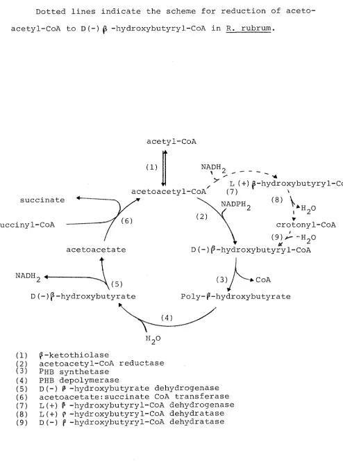
1.8 The enzymes of poly-^-hydroxybutyrate biosynthesis
The most detailed study of PHB biosynthesis has come from work with Azotobacter beijerinckii which under certain conditions can accumulate up to 70% of its weight as PHB
(Dawes and Senior, 1973). The following enzymes were found to catalyse the formation of PHB from acetyl-CoA in this bacterium: P-ketothiolase, acetoacetyl-CoA reductase and
poly-^-hydroxybutyrate synthetase (Fig. 1.6). PHB synthetase is granule-bound and has been found in H. eutropha (Schegel, Lafferty and Krauss, 1970), Rhodospirillum rubrum and
Bacillus megaterium (Greibel, Smith and Merrick, 1968) as well as A. beijerinckii. In the latter two organisms, the enzyme has a functional thiol group and in all cases the substrate is D(-) ^-hydroxybutyryl-CoA.
Dawes, 1971) while the product of the enzyme from R. rubrum is L(+)p -hydroxybutyryl-CoA.
Two separate p-hydroxybutyryl-CoA dehydratases have
been demonstrated in R. rubrum (Moskowitz and Merrick, 1969), one specific for the D(-) isomer, and the other for the
L (+) isomer. Crotonyl-CoA was incorporated into PHB in the presence of the dehydratase specific for D(-)P -hydroxy-butyryl-CoA and PHB synthetase, but substitution of the
L (+) isomer dehydratase for the D(-) enzyme in this system did not lead to incorporation. It was therefore proposed that in R. rubrum, L(+)P-hydroxybutyry1-CoA from the reduct-ion of acetoacetyl-CoA was first converted to the D(-)
isomer through the intermediacy of crotonyl-CoA before being incorporated into PHB (Fig. 1.6).
The first enzyme of PHB synthesis from acetyl-CoA is |5-ketothiolase which is present in all PHB-producing
bact-eria. The enzyme has been examined in detail from A.
beijerinckii (Senior and Dawes, 1973) and H. eutropha (Ceding and Schlegel, 1973) and possesses several features of inter-est. The thiolysis reaction is inhibited by high concentr-ations of acetoacetyl-CoA (substrate inhibition) which is relieved by increasing the concentration of the second sub-strate, CoASH. In the direction of acetyl-CoA condensation, the reaction is inhibited by CoASH. The significance of these properties in relation to the regulation of PHB meta-bolism is discussed in Section 1.10.
1.9 The enzymes of poly-P-hydroxybutyrate degradation a) Extracellular PHB degradation
— 37 —
Fig. 1.6 The metabolism of poly-P-hydroxybutyrate in Azotobacter beijerinckii (modified from Senior and Dawes, 1973) Dotted lines indicate the scheme for reduction of aceto-acetyl-CoA to D(-) ^ -hydroxybutyry1-CoA in R. rubrum.
succinate succiny1-CoA
acetyl-CoA
(1) NADH.
V
^ L (+) 6-hydroxybutyryl-Coii acetoacetyl-CoA (7) \
( 8 ) V NADPH.
acetoacetate
crotonyl-CoA
D(-)f-hydroxybutyryl~CoA
nadhg.^
(5) D(-)p-hydroxybutyrate
(3)/ ^-tCoA Poly-^-hydroxybutyrate
(1)
Si!
(4) (5)
( 6 )
(7)
( 8 )
(9)
p-ketothiolase
acetoacetyl-CoA reductase Phb synthetase
PHB depolymerase
[image:48.553.43.537.118.781.2]substance in the soil, liberated by the death and lysis of
organisms such as Azotobacter and Hydrogenomonas which
accumulate substantial amounts of the polymer. It is not
surprising therefore to find that a number of bacteria have
been isolated which are capable of utilising extracellular
PHB as a sole source of carbon and energy. Several species
of Hydrogenomonas and Bacillus have been reported to have
this property (Dawes and Senior, 1973) as well as a number
of Pseudomonads (Delafield, 1965b). The enzyme which confers
this ability is an extracellular PHB depolymerase which in
most cases degrades PHB to a mixture of monomeric and dimeric
D (-) -hydroxybutyrate which are transported into the cell.
The dimer is then further hydrolysed to D(-)P -hydroxybutyrate
by an intracellular dimer hydrolase (Delafield, 1965a).
b) Intracellular PHB degradation
The catabolism of intracellular PHB reserves is probably
not initiated until all available exogenous carbon and energy
sources are exhausted. The first degradation step is
catal-ysed by a soluble PHB depolymerase. The sole product of PHB
hydrolysis in Hydrogenomonas was D(-)f -hydroxybutyrate
(Hippe and Schlegel, 1967.) whereas the products of purified
PHB depolymerase from B. megaterium were identified as a
mixture of dimer and monomer units. A dimer hydrolase has
also been isolated from this organism which catalyses the
hydrolysis of dimeric ^-hydroxybutyrate to monomer units
(Dawes and Senior, 1973).
In every system so far studied, D(-) P -hydroxybutyrate
is further metabolised by an NAD -specific P-hydroxybutyrate
39
Ceding and Schlegel (1973) have demonstrated that the enzyme
from H. eutropha is competitively inhibited by NADH, pyruvate
and oxaloacetate. The enzyme from A. beijerinckii is also
competitively inhibited by NADH and pyruvate but differs in
that oxaloacetate does not inhibit whereas 2-oxoglutarate
does (Senior and Dawes, 1973). The significance of these
regulatory properties are discussed in Section 1.10.
Further oxidation of acetoacetate involves activation
to acetoacetyl-CoA followed by cleavage of this molecule
to two molecules of acetyl-CoA. In A. beijerinckii
aceto-acetate is activated by transfer of CoA from succinyl-CoA
in a reaction catalysed by acetoacetate:succinate CoA
transferase (thiophorase):
succinyl-CoA ^ ^ succinate
acetoacetate acetoacetyl-CoA
When assayed in the direction of CoA transfer from
acetoacetyl-CoA to succinate, acetoacetate was found to be
a potent inhibitor of the enzyme (Senior and Dawes, 1973).
^-ketothiolase, the enzyme catalysing the first
reaction of biosynthesis, catalyses the final degradation
reaction of PHB to acetyl-CoA. There is no evidence to
suggest that more than one enzyme is responsible for these
two activities in A. beijerinckii or H. eutropha.
The overall scheme for the metabolism of PHB by
A. beijerinckii is shown in Fig. 1.6. Ceding and Schlegel
succinate CoA transferase, have concluded that PHB metabolism
by H. eutropha proceeds by a process much the same as for
A. beijerinckii and with almost identical regulatory controls
(Fig. 1.7).
1.10 Regulation of poly-?-hydroxybutyrate metabolism
Dawes and Senior (1973) have pointed out that any
regul-atory mechanism of PHB metabolism must take into account the
observed physiological functions of the polymer.
The primary function of the polymer is that of a carbon
and/or energy source during starvation. This role has been
demonstrated in several bacteria including Micrococcus
halodenitrificans (Sierra and Gibbons, 1962), Bacillus
megaterium (Macrae and Wilkinson, 1958) and Hydrogenomonas
eutropha (Hippe, 1967). In some bacteria the polymer serves
as a reserve of carbon and energy during specialised
activ-ities such as sporulation and encystement. In Azotobacter
yinelandii PHB accumulates prior to encystement and
sub-sequently disappears when encystement occurs (Stevenson and
Socolofsky, 1966). Similiarly, in B. cereus the polymer
accumulates at the end of exponential growth and is degraded
at the onset of sporulation (Kominek and Halvorson, 1965).
In the Azotobacteriaeceae it has been proposed that PHB
functions as both a storage compound and as a means of
reg-ulating the oxygen environment of their natural habitat,
the soil. The nitrogenase of Azotobacter is inhibited by
oxygen concentrations in excess of about 20% air saturation.
This inhibitory effect may be countered by the organism
41
environmental partial pressure to a more acceptable value; a process known as 'respiratory protection' (Dalton and Postgate, 1969). The possession of large amounts of PHB permits the organism to increase its oxidative activity even in the absence of an exogenous substrate. It is not sur-prising therefore to fimi Wiat polymer accumulates in Azotobacter beljerinckll during atmospheric nitrogen fixa-tion under condifixa-tions of oxygen limitafixa-tion (Senior, Beech, Ritchie and Dawes, 1972) when 'respiratory protection' is unnecessary.
The regulation of PHB metabolism has been studied in detail in two organisms: A. beiierinckii (Senior and Dawes,
1973) and H. eutropha H. 16 (Ceding and Schlegel, 1973) (Fig. 1.7). Senior and Dawes have proposed the following explanation of their findings.
Under conditions of unrestricted growth, enzymes of the TCA cycle are operating maximally and citrate synthase acts as a sink for acetyl-CoA with the simultaneous release of free CoASH. Consequently the acetyl-CoA concentration in the cell is low and the CoASH concentration high, a com-bination which leads to inhibition of P-ketothiolase in the direction of acetoacetyl-CoA synthesis. Oxygen limit-ation would lead to a build up of NADH which inhibits the citrate synthase of Azotobacter beiierinckii (Senior and
Dawes, 1971). With few exceptions this inhibition of citrate synthase by NADH is common to Gram-negative bacteria (Section 1.13). The inhibition of this enzyme would lead to a
Fig. 1.7 The regulation of poly-^-hydroxybutyrate metabolism
in A. beijerinckii (from Senior and Dawes, 1973)
acetyl-CoA
CoA-acetoacetyl-CoA
+-acetoacetyl-CoA
succinate
succinyl-CoA
acetoacetate
CoASH
4-NADH,
I
Pyruvate2-oxoglutarate| \
D(-)p-hydroxybutyrate
acetoacfetyl-CoA
D(-)P -hydroxybutyryl-CoA
poly-f-hydroxybutyrate
[image:53.553.66.516.190.540.2]— 43
-P > - k e t o t h i o l a s e (Km = 0.9 mM) and the simultaneous decrease
in the concentration of free CoASH would relieve the
inhib-ition by this compound on the condensation reaction of
p"-ketothiolase. The high NADH concentration would, in turn,
inhibit degradation of the polymer at the level of
^-hydroxy-butyrate dehydrogenase and prevent unrestricted cycling of
metabolism.
Conditions favouring a high intracellular concentration
of NAD(P)^ resulting from relaxation of oxygen limitation
would stimulate degradation of the polymer only when the
steady-state concentration of acetyl-CoA decreased and that
of CoASH increased as a result of the supply of carbon
becoming restricted. The increasing CoASH concentration
would enable thiolysis of acetoacetyl-CoA to proceed by
relieving the inhibition of p-ketothiolase by its substrate
acetoacety1-CoA.
Similar considerations apply to the regulation of PHB
metabolism in organisms such as H. eutropha which accumulate
the polymer under nitrogen limitation. Nitrogen limitation
would cause a cessation of protein synthesis; pyruvate and
TCA cycle intermediates would not flow into anabolic
path-ways resulting in high acetyl-CoA and low CoASH concentrations.
Once again ^-ketothiolase condensation would be uninhibited
and PHB synthesis unimpaired.
In conclusion, poly-^-hydroxybutyrate is thus a highly
reduced carbon and energy storage compound which, according
to organism, may additionally play a role in spore or cyst
formation. In the case of nitrogen-fixing organisms in the
soil the possession of PHB could afford 'respiratory protection'