RESEARCH ARTICLE
SCREENING OF ACTINOMYCETES FROM INDIGENOUS SOIL FOR PRODUCTION OF
EXTRACELLULAR METABOLITES
*Syed Abdus Subhan, Abdul Wahab, Talat Yasmeen Mujahid, Tanveer Abbas, Ishrat Khan and
Samia Idrees
Department of Microbiology, University of Karachi, Karachi, Pakistan
ARTICLE INFO ABSTRACT
Actinomycetes are abundantly present in soil and they produce a variety of antimicrobial compounds that can be used as chemotherapeutic agent in order to limit the infection. In present study, actinomycetes were isolated from different ground soils. Primarily these isolates were screened for extracellular metabolites production by conventional methods. Cross streak method and double agar overlay methods were used in this screening. Initially we have isolated 33 actinomycetes strains from different soil samples and screened them for antimicrobial potential. About 51.51 % of isolated strains showed the antagonistic properties against one or two tested gram positive bacteria. The best strain IAS 1, IAS 7, IAS 10, IAS 11, showed maximum zone of inhibition against M.luteus. The chemical nature of ISA 10 was assessed by simply heating the supernatant and we found that the extra cellular metabolite activity was absent in heated sample suggesting the protein nature of it
Copyright © 2015 Ishrat Khan et al. This is an open access article distributed under the Creative Commons Attribution License, which permits unrestricted use, distribution, and reproduction in any medium, provided the original work is properly cited.
INTRODUCTION
Actinomycetes are Gram positive bacteria and an important source of antibiotics (Valli et al., 2012). They have high G+C content 55-77% (Lo et al., 2002; Ningthoujam et al., 2009; Lam et al., 2006 and Ndonde et al., 2000). They belong to order actinomycetales and composed of around eighty genera (Stackebrandt et al., 1997 and Goodfellow et al., 1983) .They are distributed ubiquitously including water, soil, and marine (Gebreyohannes et al., 2013). Actinomycetes are economically and biotechnologically feasible prokaryotes. They produced a variety of bioactive compounds that can be used to treat infections, they include antitumor, antifungal, antibacterial agents (Bizuye et al., 2013) and enzymes (Jeyadharshan et al., 2013). Among all genus, Streptomyces are the best secondary metabolites producers (Valli et al., 2012). First antibiotic, streptomycin has been isolated from Streptomyces in 1945 by A. Waksman (Atta et al., 2010). Up till now, a number of antibiotics have been isolated from actinomycetes including anthracyclines, peptides, macrolides β-lactams, actinomycins and tetracyclines etc. Variability among genus of actinomycetes is of great significance in many areas of science especially in antibiotics (Magarvey et al., 2004). Now a days multi-drug resistant pathogenic bacteria is an issue of extreme concern in the world whose numbers are continuously
*Corresponding author: Syed Abdus Subhan,
Department of Microbiology, University of Karachi, Karachi, Pakistan.
increasing day by day and resulting in rapid spread of infectious diseases, leading to high morbidity and mortality (Hong et al., 2009 and Alanis et al., 2005). New antibiotics are also frequently in use for pathogens that seems to be responsible for emergence of resistant pathogens in clinical cases (Lewis, 2013). However, there are some microbes which are easily destroyed by selective antibiotics are not frequently available, furthermore, antibiotics that are discovered yet are expensive and have more side effects (Bizuye et al., 2013). Most of the Actinomycetes do not cultivate in lab condition that are the important source of most of the antimicrobial drugs, that’s why we are unable to examine their potential of producing novel antibiotics (Schatz et al., 1945).
Over the past few years, actinomycetes that have been isolated and screened for antibiotics were found to be previously reported or are found to be re-isolated strains, however, unexplored ecosystem or less explored ecosystem, like marine, desert, forest, caves and hills has been found to be a more promising source for isolating new bioactive novel compounds from Actinomycetes (B´erdy et al., 2012 and Nachtigall et al., 2011). Because of the increasing resistance of microorganisms towards discovered antibiotics there is a need to explore the potential of novel strains of actinomycetes for their new secondary metabolites and to study its role in the field of antibiotics.
ISSN: 0975-833X
International Journal of Current Research
Vol. 7, Issue, 01, pp.12078-12083, January, 2015
OF CURRENT RESEARCH
Article History:
Received 26th October, 2014
Received in revised form
30th November, 2014
Accepted 07th December, 2014
Published online 31st January,2015
Key words:
Actinomycetes, Extracellular metabolites, Soil samples,
MATERIALS AND METHODS
Sample collection
Soil samples were collected from different grounds of University Of Karachi at a depth of 10-15 cm.
Chemicals
Chemicals include ethanol, NaCl, Na2HPO4, KH2PO4, H2SO4
and BaCl2. Different microbiological media including Nutrient
agar, Nutrient broth, Agar technical, and Heart infusion agar procured from Oxoid.
Sample processing and isolation of Actinomycetes
Samples were placed in an empty petri dish for two days (Jeffrey, 2008). Dilution series were begin by adding 1 gm of soil sample in 100 mL of saline and then serially diluted till 10-5 according to the protocol used byRahman et al. (2011) with slight modification. 100µl from 10-3, 10-4and 10-5 were transferred onto half strength nutrient agar (Taha et al., 2007) and allowed to dry. Plates were incubated at room temperature for 3 days.
Colonial and morphological characterization
Actinomycetes were identified by their morphological and colonial characteristics. (Gurung et al., 2009). Their morphologies were identified through Gram staining and were differentiated according to their shape, size and color of colonies.
Tested cultures
Clinical isolates were selected to evaluate antibacterial activity of Actinomycetes strains. These include: Staphylococcus aureus, Staphylococcus epidermidis, Bacillus subtilis, Enterococcus fecalis, Micrococcus luteus, Salmonella typhi, E.coli, Pseudomonas aeruginosa, Acinetobacter and Proteus mirabilus.
Primary screening of metabolites
Primary screening of metabolites were performed by two methods:
1. Cross streaking
Actinomycetes strains IAS 26 to IAS 33 were analyzed by cross streak method (Mohseni et al., 2013). These strains were streaked at the corner of plate and incubated for 3 days at room temperature. Overnight fresh cultures of test organism were selected and their suspension was prepared in PBS until the turbidity was matched to 0.5 McFarland standards. These test organisms were streaked perpendicular to the Actinomycetes strains by dipping the sterile cotton swab in suspension of test cultures. Plates were incubated at 37 °C for 24 hours.
2. Double agar overlay method
For preliminary screening of antibacterial activity of Actinomycetes, double agar overlay method was used (Shetty et al., 2014). Actinomycetes strains were stabbed on half
strength nutrient agar and incubated at room temperature for 2-3 days. After appearance of growth 5mL 1% soft agar containing 100 µL of M.luteus (which was matched with 0.5 McFarland standards) were overlayed over half strength nutrient agar and incubated at 37°C for 24 hours. After a day, zone of inhibition was measured. Strains that showed antibacterial activity against M.luteus were further analyzed with other test organisms. Concentration of isolates and test culture was maintained by matching the turbidity with 0.5 McFarland standards. For evaluation of antibacterial activity 8 µL from isolate suspension was inoculated on half strength nutrient agar. Plates were incubated at room temperature for 2 days. When growth appeared on plate, 5mL 1% soft agar containing 100 µL of test culture (which was matched with 0.5 McFarland standards) was poured over it. Plates were incubated for 24 hours at 37°C.
Determination of nature of bioactive compound
Actinomycetes strains that showed activity against test cultures were inoculated in 50 mL of half strength nutrient broth and incubated for 2 days at room temperature; adapted and modified Mohseni et al. (2013). To extract the bioactive compounds, inoculated broth was centrifuged at 5000 rpm for 10 min at 4°C modified from Valli et al. (2012). Supernatant were passed through membrane filter of 0.45 µm size. The filtered supernatants were transferred into two aliquots. 1 was heated at 100ºC for 1 min and other remained unheated. To determine the nature of bioactive compound, Agar well diffusion method was used. M.luteus was used as the test culture. 100 µL from both aliquots were transferred into wells. Plates were incubated at 37 ºC for 24 hours.
RESULTS AND DISCUSSION
The purpose of this study was to evaluate antimicrobial activity of Actinomycetes. Due to emergence of multidrug resistant human pathogens there is a need to discover new antibiotics which are effective against these pathogens. (Mohseni et al., 2013), to overcome this problem we can use the potential of Actinomycetes, that are able to produce bioactive compounds and an important source of secondary metabolites (Suthindhiran et al., 2009)
Isolation of Actinomycetes from ground soil
[image:2.595.327.542.679.731.2]We have isolated 33 Actinomycetes strains from various ground soils of University of Karachi as shown in Table 1. Half strength nutrient agar (Taha et al., 2007) was used for their isolation. They showed their optimum growth at room temperature after incubation of 3 days.
Table 1. Actinomycetes strains isolated from ground soil from University of Karachi
S. No. Places at University of Karachi No. of isolates
1 Valika ground 6
2 N.B.P ground 14
3 HBL ground 6
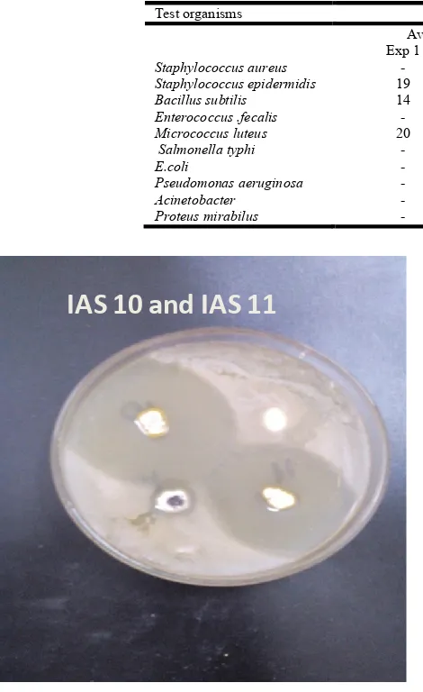
IAS 10 and IAS 11
Fig. 1. Antibacterial activity of IAS 10 and IAS 11 against
M.luteus
S.fecalis
S.epidermidis
S.aureus
M.luteus
S.fecalis
S.epidermidis
S.aureus
M.luteus
IAS 29
[image:3.595.67.530.219.316.2]IAS 28
[image:3.595.54.532.349.733.2] [image:3.595.53.288.354.738.2]Fig. 2. Antibacterial activity of Actinomycetes strains against gram positive bacteria by cross streak method Table 2. Antibacterial activity of Actinomycetes against M.luteus
Isolate No. Activity against M.luteus (mm) Isolate No Activity against M.luteus (mm) Isolate No Activity against M.luteus (mm)
IAS 1 30 IAS 11 40 IAS 21 -
IAS 2 28 IAS 12 - IAS 22 -
IAS 3 23 IAS 13 - IAS 23 -
IAS 4 - IAS14 - IAS 24 -
IAS 5 24 IAS 15 - IAS 25 -
IAS 6 20 IAS 16 -
IAS 7 30 IAS 17 -
IAS 8 20 IAS 18 -
IAS 9 - IAS 19 -
[image:3.595.322.532.354.730.2]IAS 10 45 IAS 20 -
Table 3. Antibacterial activity of Actinomycetes strains against gram positive bacteria by cross streak method
Isolate No. Growth pattern
S.epidermidis S.fecalis S.aureus M.luteus
IAS 26 + + + +
IAS 27 GI + + +
IAS 28 + GI + +
IAS 29 + GI + +
IAS 30 + GI + +
IAS 31 + GI + +
IAS 32 + GI + +
IAS 33 + + + +
Keys: + show no inhibition of growth GI shows growth inhibition
Table 2. Antibacterial activity of IAS 10 against clinical isolates – shows no zone of inhibition
Test organisms IAS 10
Average diameter of zone of inhibition (mm)
Exp 1 Exp 2 Exp 3
Staphylococcus aureus - - -
Staphylococcus epidermidis 19 19 20
Bacillus subtilis 14 15 14
Enterococcus .fecalis - - -
Micrococcus luteus 20 19 19
Salmonella typhi - - -
E.coli - - -
Pseudomonas aeruginosa - - -
Acinetobacter - - -
IAS 10
[image:4.595.58.270.52.209.2]Staphylococcus
epidermidis
Fig. 3. Antibacterial activity of IAS 10 against S.epidermidis
IAS 10
[image:4.595.339.529.141.485.2]M.luteus
Fig. 4. Antibacterial activity of IAS 10 against M.luteus
Fig. 5. Antibacterial activity of IAS10 against test cultures
Colonial and morphological characterization
All of the isolated strains were found to be gram positive, having fine thread like morphology along with spores (Gurung et al., 2009). Their colonies on half strength nutrient agar were chalky white, dry, nodular and sticky to agar.
Primary screening of metabolites
We have isolated 33 Actinomycetes strains from different soil samples. Out of 33 Actinomycetes strains 17 showed antibacterial activity against tested organisms. IAS 10 showed
maximum activity against test cultures while least activity was shown by IAS 6. In this research two different screening
methods were used including cross streak method (Mohseni et al., 2013) and double layer overlay method (Shetty et al.,
2014). Antibacterial activity of isolated strains was screened against different clinical isolates among which, M luteus was found to be the most sensitive organism
(a)
(b)
Fig. 6. IAS 10 filtrate heated (a) and unheated (b)
1. Cross streaking method
Cross streaking method was performed by using four bacterial cultures i.e., S.epidermidis, S.fecalis, S.aureus, M.luteus. Out of 8 strains five of them were found to have antimicrobial properties.
2. Double layer overlay method
[image:4.595.61.267.236.431.2] [image:4.595.41.286.454.607.2]reliability of the results. After maintaining inoculum size of isolate, double agar overlay method was re-performed and we saw IAS 10 showed activity against different clinical isolates as well.
Determination of nature of metabolites
In order to determine the nature of metabolites produced by Actinomycetes, an experiment was performed by using IAS 10. Extraction of metabolites was done by using the method as performed by Valli et al. (2012) with slight modifications. The well onto which unheated filtrate was added showed inhibition, while the well onto which heated filtrate was added, did not give any activity, this shows that the nature of metabolites produced by IAS 10 is most likely to be protein, as proteins gets denatured when heated at a high temperature.
Conclusion
The present research highlights the importance of soil actinomycetes which are quite active in producing antagonistic metabolites. In this study we found 17 out of 33 isolates (51.5%) are efficient in producing antimicrobial substances which are effective against S.epidermidis, S.fecalis, S.aureus and M.luteus. Further, one of the strain IAS 10, was found to produce extra cellular metabolites that are easily inactivated by heating, suggesting the protein nature of metabolite. Future research will be carried out in determining the molecular nature of this bioactive metabolite, produced from soil actinomycetes.
REFERENCES
Alanis, A. J. 2005. Resistance to antibiotics: are we in the post-antibiotic era? Arch. Med. Res., 36: 697-705.
Atta, H. M., Bayoumi, R., El-Sehrawi, M., Aboshady, A. and Al-Humiany, A. 2010. Biotechnological application for producing some antimicrobial agents by actinomycetes isolates from Al-khurmah Governorate. Eur. J. Appl. Sci., 2: 98-107.
Bérdy, J. 2012. Thoughts and facts about antibiotics: where we are now and where we are heading. J. Antibiot., 65(8): 385-395.
Bizuye, A., Moges, F. and Andualem, B. 2013. Isolation and screening of antibiotic producing actinomycetes from soils in Gondar town, North West Ethiopia. Asian Pac. J. Trop. Dis., 3(5): 375-381.
Das, A., Bhattacharya, S., Mohammed, A. Y. H. and Rajan, S. S. 2014. In vitro antimicrobial activity and characterization of mangrove isolates of streptomycetes effective against bacteria and fungi of nosocomial origin. Braz. Arch. Biol. Technol., 57(3): 349-356.
Gebreyohannes, G., Moges, F., Sahile, S. and Raja, N. 2013. Isolation and characterization of potential antibiotic producing actinomycetes from water and sediments of Lake Tana, Ethiopia. Asian Pac. J. Trop. Biomed., 3(6): 426-435.
Goodfellow, M. and Williams, S. T. 1983. Ecology of Actinomycetes. Annual Reviews in Microbiology, 37(1): 189-216.
Gurung, T. D., Sherpa, C., Agrawal, V. P. and Lekhak, B. 2009. Isolation and characterization of antibacterial
actinomycetes from soil samples of Kalapatthar, Mount Everest Region. Nepal J. Sci. Technol., 10: 173-182. Hong, K., Gao, A. H., Xie, Q. Y., Gao, H. G., Zhuang, L., Lin,
H. P., Yu, H. P., Jia, L., Yao, X. S., M, G. and Ruan, J. S. 2009. Actinomycetes for marine drug discovery isolated from mangrove soils and plants in China. Mar. Drugs, 7(1): 24-44.
Jeffrey, L. S. H. 2008. Isolation, characterization and identification of actinomycetes from agriculture soils at Semongok, Sarawak. Afr. J. Biotechnol., 7(20): 3697-3702.
Jeyadharshan, V. N. 2013. Production and Partial Purification of Protease by Actinomyces Species. Int. J. Sci. Res. Publ., 3(4): 1-3.
Lam, K. S. 2006. Discovery of novel metabolites from marine actinomycetes. Curr. Opin., 9: 245-251
Lewis, K. 2013. Platforms for antibiotic discovery. Nat. Rev. Drug Discov., 12(5): 371-387.
Lo C., Lai, N., Ho C., Cheah, H. and Wong, N. 2002. Actinomycetes isolated from soil samples from the Crocker range Sabah. ASEAN Rev. Biodivers. Environ. Conserv., 9: 1-7.
Magarvey, N. A., Keller, J. M., Berman, V., Dworkin, M. and Sherman, D. H. 2004. Isolation and characterization of novel marine-derived actinomycete taxa rich in bioactive metabolites. Appl. Environ. Microbiol., 70(12): 7520-7529.
Mohseni, M., Norouzi, H., Hamedi, J. and Roohi, A. 2013. Screening of antibacterial producing actinomycetes from sediments of the Caspian Sea. Int. J. Mol. Cell. Med., 2(2): 64-71.
Nachtigall, J., Kulik, A., Helaly, S., Bull, A. T., Goodfellow, M., Asenjo, J. A., Maier, A., Wiese, J., Imhoff, J. F., Sussmuth, R. D. and Fiedler, H. P. 2011. Atacamycins A– C, 22-membered antitumor macrolactones produced by Streptomyces sp. C38*. J. Antibiot., 64(12): 775-780. Ndonde, M. J. M. and Semu, E. 2000. Preliminary
characterization of some Streptomyces species from four Tanzanian soils and their antimicrobial potential against selected plant and animal pathogenic bacteria. World J. Microbiol. Biotechnol., 16: 595-599.
Ningthoujam, D. S., Sanasam, S. and Nimaichand, S. 2009. Screening of actinomycete isolates from niche habitats in Manipur for antibiotic activity. Am. J. Biochem. Biotechnol., 5(4): 221.
Parungao, M. M., Maceda, E. B. G. and Villano, M. A. F. 2007. Screening of antibiotic-producing actinomycetes from marine, brackish and terrestrial sediments of Samal Island, Philippines. J. Res. Sci. Comput. Eng., 4(3): 29-38. Rahman, M. A., Islam, M. Z. and Islam, M. A. U. 2011.
Antibacterial activities of Actinomycete isolates collected from soils of Rajshahi, Bangladesh. Biotechnol. Res. Int., doi:10.4061/2011/857925
Schatz, A. and Waksman, S. A. 1945. Strain Specificity and Production of Antibiotic Substances: IV. Variations Among Actionomycetes, with Special Reference to Actinomyces Griseus. Proc. Natl. Acad. Sci. USA, 31(5): 129-137.
activity. Braz. J. Microbiol., 45(1): doi.org/10.1590/ S1517-83822014005000022
Stackebrandt, E., Rainey, F. A. and Ward-Rainey, N. L. 1997. Proposal for a new hierarchic classification system, Actinobacteria classis nov. Int. J. Sys. Bacteriol., 47:479– 491.
Suthindhiran, K. and Kannabiran, K. 2009. Cytotoxic and Antimicrobial Potential of Actinomycete Species Saccharopolyspora salina VITSDK4 Isolated from the Bay of Bengal Coast of India. Am. J. Infect. Dis., 5(2): 90-98.
Taha, M. P. M., Drew, G. H., Tamer Vestlund, A., Aldred, D., Longhurst, P. J. and Pollard, S. J. 2007. Enumerating actinomycetes in compost bioaerosols at source—use of soil compost agar to address plate ‘masking’. Atmos. Environ., 41(22): 4759-4765.
Valli, S., Suvathi, S. S., Aysha, O. S., Nirmala, P., Vinoth, K. P. and Reena, A. 2012. Antimicrobial potential of Actinomycetes species isolated from marine environment. Asian Pac. J. Trop. Biomed., 2(6): 469-473.