http://www.scirp.org/journal/jbise J. Biomedical Science and Engineering, 2018, Vol. 11, (No. 9), pp: 254-262
https://doi.org/10.4236/jbise.2018.119021 254 J. Biomedical Science and Engineering
Comparative Study of the Effects of Metformin and Garlic
Extract on Hippocampal Na
+/K
+ATPase, Ca
2+ATPase and
Glutamine Synthetase Activities in Type II Diabetic
Wistar Rat
Abass Alao Safiriyu1, Ibrahim Semuyaba1, Sodiq Kolawole Lawal2,
Muhammad Olanrewaju Buhari3, Emmanuel Ayikobua Tiyo1, Muhamudu Kalange1,
Alfred Omachonu Okpanachi1, Miriam Nansunga1,4
1Department of Physiology, Faculty of Biomedical Sciences, Kampala International University, Ishaka, Uganda;
2Department of Anatomy, St. Francis University College of Health and Applied Sciences, Ifakara, Tanzania;
3Department of Anatomy, Faculty of Biomedical Sciences, Kampala International University, Ishaka, Uganda;
4Department of Physiology, Faculty of Biomedical Sciences, St. Augustine International University, Kampala, Uganda
Correspondence to: Abass Alao Safiriyu,
Keywords: ATPase, Diabetic Mellitus, Garlic, Glutamine Synthetase, Hippocampus, Metformin, Na+/K+ ATPase Received: April 5, 2018 Accepted: September 26, 2018 Published: September 29, 2018
Copyright © 2018 by authors and Scientific Research Publishing Inc.
This work is licensed under the Creative Commons Attribution International License (CC BY 4.0). http://creativecommons.org/licenses/by/4.0/
ABSTRACT
Diabetes mellitus has not ceased to be on rise in spite of the continuous research on its management. Brain dysfunction associated with Diabetes mellitus especially Type II has been the great concern. The aim of this study was to investigate the effect of insulin
sensi-tizing drug metformin and ethanolic extract of garlic on membrane bound enzymes Na+/K+
ATPase, Ca2+ ATPase and glutamate-glutamine cycle enzyme, Glutamine Synthetase
activi-ties in the hippocampus of streptozotocin-Nicotinamide induced Type II Diabetic rats. Twenty four male wistar rats weighted 120 - 150 g were used and divided into four groups with six rats in each group. Group A was non-diabetic (Control) and Groups B, C and D were diabetic. Group B received no treatment (DNT) while Groups C and D were treated with 1000 mg/kg of ethanolic garlic extract (EGE) and 50 mg/kg of metformin (MET) re-spectively orally for three weeks. All the groups were fed on standard rat chow with water ad libitum. Blood glucose was monitored weekly. Animals were sacrificed and the brains were removed and hippocampi were carefully excised and homogenate were obtained.
Homoge-nate was analyzed for Na+/K+ ATPase, Ca2+ ATPase and Glutamine Synthetase (GS)
activi-ties. MET and EGE significantly reduced the blood glucose levels. There was a significant
increase in the activities of hippocampal Na+/K+ ATPase, Ca2+ ATPase and GS in MET and
https://doi.org/10.4236/jbise.2018.119021 255 J. Biomedical Science and Engineering EGE when compared to DNT. The results suggest that both MET and EGE increase the
ac-tivities of hippocampal Na+/K+ ATPase, Ca2+ ATPase and GS which were reduced by
di-abetes mellitus, thus garlic and metformin administration exhibiting neuroprotective effect during hippocampal-related disorders associated with diabetes mellitus.
1. INTRODUCTION
Diabetes has become a global epidemic in the general population. The estimated number of African population that would be diabetic by 2030 according to World Health Organisation is approx. 18 million. Diabetes mellitus as a metabolic disease posed neuronal impairment, cognitive deficits and promote
pro-gressive neurodegeneration [1]. Insulin is one of the hormones that regulate the activity of Na+/K+ ATPase.
Complications associated with diabetes has been shown to alter the activity Na+/K+ ATPase [2]. Study has
shown decreased Na+/K+ ATPase activity in animal models of T2DM [3]. Diabetes has been shown to
af-fect neurotransmitter synthesis or release in several brain regions [4]. Ca2+ ATPase regulates Ca2+ pump
activity which acts as a second messenger in the control of cellular processes that plays a central role in
mediating neurosecretion. Inhibition of Ca2+ ATPase activity has shown to increase intracellular
concen-tration of Ca2+ which in turn altered the signal transduction pathways and cellular fluidity and eventually
resulted in cell death [5]. A decrease in Ca2+ ATPase activity of diabetic brain has been reported [6].
Glu-tamatergic neurotransmission is important for hippocampal modulation of learning and memory
processing [7]. The conversion of glutamate to glutamine by glutamine synthetase that takes place within
the astrocytes, represents a key mechanism in the regulation of excitatory neurotransmission [8]. Study
has shown that healthy neuronal cells require both intracellular and extracellular glutamine [9].
Develop-ment of diabetes-associated cognition decline in db/db mice has been suggested to be due to disturbance
of glutamate-glutamine shuttling between neurons and astrocytes in hippocampus [10]. Metformin is an
antidiabetic agent that is being widely used in the management of diabetes mellitus especially Type II. Several studies have shown that metformin improved learning and memory behaviours in diabetic model
[11, 12], by protecting hippocampal neurons [13, 14], by activating AMPK and Na+/K+ ATPase system
[15]. Metformin treatment is neuroprotective against the detrimental effects of β-Amyloid and high fat
diets on hippocampal synaptic plasticity [16]. Several studies have investigated the anti-diabetic potential
of both fresh and aged garlic extract and reported it to have antihyperglycemia, hypolipidemic, renopro-tective effects and its ability to scavenging many reactive oxygen species (ROS) in both animal models and
humans [17-21]. Fresh and cooked but not aged garlic extracts has been shown to increase both short and
long term memory in both diabetic male and female rats [22]. Ethanolic extract of garlic has previously
been reported to enhance spatial working memory by increasing hippocampal Na+/K+ and Ca2+ ATPases
activities in wistar rats [23] and improve memory in Type II diabetic rats by increasing Na+/K+,
Ca2+ATPases and glutamine synthetase activities in the hippocampus [24]. This study aimed to compare
the effect of metformin, a standard anti-diabetic drug and ethanolic extract of garlic on hippocampal
Na+/K+ ATPase, Ca2+ ATPase and Glutamine synthetase activities in Type II Diabetic rat model. Our
findings showed that both metformin and garlic increased the activities of these enzymes, though in-creased effect posed by metformin was more than that of garlic thus metformin may be a better an-ti-diabetic drug, among others in managing hippocampal-dependent dysfunction associated with di-abetes mellitus.
2. MATERIALS AND METHODS
2.1. Chemicalhttps://doi.org/10.4236/jbise.2018.119021 256 J. Biomedical Science and Engineering
2.2. Ethanol Extraction of Garlic
Extraction was done using cold maceration at the laboratory in the department of Pharmacology, Kampala International University Western Campus Uganda. Peeled garlic weighing 500 g was cut into small pieces and homogenized in 70 ml of cold sterile 0.9% NaCl. The paste material was suspended in 80% ethanol for 48 hours in air tight glass jar using a rubber stopper, and the suspension was shaken pe-riodically for three times a day at 5 minute interval. After 2 days, the suspension was filtered using Whit-man filter paper to remove residue. Filtration was repeated 3 times and clear filtrate was obtained. The fil-trate was concenfil-trated using rotary evaporator at a bath temperature of 40˚C. The extract concenfil-trate ob-tained was then transferred to a cornical flask and further evaporated in oven drier at 50˚C to obtain ulti-mately a gel like mass for the study [23, 25].
2.3. Animals
Male Wistar rats weighing (120 - 150 g) were used in this experiment. The animals were obtained from the Animal House of College of Medicine, Mbarara University of Science and Technology, Uganda. The animals were housed in a well-ventilated room maintained under standard conditions of light, feeding and temperature of research laboratory of Kampala International University Western Campus Uganda. The study was conducted in accordance with the standards established by the Guide for the Care and Use of Laboratory Animals.
2.4. Induction of Diabetes
After acclimatization of animals for five days, diabetes mellitus type II was induced in rats fasted overnight on day six with a single injection of 60 mg/kg streptozotocin dissolved in 0.05 ml of citrate buf-fer (pH 4.5) intraperitoneally which was followed by administration of 120 mg/kg of nicotinamide dis-solved in 0.5 ml of normal saline intraperitoneally 15 min later. Elevated levels of blood glucose (Hyper-glycemia) was measured from a drop of tail blood of rats 3 days and a week after injection using One-Touch Ultra-Easy Glucometer (LifeScan, UK). Rats confirmed hyperglycemic with blood glucose concentration (>250 mg/dl) was used as diabetic rats for this study [24, 26].
2.5. Grouping
The rats were divided into 4 groups (n = 6) as follows:
Group A: Non-diabetic Control-received normal saline 1 ml/kg body weight Group B: Diabetic non-treated (DNT)-received normal saline 1 ml/kg body weight
Group C: Diabetic + garlic (EGE) received 1000 mg/kg body weight of ethanolic extract of garlic Group D: Diabetic + metformin (MET)-received 50 mg/kg body weight of metformin
Administration orally was carried out daily between 8 - 10 am for period of three weeks.
2.6. Measurement of Na+/K+ ATPase Activity
The hippocampal homogenates was analyzed for Na+/K+ ATPase according to the method of Tirri et
al. [27]. Assay medium used consist of (in mM) 30 Tris-HCl buffer (pH 7.4), 50 NaCl, 6 MgCl2, 5 KCl and
50 μg of protein in the presence and absence of ouabain, 0.1 EGTA, in a final volume of 350 μL. The tion was started by the addition of ATP to a final concentration of 3 mM. After 30 min at 37˚C, the reac-tion was stopped by the addireac-tion of 50% (w/v) trichloroacetic acid (70 μL). The saturating substrate con-centrations was used, and reaction was in linear with protein and time. Some controls was included in the assays for non-enzymatic hydrolysis of ATP. The Pi (amount of inorganic phosphate) released was
quanti-fied calorimetrically, as described Fiske and Subba [28], using 300 KH2PO4 as reference standard. Specific
Na+/K+ ATPase activity was calculated from the overall activity (in the absence of ouabain) and was
https://doi.org/10.4236/jbise.2018.119021 257 J. Biomedical Science and Engineering
2.7. Measurement of Ca2+ ATPase Activity
The method of Desaiah and Ho [29] was used to assay Ca2+ ATPase in hippocampal homogenates. Pi
(Inorganic phosphates) was estimated by the method of [28]. The assay medium had a final volume of 200
μL. It consisted of (in mM), 30 Tris-HCl, 100 μg of protein in the presence or absence of 0.4 CaCl2, buffer
(pH 7.4), 3 MgCl2 and 0.1 EGTA. The reaction was started by the addition of ATP to a final volume of 3
mM. 60 min after at 37˚C, the reaction was stopped by the addition of 50% (w/v), 70 μL of trichloroacetic acid. Substrate concentrations was used, and reaction was in linear with time and concentration of protein. Some controls was included in the assays to assess non-enzymatic ATP hydrolysis. The Pi (concentration
of inorganic phosphate) released was quantified colorimetrically, as described [28], using KH2PO4 as a
ref-erence standard. The Ca2+ ATPase activity was determined by subtracting the activity measured from
ab-sence of Ca2+ (no added 0.1 mM EGTA and Ca2+) and expressed as Pi/min/mg protein in nmol [23].
2.8. Measurement of Glutamine Synthetase Activity
The method of Rowe et al. [30] was used in the enzymatic assay of glutaminesynthetase. In this
me-thod, 0.1 mL homogenates solubilized in 140 mM KCl was added to 0.1 mL of the reaction mixture in mM and incubated for 15 min (37˚C). The reaction was stopped by 0.4 mL addition of a solution containing (in mM): 370 ferric chloride, 200 TCA and 670 HCl. The absorbance of the supernatant was measured at 530 nm after centrifugation and standard quantities of ferric chloride reagent treated with c-glutamylhy-droxamate was compared to the absorbance generated. Results were expressed as percentages of the control condition [24].
2.9. Statistical Analysis
All statistical analyses were performed using SPSS version 20 and GraphPad Prism software version 6 was for graphs. All values were presented as means ± SEM (standard error of mean). ANOVA which was
followed by Post Hoc Bonferroni test was done and statistically significant differences were accepted at p <
0.05.
3. RESULTS
3.1. Effect of Metformin and Ethanolic Extract of Garlic on Hippocampal Na+/K+ ATPase Activity
in Diabetic Rats
There was a decrease activity of hippocampal Na+/K+ ATPase (µmol of pi liberated/min/mg protein)
in diabetic control DNT when compared with normal rats. Na+/K+ ATPase activity in the hippocampus of
diabetic rats in groups C and D i.e. EGE and MET were found to be significantly (p < 0.001) higher when
compared with the diabetic control rats. However, there was a significant (p < 0.05) increase in the activity
of hippocampal Na+/K+ ATPase in MET group when compared with EGE group (Figure 1).
3.2. Effect of Metformin and Ethanolic Extract of Garlic on Hippocampal Ca2+ ATPase Activity in
Diabetic Rats
There was a decrease activity of hippocampal Ca2+ ATPase (µmol of pi liberated/min/mg protein) in
diabetic control DNT when compared with normal rats. Ca2+ ATPase activity in the hippocampus of
di-abetic rats in groups C and D i.e. EGE and MET were found to be significantly (p < 0.001) higher when
compared with the diabetic control rats. There was also a significant (p < 0.05) increase in the activity of
hippocampal Ca2+ ATPase in MET group when compared with EGE group (Figure 2).
3.3. Effect of Metformin and Ethanolic Extract of Garlic on Hippocampal Glutamine Synthetase Activity in Diabetic Rats
https://doi.org/10.4236/jbise.2018.119021 258 J. Biomedical Science and Engineering
Figure 1. Effect of Metformin (MET) and ethanolic garlic extract (EGE) on
hippocampal Na+/K+ ATPase activity in diabetic rats. Data are expressed as mean ±
SEM (n = 6). *P < 0.001 when compared with diabetic non-treated (DNT) group. β
[image:5.595.213.384.295.419.2]denotes significant difference when compared with EGE group.
Figure 2. Effect of Metformin (MET) and ethanolic garlic extract (EGE) on
hippocampal Ca2+ ATPase activity in diabetic rats. Data are expressed as mean ±
SEM (n = 6). *P < 0.001 when compared with diabetic non-treated (DNT) group. β
denotes significant difference when compared with EGE group.
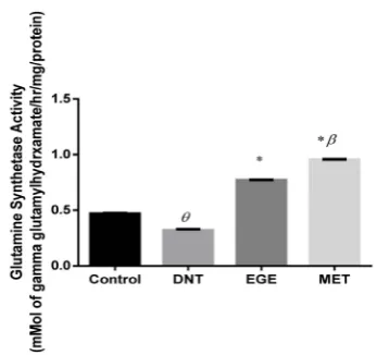
Figure 3. Effect of Metformin (MET) and ethanolic garlic extract (EGE) on hippocampal Glutamine
Synthetase activity in diabetic rats. Data are expressed as mean ± SEM (n = 6). *P < 0.001 when
compared with diabetic non-treated (DNT) group. β denotes significant difference when compared
[image:5.595.211.385.497.660.2]https://doi.org/10.4236/jbise.2018.119021 259 J. Biomedical Science and Engineering gamma glutamylhydroxamate/hr/mg/protein) of hippocampus of diabetic non-treated group when
com-pared with normal control group. Hippocampal glutamine synthetase activity was significantly (p < 0.001)
higher in EGE and MET groups when compared with diabetic control group. Activity of glutamine
syn-thetase in hippocampus was found to be significantly (p < 0.05) higher in MET group when compared
with EGE group.
4. DISCUSSION
In the present study, we investigated the effect of metformin, an insulin sensitizing drug and
ethanol-ic extract of garlethanol-ic on hippocampal membrane bound enzymes Na+/K+ ATPase, Ca2+ ATPase activities and
glutamate-glutamine cycle enzyme, glutamine synthetase activity in Type II diabetic rat model. Both
met-formin and garlic increased the activities of Na+/K+ ATPase, Ca2+ ATPase and glutamine synthetase in the
hippocampus of diabetic rat. Diabetes mellitus showed reduction in the activities of these enzymes which
is in line with the study of Vague et al. [3] for Na+/K+ ATPase, Kamboj et al. [6] for Ca2+ ATPase and
Zheng et al. [10] for glutamine synthetase [24]. Administration of metformin and garlic to diabetic rats
raised the level of activities of these enzymes in the hippocampus more than non-diabetic rats. However, metformin posed higher effects compared with garlic extract. Our previous studies reported increased
hippocampal Na+/K+ ATPase and Ca2+ ATPase activities in normal rats [23] and in Type II diabetic rats
[24] following garlic extract consumption. Diabetes in pregnancy has been shown to induce neuronal cell
apoptosis in offspring hippocampus which could be normalized by controlling the maternal glycemic state
with insulin treatment [31]. Study has shown that garlic administration during pregnancy and lactation
protected the rat offspring against lead-induced neuronal damage in the hippocampus [32]. Metformin
has been reported to reduce the decrease loss of neurons in the hippocampus of diabetic animals [14]. The
increased activities of Na+/K+ ATPase, Ca2+ ATPase and Glutamine synthetase in the hippocampus by
metformin revealed by our present study could be mechanism through which loss of neurons in hippo-campus is prevented in diabetic rats. Metformin has been shown to recruit neural stem cells and enhance
neural function by activating an a PKC-CBP pathway [33]. Neuroprotective properties of glucagon-like
peptide-1, alone and in combination with metformin, in Type II diabetes has been shown by Lennox et al.
[34] where metformin combination enhanced learning and memory, improved long-term potentiation in
the hippocampal CA1 region with reduced hippocampal levels of 8-oxoguanine and glia fibriallary acidic protein. The results of our present study could be added to the established mechanisms through which metformin mediates its neuroprotective functions especially hippocampal–dependent cognitive functions.
5. CONCLUSION
In conclusion, the present study suggested that the neuroprotective effect of metformin could be
found by increasing the activities of Na+/K+, Ca2+ ATPases and glutamine synthetase in the hippocampus
of Type II diabetic rats, thus makes the metformin a better drug in the management of Type II diabetes that is associated with complication of cognitive dysfunction.
CONFLICTS OF INTEREST
The authors declare that there is no conflict of interest regarding the publication of this paper.
REFERENCES
1. Sacai, H., Sasaki-Hamada, S., Sugiyama, A., Saitoh, A., Mori, K., Yamada, M. and Oka, J. (2014) The Impair-ment in Spatial Learning and Hippocampal LTD Induced through the PKA Pathway in Juvenile-Onset Diabetes Rats Are Rescued by Modulating NMDA Receptor Function. Neuroscience Research, 81-82, 55-63.
https://doi.org/10.1016/j.neures.2014.02.002
https://doi.org/10.4236/jbise.2018.119021 260 J. Biomedical Science and Engineering Slowing of Nerve Conduction in the Streptozotocin Diabetic Rat. Journal of Clinical Investigation, 94, 853-859. https://doi.org/10.1172/JCI117406
3. Vague, P., Coste, T.C., Jannot, M.F., Raccah, D. and Tsimaratos, M. (2004) C-Peptide, Na+, K+-ATPase, and Diabetes. Experimental Diabesity Research, 5, 37-50. https://doi.org/10.1080/15438600490424514
4. Welsh, B. and Wecker, L. (1991) Effects of Streptozotocin-Induced Diabetes on Acetylcholine Metabolism in Rat Brain. Neurochemical Research, 16, 453-460. https://doi.org/10.1007/BF00965566
5. Aubier, M. and Viires, N. (1998) Calcium ATPase and Respiratory Muscle Function. The European Respiratory
Journal, 11, 758-766.
6. Kamboj, S.S., Chopra, K. and Sandhir, R. (2009) Hyperglycemia-Induced Alterations in Synaptosomal Mem-brane Fluidity and Activity of MemMem-brane Bound Enzymes: Beneficial Effect of N-Acetylcysteine Supplementa-tion. Neuroscience, 162, 349-358. https://doi.org/10.1016/j.neuroscience.2009.05.002
7. Khakpai, F., Nasehi, M., Haeri-Rohani, A., Eidi, A. and Zarrindast, M.R. (2012) Scopolamine Induced Memory Impairment Possible Involvement of NMDA Receptor Mechanisms of Dorsal Hippocampus and/or Septum.
Behavioural Brain Research, 231, 1-10. https://doi.org/10.1016/j.bbr.2012.02.049
8. Szatkowski, M. and Attwell, D. (1994) Triggering and Execution of Neuronal Death in Brain Ischaemia: Two Phases of Glutamate Release by Different Mechanisms. Trends in Neurosciences, 17, 359-365.
https://doi.org/10.1016/0166-2236(94)90040-X
9. Chen, J. and Herrup, K. (2012) Glutamine Acts as a Neuroprotectant against DNA Damage, Beta-Amyloid and H2O2-Induced Stress. PLoS ONE, 7, e33177. https://doi.org/10.1371/journal.pone.0033177
10. Zheng, Y., Yang, Y., Dong, B., Zheng, H., Lin, X., Du, Y., Li, X., Zhao, L. and Gao, H. (2016) Metabonomic Pro-file Delineate Potential Role of Glutamate-Glutamine Cycle in db/db Mice with Diabetes-Associated Cognitive Decline. Molecular Brain, 18, 9-40. https://doi.org/10.1186/s13041-016-0223-5
11. Pintana, H., Apaijai, N., Pratchayasakul, W., Chattipakorn, N. and Chattipakorn, S.C. (2012) Effects of Met-formin on Learning and Memory Behaviors and Brain Mitochondrial Functions in High Fat Diet Induced Insu-lin Resistant Rats. Life Sciences, 91, 409-414. https://doi.org/10.1016/j.lfs.2012.08.017
12. Yuan, X., Chen, Y.B., Gan, D.N., Cheng, Y.F. and Xu, J.P. (2014) Metformin Improves Learning and Memory of Rats Induced by High-Fat Diet. Military Medical Sciences, 38, 17-21.
13. Zhang, W.H., Liu, Z., Shi, F.G. and Chen, Z.M. (2012). Huanglianwendan Tang in Mice with Type 2 Diabetes and the Impact on the Learning and Memory and the Form of Hippocampal Nerve Cells. Lishizhen Medicine
and Materia Medica Research, 23, 948-949.
14. Oliveira, W.H., Nunes, A.K., Franca, M.E., Santos, L.A., Los, D.B., Rocha, S.W., Barbosa, K.P., Rodrigues, G.B. and Peixoto, C.A. (2016) Effect of Metformin on Inflammation and Short-Term Memory in Streptozoto-cin-Induced Diabetic Mice. Brain Research, 1644, 149-160. https://doi.org/10.1016/j.brainres.2016.05.013 15. Mo, W. and Qian, G.F. (2014) Metformin for High Fat Diet Induced Insulin Resistance the Effect of Space
Cog-nition, Learning and Memory and Cerebral Energy Metabolism. Chinese Journal of Gerontology, 34, 2813-2815. 16. Asadbegi, M., Yaghmaei, P., Salehi, I., Ebrahim-Habibi, A. and Komaki, A. (2016) Neuroprotective Effects of Metformin against a β-Mediated Inhibition of Long-Term Potentiation in Rats Fed a High-Fat Diaet. Brain
Re-search Bulletin, 121, 178-185. https://doi.org/10.1016/j.brainresbull.2016.02.005
17. Anwar, M. and Meki, A. (2003) Oxidative Stress in Streptozotocin-Induced Diabetic Rats: Effects of Garlic Oil and Melatonin. Comparative Biochemistry and Physiology Part A, 135, 539-547.
https://doi.org/10.1016/S1095-6433(03)00114-4
https://doi.org/10.4236/jbise.2018.119021 261 J. Biomedical Science and Engineering
Diabetes and Metabolism, 15, 108-115.
19. Drobiova, H., Thomson, M., Al-Qattan, K., Peltonen-Shalaby, R., Al-Amin, Z. and Ali, M. (2011) Garlic In-creases Antioxidant Levels in Diabetic and Hypertensive Rats Determined by a Modified Peroxidase Method.
Evidence-Based Complementary and Alternative Medicine, 2011, Article ID: 703049.
20. Liu, C.T., Hse, H., Lii, C.K., Chen, P.S. and Sheen, L.Y. (2005) Effects of Garlic Oil and Diallyl Trisulfide on Glycemic Control in Diabetic Rats. European Journal of Pharmacology, 516, 165-173.
https://doi.org/10.1016/j.ejphar.2005.04.031
21. Shiju, T.M., Rajesh, N.G. and Viswanathan, P. (2013) Renoprotective Effect of Aged Garlic Extract in Strepto-zotocin-Induced Diabetic Rats. Indian Journal of Pharmacology, 45, 18-23.
22. Sarkaki, A., Valipour, S.C., Farbood, Y., Mohammad, S., Mansouri, T., Naghizadeh, B. and Basirian, E. (2013) Effects of Fresh, Aged and Cooked Garlic Extracts on Short- and Long-Term Memory in Diabetic Rats.
Avicen-na JourAvicen-nal of Phytomedicine, 3, 45-55.
23. Safiriyu, A.A., Semuyaba, I., RemónFigueredo, N., Etibor, T.A. and Ajibola, M.I. (2017) Garlic Extract (Allium
sativum) Enhances Spatial Working Memory in Wistar Rats: Involvement of Hippocampal Na+/K+ ATPase and
Ca2+ ATPase Activities. Journal of Behavioral and Brain Science, 7, 31-40. https://doi.org/10.4236/jbbs.2017.72004
24. Semuyaba, I., Safiriyu, A.A., Tiyo, E.A. and RemónFigueredo, N. (2017) Memory Improvement Effect of Etha-nol Garlic (A. sativum) Extract in Streptozotocin-Nicotinamide Induced Diabetic Wistar Rats Is Mediated through Increasing of Hippocampal Sodium-Potassium ATPase, Glutamine Synthetase, and Calcium ATPase Activities. Evidence-Based Complementary and Alternative Medicine, 3, 1-7.
https://doi.org/10.1155/2017/3720380
25. Homayounfar, H., BaluchnejadMojarad, T., Roghani, M., Hosseini, M. and Kamalinejad, M. (2003) Effect of Aqueous Garlic (Allium sativum L.) Extract on Acetylcholine and Isosorbide-Induced Relaxation of Isolated Aorta in Rat. Iranian Biomedical Journal, 7, 23-27.
26. Marudamuthu, A.S. and Leelavinothan, P. (2008) Emerging Targets Effect of Pterostilbene on Lipids and Lipid Profiles in Streptozotocin-Nicotinamide Induced Type 2. Journal of Applied Biomedicine, 6, 31-37.
27. Tirri, R., Lagrspetz, K.Y.H. and Kohomen, J. (1973) Temperature Dependence of the ATPase Activity in Brain Homogenates during the Postnatal Development of Rat. Comparative Biochemistry and Physiology, 44, 473-480. 28. Fiske, C.H. and Subba Row, Y. (1925) The Colorimetric Determination of Phosphates. The Journal of Biological
Chemistry, 66, 375-400.
29. Desaiah, D. and Ho, I.K. (1979) Effect of Acute and Continuous Morphine Administration on Catechola-mine-Sensitive Adenosine Triphosphatase in Mouse Brain. Journal of Pharmacology and Experimental
Thera-peutics, 208, 80-85.
30. Rowe, W.B., Ronzio, R.A., Wellner, V.P. and Meister, A. (1970) Glutamine Synthetase (Sheep Brain) Methods.
Enzymology, 17, 900-910. https://doi.org/10.1016/0076-6879(71)17304-1
31. Lotfi, N., Hami, J., Hosseini, M., Haghir, D. and Haghir, H. (2016) Diabetes during Pregnancy Enhanced Neu-ronal Death in the Hippocampus of Rat Offspring. International Journal of Developmental Neuroscience, 51, 28-35. https://doi.org/10.1016/j.ijdevneu.2016.04.009
32. Sadeghi, A., Ebrahimzadeh Bideskan, A.R., Alipour, F., Fazel, A.R. and Haghir, A. (2013) The Effect of Ascorbic Acid and Garlic Administration on Lead-Induced Neural Damage in Rat Offspring’s Hippocampus. Iranian
Journal of Basic Medical Sciences, 16, 157-164.
https://doi.org/10.4236/jbise.2018.119021 262 J. Biomedical Science and Engineering Enhance Spatial Memory Formation. Cell Stem Cell, 11, 23-35. https://doi.org/10.1016/j.stem.2012.03.016 34. Lennox, R., Porter, D.W., Flatt, P.R., Holscher, C., Irwin, N. and Gault, V.A. (2014) Comparison of the
Inde-pendent and Combined Effects of Sub-Chronic Therapy with Metformin and a Stable GLP-1 Receptor Agonist on Cognitive Function, Hippocampal Synaptic Plasticity and Metabolic Control in High-Fat Fed Mice.