ISSN Online: 2165-3364 ISSN Print: 2165-3356
DOI: 10.4236/ojvm.2018.87010 Jul. 31, 2018 87 Open Journal of Veterinary Medicine
Physiological and Molecular Characterization
of
Malassezia pachydermatis
Reveals No
Differences between Canines and Their Owners
Juan Camilo Galvis Marín
1, Fernando Borda Rojas
2, Andrés Julián Gutiérrez Escobar
11Group of Biological Investigations and Applied Genetics (GIBGA), Faculty Health Sciences, Universidad de Ciencias Aplicadas y
Ambientales (UDCA), Bogotá, Colombia
2Animal Science Research Group, Faculty of Animal Sciences, Universidad de Ciencias Aplicadas y Ambientales (UDCA), Bogotá,
Colombia
Abstract
Introduction: The genus Malassezia comprises 17 species of commensal and pathogenic yeasts of homeotherms animal skin. The most common species are M. furfur, M. globosa, and M. sympodialis in humans and M. pachydermatis in animals. However, some publications have reported potentially serious human infections by M. pachydermatis in individuals with risk factors and the isolation of human species from domestic animals. Given the scarcity of in-formation about their capacity for transmission between hosts and zoonotic potential, the aim of the present study was to physiologically and molecularly
characterize Malassezia spp. isolates obtained from canines and their human
owners. Materials and Methods: An experimental study was conducted at the Veterinary Clinic of Universidad de Ciencias Aplicadas y Ambientales of Bogotá (Colombia) from July 2015 to December 2016. Phenotypic identifica-tion and molecular characterizaidentifica-tion via the amplificaidentifica-tion of the 5.8S rDNA- ITS2 and 26S rDNA gene regions, nucleic acid sequencing, and phylogenetic analyses were performed on isolates originating from canines with otitis ex-terna and from the skin of healthy owners compatible with Malassezia spp. Results: Eighty samples were cultured, of which 32 (40%) were suggestive of Malassezia spp. A total of 29 out of 46 (63%) isolates in canines and 3 out of 34 (9%) isolates in humans corresponded entirely with M. pachydermatis. Isolates from the canines and their owners presented similar behavior in bio-chemical and phospholipase activity tests, 100% molecular sequence identi-ties, and close proximity in the phylogenetic trees. Conclusion: The isolation of M. pachydermatis from humans and their dogs with identity based on bio-chemical, physiological, molecular, and phylogenetic perspectives indicate the How to cite this paper: Marín, J.C.G.,
Rojas, F.B. and Escobar, A.J.G. (2018) Phy-siological and Molecular Characterization of Malassezia pachydermatis Reveals No Differences between Canines and Their Owners. Open Journal of Veterinary Medi-cine, 8, 87-105.
https://doi.org/10.4236/ojvm.2018.87010
Received: April 24, 2018 Accepted: July 28, 2018 Published: July 31, 2018
Copyright © 2018 by authors and Scientific Research Publishing Inc. This work is licensed under the Creative Commons Attribution International License (CC BY 4.0).
DOI: 10.4236/ojvm.2018.87010 88 Open Journal of Veterinary Medicine ability of this species to adapt to new hosts and its potential for zoonotic transmission. These findings contribute to knowledge of the ecology of this important fungus in human and veterinary medicine.
Keywords
Malassezia, Transmission, Animals, Humans, Zoonosis
1. Introduction
The genus Malassezia comprises lipophilic, lipid-dependent, and non-lipid- dependent yeasts that are commensal on the skin of humans and animals. These yeasts may become pathogenic in the presence of predisposing factors, such as changes in the cutaneous microenvironment or alterations of host defense me-chanisms [1] [2].
The identification of different species within the genus has traditionally been based on phenotypic tests [3]. More recently, identification has depended on molecular biology techniques based on the polymerase chain reaction (PCR) and the sequencing of different genes, such as genes of the ribosomal DNA complex (rDNA), including the 26S and 5.8S subunits [1] [4] [5]. These molecular studies have contributed to our understanding of the biodiversity of the genus and clari-fied some aspects of its taxonomy and phylogenetic relationships [6] [7] [8], which has led to the determination of new species. Currently, 17 species are rec-ognized [9]-[17].
Species such as M. furfur, M. globose, and M. sympodialis are part of the hu-man microbiota and have the ability to produce dermatological syndromes, such as pityriasis versicolor, and to exacerbate diseases, such as seborrheic dermatitis, atopic dermatitis, and psoriasis, as well as systemic syndromes in immunocom-promised patients [2] [18]. However, these species have occasionally been re-ported in domestic animals [19]-[25].
M. pachydermatis is the species isolated most often from the skin and mucosa of mammals (mainly canines), in which it can produce otitis externa and derma-titis [26]. However, this species has been reported in potentially serious human infections of individuals with associated risk factors, including some health per-sonnel with contact with dogs [25] [27] [28] [29] [30].
Whether an ecological relationship exists between the presence of this fungus in domestic animals and their owners is not completely clear because few studies have evaluated its capacity for transmission between hosts and the zoonotic po-tential [31] [32] [33] [34]. Therefore, the objective of the present study was to physiologically and molecularly characterize Malassezia spp. isolates obtained from canines and their human owners.
2. Materials and Methods
DOI: 10.4236/ojvm.2018.87010 89 Open Journal of Veterinary Medicine Skin or external auditory canal swabs were performed on canines with pa-thologies such as dermatitis or otitis externa and on the skin of healthy owners who attended the Veterinary Clinic of Universidad de Ciencias Aplicadas y Am-bientales of Bogota (Colombia) between October 2015 and September 2016. All samples were immediately transported to the microbiology laboratory in a sterile tube containing distilled water and 0.05% Tween 40 [19] [35] at room tempera-ture. Each sample was inoculated onto modified Dixon agar medium [3] and incubated at 32˚C for 5 days.
2) Phenotypic identification:
A smear and Gram staining were performed from the inoculations in which colonies compatible with Malassezia spp. were observed to evaluate the micro-scopic morphology. Additionally, a new isolation was conducted to obtain pure colonies and to perform phenotypic identification at the species level using the following biochemical tests: urease [36], catalase, β-glucosidase [37], Cremo-phor-EL assimilation [38], Tween assimilation, growth on Sabouraud agar [38],
39], and growth on Dixon agar at 37˚C and 40˚C [38] [40]. The physiological
test for phospholipase activity on Sabouraud agar supplemented with egg yolk was also performed with determination of Pz index [41].
3) Quality control and maintenance of strains:
The following reference strains from the Central Bureau voor Schimmelcul-tures (CBS) were used: M. furfur CBS 7019, M. pachydermatis CBS 1879, M. sympodialis CBS 7222, and M. slooffiae CBS 7956. These strains and the ob-tained isolates were conserved in skim milk medium at −20˚C.
4) Extraction of genomic DNA:
The reference strains and clinical isolates were cultured on modified Dixon agar for 72 hours at 32˚C. Subsequently, DNA extraction was performed using a
Fungi/Yeast Genomic DNA Isolation Kit (Norgen) according to the
manufac-turer’s instructions. The DNA was stored at −20˚C prior to use [42]. 5) Amplification of ribosomal genes:
The primers used to amplify the 5.8S rDNA-ITS2 target sequence were ITS3 (5’-GCATCGATGAAGAACGCAGC-3’) and ITS4
(5’-TCCTCCGCTTATTGATATGC-3’) [39] [43]. The amplification was
per-formed by PCR in a 25-μL reaction volume containing 10X buffer, 2 mM MgCl2,
1 μmol of each primer, 0.2 mM dNTPs (Thermo Scientific), 1 U of Taq
poly-merase (Bioline), and 1 µL of genomic DNA under the following conditions: an
initial denaturation cycle at 95˚C for 5 minutes, 25 cycles at 95˚C for 1 minute, 55.4˚C for 30 seconds, and 72˚C for 1 minute, and a final extension cycle at 72˚C for 5 minutes. The 5’-TAACAAGGATTCCCCTAGTA-3’ and
5’-ATTACGCCAGCATCCTAAG-3’ primers were used to amplify the 26S rDNA target sequence [4] [39]. The amplification was performed by PCR in a
25-µL reaction volume containing 10X buffer, 1.5 mM MgCl2, 1 μmol of each
primer, 0.2 mM dNTPs (Thermo Scientific), 1 U of Taq polymerase (Bioline),
denatura-DOI: 10.4236/ojvm.2018.87010 90 Open Journal of Veterinary Medicine tion cycle at 95˚C for 5 minutes, 25 cycles of 95˚C for 1 minute, 51.7˚C for 30 seconds, and 72˚C for 1 minute, and one final extension cycle at 72˚C for 5 mi-nutes. The amplification products were examined by electrophoresis on 1.5% agarose gels with 1X TAE buffer and stained with EZ-Vision.
6) Sequencing and phylogenetic analysis:
The amplification products corresponding to the 5.8S rDNA-ITS2 and 26S
rDNA regions were sent to Macrogen, Inc. (Korea) for purification and
se-quencing. The DNA sequences were edited and assembled manually using Geneious v7.0.6 software (created by Biomatters, available at
http://www.geneious.com) and subsequently compared with GenBank sequences using the National Center for Biotechnology Information (NCBI) BLASTn tool for identification at the species level. Each data set was aligned using the MUSCLE v3.8.31 algorithm [44] using default parameters, and conserved blocks were selected using Gblocks v0.91b [45]. Phylogenetic reconstruction from the alignments of the conserved blocks was performed using MEGA v7.0.21 software [46]. First, we determined that the evolutionary models that best explained the alignments were T92+G+I and K2. The maximum likelihood methodology was used for the reconstruction of trees under the selected models, and statistical robustness was achieved with 2000 bootstrap replicates. The trees were edited using the iTOL v3. tool [47].
3. Results
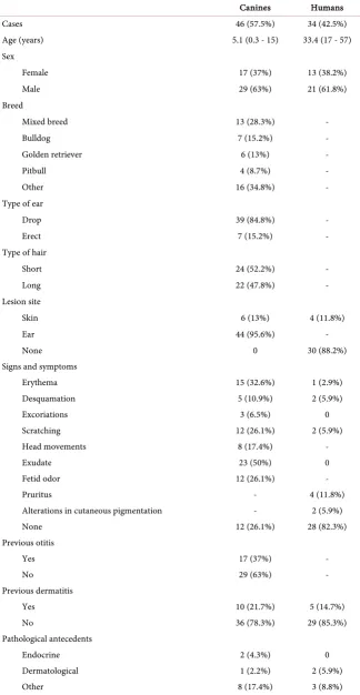
A total of 80 samples were collected, including 46 samples of canine origin and 34 of human origin. The average age was 5 years for the canines and 33 years for the humans. A total of 37% of the canines were females and 63% were males, whereas 38% of the humans were females and 62% were males. The demograph-ic and clindemograph-ical characteristdemograph-ics of the canines and humans included in the study are shown in Table 1.
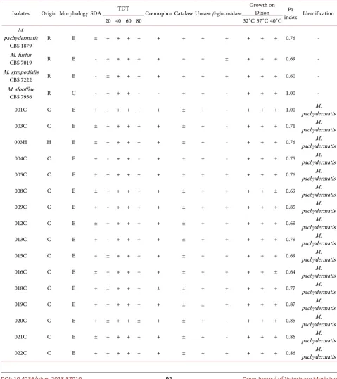
Thirty-two positive cultures were obtained for Malassezia spp., of which 29 were of canine origin and 3 were of human origin. Of these isolates, 90.6% were identified as M. pachydermatis by phenotypic tests. The remaining isolates were not conclusive for identification at the species level. The phospholipase activity of the studied isolates was high in 87.5% of the cases, very high in 6.25% of the cases, and null in 6.25% of the cases as shown by Pz indexes (Table 2).
DOI: 10.4236/ojvm.2018.87010 91 Open Journal of Veterinary Medicine
Table 1. Demographic and clinical characterization of the study population.
Canines Humans
Cases 46 (57.5%) 34 (42.5%)
Age (years) 5.1 (0.3 - 15) 33.4 (17 - 57)
Sex
Female 17 (37%) 13 (38.2%)
Male 29 (63%) 21 (61.8%)
Breed
Mixed breed 13 (28.3%) -
Bulldog 7 (15.2%) -
Golden retriever 6 (13%) -
Pitbull 4 (8.7%) -
Other 16 (34.8%) -
Type of ear
Drop 39 (84.8%) -
Erect 7 (15.2%) -
Type of hair
Short 24 (52.2%) -
Long 22 (47.8%) -
Lesion site
Skin 6 (13%) 4 (11.8%)
Ear 44 (95.6%) -
None 0 30 (88.2%)
Signs and symptoms
Erythema 15 (32.6%) 1 (2.9%)
Desquamation 5 (10.9%) 2 (5.9%)
Excoriations 3 (6.5%) 0
Scratching 12 (26.1%) 2 (5.9%)
Head movements 8 (17.4%) -
Exudate 23 (50%) 0
Fetid odor 12 (26.1%) -
Pruritus - 4 (11.8%)
Alterations in cutaneous pigmentation - 2 (5.9%)
None 12 (26.1%) 28 (82.3%)
Previous otitis
Yes 17 (37%) -
No 29 (63%) -
Previous dermatitis
Yes 10 (21.7%) 5 (14.7%)
No 36 (78.3%) 29 (85.3%)
Pathological antecedents
Endocrine 2 (4.3%) 0
Dermatological 1 (2.2%) 2 (5.9%)
DOI: 10.4236/ojvm.2018.87010 92 Open Journal of Veterinary Medicine
Continued
None 36 (78.3%) 29 (85.3%)
Previous treatments
Antimicrobials 7 (15.2%) 0
Immunosuppressants 5 (10.9%) 1 (2.9%)
Other 11 (23.9%) 3 (8.8%)
[image:6.595.57.537.207.747.2]None 33 (71.7%) 31 (91.2%)
Table 2. Results of biochemical tests for phenotypic identification.
Isolates Origin Morphology SDA TDT Cremophor Catalase Urease β-glucosidase
Growth on Dixon Pz
index Identification
20 40 60 80 32˚C 37˚C 40˚C
M. pachydermatis
CBS 1879 R E ± + + + + + + + + + + + 0.76 -
M. furfur
CBS 7019 R E - + + + + + + + ± + + + 0.69 -
M. sympodialis
CBS 7222 R E - ± + + + + + + + + + + 0.60 -
M. slooffiae
CBS 7956 R C - + + + - - + + - + + + 1.00 -
001C C E + + + + + + ± + - + + + 1.00 pachydermatis M.
003C C E ± + + + + + ± + - + + + 0.71 pachydermatis M.
003H H E ± + + + + + ± + - + + + 0.76 pachydermatis M.
004C C E + - + + - + ± + - + + ± 0.75 pachydermatis M.
005C C E ± + + + + + ± ± ± + + + 0.76 pachydermatis M.
008C C E ± + + + + + ± + + + + ± 0.69 pachydermatis M.
009C C E + - + + + + ± + + + + + 0.85 pachydermatis M.
012C C E ± + + + + + ± + + + + + 0.69 pachydermatis M.
013C C E + - + + + + ± + + + + + 0.79 pachydermatis M.
015C C E + ± + + + + ± + + + + + 0.69 pachydermatis M.
016C C E ± + + + + + ± + + + + ± 0.64 pachydermatis M.
018C C E + ± + + + ± ± + + + + + 0.77 pachydermatis M.
019C C E + + + + + + ± ± + + + + 0.87 pachydermatis M.
020C C E + ± + + ± + ± + - + + + 0.85 pachydermatis M.
021C C E ± + + + + + ± + - + + + 0.86 pachydermatis M.
DOI: 10.4236/ojvm.2018.87010 93 Open Journal of Veterinary Medicine
Continued
025C C E + - ± + + + ± + + + + ± 0.77 pachydermatis M.
026C C E + - ± + + + ± + + + + ± 0.77 pachydermatis M.
029C C E + - + + + ± ± + + + + ± 0.63 pachydermatis M.
031C C E + - + + + + ± + + + + + 0.73 pachydermatis M.
036C C E + - - + + - ± + + + + + 0.86 Malassezia sp.
037C C E + - + + + + ± + ± + + + 0.81 pachydermatis M.
038C C E + - + + + - ± + + + + + 0.76 Malassezia sp.
039C C E + - + + + + ± + + + + + 0.82 pachydermatis M.
040C C E + - + + + + ± + + + + + 0.76 pachydermatis M.
041H H E + - + + + + + + - + + + 1.00 pachydermatis M.
042C C E + - + + + + ± + - + + + 0.76 pachydermatis M.
043H H E + + + + + + + + - + + + 0.59 pachydermatis M.
044C C E + ± + + + + ± + - + + + 0.76 pachydermatis M.
046C C E + + + + + ± ± + - + - - 0.69 Malassezia sp.
047C C E + ± + + + + ± + - + + ± 0.84 pachydermatis M.
048C C E + ± + + ± + ± + - + + + 1.00 pachydermatis M.
Reference strain (R); canine (C); human (H); ellipsoidal (E); cylindrical (C); Sabouraud dextrose agar (SDA); weakly positive (±); Tween diffusion test (TDT).
[image:7.595.216.536.484.641.2](a) (b)
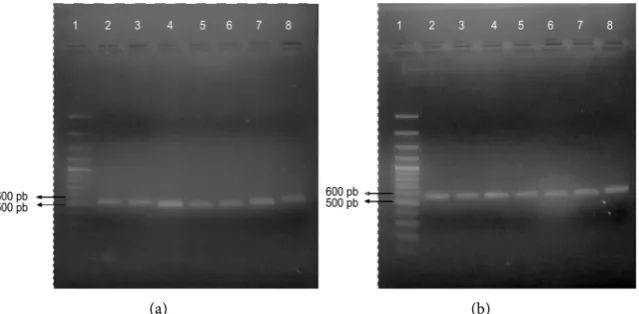
Figure 1. Amplification of ribosomal genes. (a) Amplification of the 5.8S ADNr-ITS2 re-gion: lane 1, HyperLadder II molecular size marker; lanes 2 - 4, canine isolates; lanes 5 - 6: human isolates; lane 7, M. pachydermatis CBS 1879; lane 8: M. furfur CBS 7019; (b) Am-plification of the 26S rDNA region: lane 1, HyperLadder II molecular size marker; lanes 2-4, canine isolates; lanes 5-6: human isolates; lane 7, M. pachydermatis CBS 1879; lane 8:
DOI: 10.4236/ojvm.2018.87010 94 Open Journal of Veterinary Medicine However, the consensus sequences of the 5.8S rDNA-ITS2 and 26S rDNA re-gions were obtained, a BLASTn analysis was performed, and these sequences were compared with the sequences deposited in the databases. One hundred
percent (100%) identification of the isolates (i.e., M. pachydermatis) was
achieved using the two genetic markers, with identity percentages of 95.6% - 100% and E-values of 0.0. Thus, the occurrence of M. pachydermatis was con-firmed in 63% of the canines and 9% of the humans included in the study.
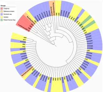
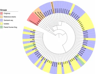
Phylogenetic analyses were performed with the two genetic regions (Figure 2 and Figure 3). All isolates of both canine and human origin clustered together in the same clade with sequences corresponding to M. pachydermatis identified with the GenBank accession numbers KY272204 to KY272250 and KU757185 to KU757234 and the reference strain M. pachydermatis CBS 1879. The reference
strains of other species of the genus Malassezia and Cryptococcus neoformans
CBS 132 were used as outgroups in addition to the sequences of three isolates identified as Cryptococcus spp. In these trees, we can observe the phylogenetic proximity of the isolates of canine and human origin.
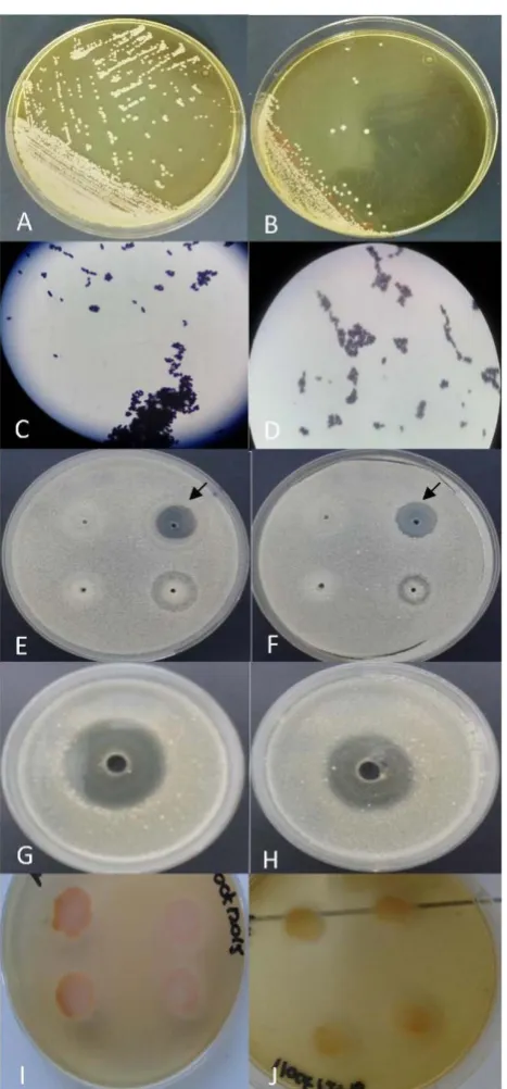
[image:8.595.208.540.390.685.2]Specifically, the 003H isolate of human origin and the 003C isolate of the ca-nine owned by this individual showed almost identical biochemical and phos-pholipase activity profiles (Figure 4), as well as 100% sequence identity and a close proximity in the phylogenetic reconstructions (Figure 2 and Figure 3).
DOI: 10.4236/ojvm.2018.87010 95 Open Journal of Veterinary Medicine
Figure 3. Phylogenetic analysis by maximum likelihood for the 26S rDNA region. Hu-man isolates have the letter H and canine isolates have the letter C.
4. Discussion
In the present study, M. pachydermatis was isolated from 63% of the canine subjects with otitis externa or dermatitis, which coincided with the results re-ported by several authors who claimed that this yeast was most commonly asso-ciated with otic and dermatologic pathologies in canines [19] [20] [23] [48] [49]
[50] [51] [52]. M. pachydermatis was isolated from animals of all ages, ranging
from 3-month-old puppies to older dogs (15 years). Similar results were found in a Colombian study in which no significant differences were found in the ages of canine patients with otitis caused by Malassezia [19].
When analyzing the canine breeds from which this yeast was isolated, we ob-served that less than 30% of the canines corresponded to mixed-breed dogs. This finding agreed with a previous publication that showed that pure breeds were more susceptible to suffering pathologies caused by M. pachydermatis [53]. Moreover, the majority of dogs in which the fungus was isolated belonged to breeds with drop ears, which seemed to be a predisposing factor for otitis caused by Malassezia [26]. The main clinical manifestations of the affected canines were scratching, erythema, exudate, and a fetid odor; this description coincides with reports by other researchers [50] [53] and is the result of the inflammatory re-sponse triggered by the fungus and associated bacteria, such as Staphylococcus spp. [54].
DOI: 10.4236/ojvm.2018.87010 96 Open Journal of Veterinary Medicine
DOI: 10.4236/ojvm.2018.87010 97 Open Journal of Veterinary Medicine to the use of immunosuppressive and antimicrobial drugs [53]. However, most of the animals that participated in the study did not have a history of dermatitis or other conditions that could favor the growth and pathogenicity of this op-portunistic yeast. Therefore, the majority of canines presented a primary der-matomycosis. This observation suggests that there may be companion animals with dermatomycosis, but without underlying diseases. This possibility may faci-litate transmission to their owners because the probability of a timely diagnosis and adequate treatment and prevention measures decreases in the absence of significant signs of illness.
M. pachydermatis was isolated from the skin of 9% of the healthy humans participating in this study. The first publications describing the presence of M. pachydermatis on human skin dated back to the 1970s in patients with chronic cutaneous diseases, in which this yeast was isolated in 16% of the cases [56] and in one patient with a primary immunodeficiency [57]. Later, the isolation of M. pachydermatis from skin and other anatomical sites was reported in 47% of pre-term infants and in adults with various syndromes, although whether this yeast actually caused the infections or was a colonizer was unclear [58]. This species was also found in 6% of healthy patients from cultures of skin smears and con-ventional biochemical tests, indicating that in most cases, the presence of M. pachydermatis on human skin was rare and transient [59].
An additional finding suggesting the exogenous origin of M. pachydermatis in human infections is that cutaneous colonization by Malassezia species in healthy full-term infants begins at birth and increases in the first weeks of life for M. sympodialis and M. globosa, whereas M. pachydermatis has not been isolated from the skin of newborns or their mothers [60]. In more recent studies, M. pa-chydermatis was identified in less than 1% and up to 5% of patients with der-matological lesions and healthy controls, respectively, with the isolation of M. globosa, M. sympodialis, and M. furfur being more common [30] [35] [61]. However, in the present work, these species were probably not isolated due to their more demanding nutritional requirements.
Using molecular techniques, M. pachydermatis was found in only 0.4% of pa-tients with pityriasis versicolor [62] and in 0.7% of papa-tients with seborrheic dermatitis [63]. Studies attempting to determine the species of the genus most common in patients with pityriasis versicolor noted that M. pachydermatis has only been found in one person with contact with domestic animals [64]. These data support the hypothesis that M. pachydermatis is not a member of the nor-mal human microbiota and that its presence on the skin indicates transmission from an external source, such as contact with companion animals.
DOI: 10.4236/ojvm.2018.87010 98 Open Journal of Veterinary Medicine furfur. Infection with this fungus was associated with the lipid-rich parenteral nutrition in immunocompromised patients due to the lipophilicity shared by all species within the genus and their opportunistic behavior because most cases were isolated from the blood and other body fluids of preterm infants in neonat-al intensive care units (NICUs) [65]. Thus, M. pachydermatis was found to co-lonize several anatomical sites in hospitalized patients [66].
The use of molecular biology tests and genetic typing of clinical isolates clearly demonstrated the nosocomial nature of the epidemics caused by M. pachyder-matis in the NICUs because all isolates recovered from both the patients and the incubator surfaces were genetically comparable and persisted for several months despite regular cleaning [67]. Moreover, identical genetic profiles were generated among strains of M. pachydermatis isolated from hospitalized neonates [68]. The data from these studies supported the theory that M. pachydermatis could cause outbreaks of nosocomial fungemia and that children with low birth weights, who were severely ill, and who had arterial catheters in place for several days might be at greater risk. Furthermore, nosocomial dissemination from health personnel to patients was thought to be facilitated by inappropriate asep-tic and antisepasep-tic techniques of surfaces, and improper hand washing. Thus, me-ticulous measures of personal hygiene should be implemented for the doctors and nurses who handle newborns, and modifications in cleaning procedures should be implemented [32] [69].
The characterization of isolates of canine and human origin recovered in the present work suggests genetic identity and a possible route of zoonotic transmis-sion. We perform these phenotypic tests because they have been used tradition-ally in the identification of the species of the genus [1] and to observe if the yeasts recovered from dogs and their owners had similar biochemical and physi-ological behaviors. The zoonotic association of pets with M. pachydermatis in-fections was speculated in an outbreak described in a NICU whose origin ap-parently was from the hands of a nurse who had dogs at home. This conclusion was reached by molecular typing because a common strain colonized the dogs, the health worker, and the newborns [32] [70]. The carrier status of M. pachy-dermatis in dog owners was shown from cultures. This species was found in 38.7% of canine owners with dermatitis or otitis attributed to Malassezia and in only 6% of healthy dog owners. Conversely, the diagnostic yield of the PCR-based tests was much higher (up to 94%) [33].
DOI: 10.4236/ojvm.2018.87010 99 Open Journal of Veterinary Medicine In this study, molecular and phylogenetic characterizations of M. pachyder-matis isolates from humans and canines were performed. Two genetic markers have been described for the systematic analysis of yeasts of the genus Malassezia: the 5.8S rDNA-ITS2 region, which includes the internal transcribed spacers (ITS), whose length differs between species and is usually identical between strains of the same species, and the 26S rDNA region, which includes the D1/D2 domains, whose lengths are identical in all species of the genus and the similarity of the sequences within the same species is greater than 99% [71]. The amplifica-tion of these regions showed similar results to the literature, such as a 483-bp product using the 5.8S rDNA-ITS2 region for M. pachydermatis isolates from the lesions of canines with otitis [43] and a 550-bp product for eight Malassezia species using the 26S rDNA region [12]. Sequencing of these products confirmed that all of the isolates corresponded to M. pachydermatis.
Phylogenetic trees constructed with both markers showed the inclusion of isolates of human and canine origin within the same clade. A study in the late twentieth century classified isolates recovered from humans in the sequevar with the most common type sequences of strains isolated from dogs, which suggested the capacity of this canine-specific yeast to adapt to new hosts [31]. Subsequent-ly, the molecular characterization of lipid-dependent isolates from different spe-cies of domestic animals was performed. Although that study did not include human strains, four independent clusters as a function of the origin of the yeast were obtained. Thus, whether the value of the genetic differences observed was important for the definition of species or for the process of adaptation to specific hosts was unclear [72].
The observation of genetic variants of M. pachydermatis from canine skin and their relationship with corporal distribution and phospholipase activity has also been evaluated. Higher activity is associated with isolates recovered from cuta-neous lesions compared to healthy skin, suggesting that the presence of particu-lar genotypes of M. pachydermatis on the skin of canines is related to the affinity of the yeast for the host and specific cutaneous sites [73]. In the present work, the relationship between phospholipase activity and anatomical sites was not studied. However, the activity indexes were similar between isolates of canine and human origin, which indicated similar virulence patterns in both host types. The characterization of atypical M. furfur isolates from skin lesions of one ca-nine showed the presence of genetic and physiological polymorphisms that could be the result of the adaptation process of this human-specific species to a new host [21]. However, in the present study, the lipid-dependent species spe-cific to humans were not isolated from the canines.
5. Conclusions
DOI: 10.4236/ojvm.2018.87010 100 Open Journal of Veterinary Medicine
known, but few reports of human infections by M. pachydermatis are available,
and the risk factors have not been well studied. Currently, this yeast is consi-dered rare and transient on human skin and is not part of the normal microbi-ota, so its isolation suggests an exogenous origin.
Although more robust molecular epidemiology studies are required to con-firm the transmission of this canine microorganism to its owners and catalog human infections caused by M. pachydermatis as a true emerging zoonosis, finding the yeast in people living with dogs with otitis or dermatitis suggests its capacity to adapt to new hosts. These new hosts will behave as asymptomatic carriers in the case of healthy patients or become ill if they have predisposing factors. All of these findings will contribute to knowledge of the ecology and ep-idemiology of this fungus of importance in human and veterinary medicine.
Acknowledgements
The authors thank Universidad de Ciencias Aplicadas y Ambientalesfor financial support of the project and especially the University Veterinary Clinic for their collaboration in the collection of the samples. We also thank the Laboratory of Mycology and Phytopathology at Universidad de Los Andes for kindly providing the reference strains.
Author’s Disclosure Statement
There are no financial interests.
References
[1] Cafarchia, C., Gasser, R., Figueredo, L., Latrofa, M. and Otranto, D. (2011) Ad-vances in the Identification of Malassezia. Molecular and Cellular Probes, 25, 1-7.
https://doi.org/10.1016/j.mcp.2010.12.003
[2] Gaitanis, G., Magiatis, P., Hantschke, M., Bassukas, I. and Velegraki, A. (2012) The
Malassezia Genus in Skin and Systemic Diseases. Clinical Microbiology Reviews, 25, 106-141. https://doi.org/10.1128/CMR.00021-11
[3] Guillot, J., Breugnot, C., De Barros, M. and Chermette, R. (1998) Usefulness of Modified Dixon’s Medium for Quantitative Culture of Malassezia Species from Ca-nine Skin. Journal of Veterinary Diagnostic Investigation, 10, 384-386.
https://doi.org/10.1177/104063879801000418
[4] Mirhendi, H., Makimura, K., Zomorodian, K., Yamada, T., Sugita, T. and Yamaguchi, H. (2005) A Simple PCR-RFLP Method for Identification and Differen-tiation of 11 Malassezia Species. Journal of Microbiological Methods, 61, 281-284.
https://doi.org/10.1016/j.mimet.2004.11.016
[5] Gaitanis, G., Robert, V. and Velegraki, A. (2006) Verifiable Single Nucleotide Po-lymorphisms of the Internal Transcribed Spacer 2 Region for the Identification of 11 Malassezia Species. Journal of Dermatological Science, 43, 214-217.
https://doi.org/10.1016/j.jdermsci.2006.03.013
DOI: 10.4236/ojvm.2018.87010 101 Open Journal of Veterinary Medicine
https://doi.org/10.1016/S0723-2020(89)80067-0
[7] Begerow, D., Bauer, R. and Boekhout, T. (2000) Phylogenetic Placements of Ustila-ginomyceteous Anamorphs as Deduced from LSU rDNA Sequences. Mycological Research, 104, 53-60. https://doi.org/10.1017/S0953756299001161
[8] Fell, J., Boekhout, T., Fonseca, A., Scorzetti, G. and Statzell, A. (2000) Biodiversity and Systematic of Basidiomycetous Yeast as Determined by LSU rDNA D1/D2 Domain Sequence Analysis. International Journal of Systematic and Evolutionary Microbiology, 50, 1351-1371. https://doi.org/10.1099/00207713-50-3-1351
[9] Sugita, T., Takashima, M., Shinoda, T., Suto, H., Unno, T., Tsuboi, R., Ogawa, H. and Nishikawa, A. (2002) New Yeast Species Malassezia dermatis Isolated from Pa-tients with Atopic Dermatitis.Journal of Clinical Microbiology, 40, 1363-1367.
https://doi.org/10.1128/JCM.40.4.1363-1367.2002
[10] Sugita, T., Takashima, M., Kodama, M., Tsuboi, R. and Nishikawa, A. (2003) De-scription of a New Yeast Species Malassezia japonica and Its Detection in Patients with Atopic Dermatitis and Healthy Subjects. Journal of Clinical Microbiology, 41, 4695-4699. https://doi.org/10.1128/JCM.41.10.4695-4699.2003
[11] Gupta, A., Boekhout, T., Theelen, B., Summerbell, R. and Batra, R. (2004) Identifi-cation and Typing of Malassezia Species by AFLP and Sequence Analysis of the ITS and LSU Regions of rDNA. Journal of Clinical Microbiology, 42, 4253-4260.
https://doi.org/10.1128/JCM.42.9.4253-4260.2004
[12] Hirai, A., Kano, R., Makimura, K., Duarte, E., Hamdan, J., Lachance, M., Yamagu-chi, H. and Hasegawa, A. (2004) Malassezia nana, a Novel Lipid-Dependent Yeast Species Isolated from Animals. International Journal of Systematic and Evolutionary Microbiology, 54, 623-627. https://doi.org/10.1099/ijs.0.02776-0
[13] Sugita, T., Tajima, M., Takashima, M., Amaya, M., Saito, M., Tsuboi, R. and Nishi-kawa, A. (2004) A New Yeast Malassezia yamatoensis Isolated from Patients with Seborrheic Dermatitis and Its Distribution in Patients and Healthy Subjects.
Microbiology and Immunology, 48, 579-583.
https://doi.org/10.1111/j.1348-0421.2004.tb03554.x
[14] Cabañes, F., Theelen, B., Castellá, G. and Boekhout, T. (2007) Two New Li-pid-Dependent Malassezia Species from Domestic Animals. FEMS Yeast Research, 7, 1064-1076. https://doi.org/10.1111/j.1567-1364.2007.00217.x
[15] Cabañes, F., Vega, S. and Castellá, G. (2011) Malassezia cuniculi sp. nov., a Novel Yeast Species Isolated from Rabbit Skin. Medical Mycology, 49, 40-48.
[16] Cabañes, F., Coutinho, S., Puig, L., Bragulat, M. and Castellá, G. (2016) New Li-pid-Dependent Malassezia Species from Parrots. Revista Iberoamericana de Micología, 33, 92-99. https://doi.org/10.1016/j.riam.2016.03.003
[17] Honnavar, P., Prasad, G., Ghosh, A., Dogra, S., Handa, S. and Rudramurthy, S. (2016) Malassezia arunalokei sp. nov., a Novel Yeast Species Isolated from Seborr-heic Dermatitis Patients and Healthy Individuals from India. Journal of Clinical Microbiology, 54, 1826-1834. https://doi.org/10.1128/JCM.00683-16
[18] Tragiannidis, A., Bisping, G., Koehler, G. and Groll, A. (2009) Malassezia Infections in Immunocompromised Patients. Mycoses, 53, 187-195.
https://doi.org/10.1111/j.1439-0507.2009.01814.x
[19] Pulido, A., Castañeda, R., Linares, M. and Mercado, M. (2010) Clinical-Microbiological Diagnostic of External Otitis in Canines in Bogotá, Colombia. Revista MVZ Córdoba, 15, 2215-2222. https://doi.org/10.21897/rmvz.308
Pra-DOI: 10.4236/ojvm.2018.87010 102 Open Journal of Veterinary Medicine
pasarakul, N. (2011) Comparative Analysis of the Frequency, Distribution and Pop-ulation Sizes of Yeasts Associated with Canine Seborrheic Dermatitis and Healthy Skin. Veterinary Microbiology, 148, 356-362.
https://doi.org/10.1016/j.vetmic.2010.09.020
[21] Cafarchia, C., Latrofa, M., Figueredo, L., Da Silva, M., Ferreiro, L., Guillot, J., Boekhout, T. and Otranto, D. (2011) Physiological and Molecular Characterization of Atypical Lipid-Dependent Malassezia Yeasts from a Dog with Skin Lesions: Adaptation to a New Host? Medical Mycology, 49, 365-374.
[22] Brigitta, B., Edina, B., Nocoletta, S. and Gyory, H. (2014) Isolation of Malassezia furfur from Canine Skin Scrapings. Magyar Allatorvosok Lapja, 136, 41-44. [23] Pulido, A., Castañeda, R., Linares, M. and Mercado, M. (2015) Concordance
be-tween Otic Cytology and Culture in Diagnosis of External Otitis Canine by Malas-sezia spp. Revista MVZ Córdoba, 20, 4720-4724. https://doi.org/10.21897/rmvz.42
[24] Shokri, H. and Khosravi, A. (2016) An Epidemiological Study of Animals Derma-tomycosis in Iran. Journal de Mycologie Médicale, 2, 170-177.
https://doi.org/10.1016/j.mycmed.2016.04.007
[25] Galvis, J. and Borda, F. (2016) Infecciones zoonóticas por levaduras del género Ma-lassezia: Una revisión. Revista U.D.C.A Actualidad & Divulgación Científica, 19, 381-393.
[26] Bond, R., Guillot, J. and Cabañes, F. (2010) Malassezia Yeasts in Animal Disease. In: Boekhout, T., Guého, E., Mayser, P. and Velegraki, A., Ed., Malassezia and the Skin, Springer-Verlag, Berlin, 271-299. https://doi.org/10.1007/978-3-642-03616-3_10
[27] Guillot, J. and Bond, R. (1999) Malassezia pachydermatis: A Review. Medical My-cology, 37, 295-306. https://doi.org/10.1046/j.1365-280X.1999.00237.x
[28] Chitty, J. and Hendricks, A. (2007) Zoonotic Skin Disease in Small Animals. In Practice, 29, 92-97. https://doi.org/10.1136/inpract.29.2.92
[29] Mani, I. and Maguire, J. (2009) Small Animal Zoonoses and Immunocompromised Pet Owners. Topics in Companion Animal Medicine, 24, 164-174.
https://doi.org/10.1053/j.tcam.2009.07.002
[30] Prohic, A., Jovovic, T., Krupalija, M. and Kuskunovic, S. (2016) Malassezia Species in Healthy Skin and in Dermatological Conditions. International Journal of Dermatology, 55, 494-504. https://doi.org/10.1111/ijd.13116
[31] Guillot, J., Guého, E., Chévrier, G. and Chermette, R. (1997) Epidemiological Anal-ysis of Malassezia pachydermatis Isolates by Partial Sequencing of the Large Subunit Ribosomal RNA. Research in Veterinary Science, 62, 22-25.
https://doi.org/10.1016/S0034-5288(97)90174-0
[32] Chang, H., Miller, H., Watkins, N., Arduino, M., Ashford, D., Midgley, G., Agüero, S., Pinto, R., von Reyn, C., Edwards, W., McNeil, M. and Jarvis, W. (1998) An Epi-demic of Malassezia pachydermatis in an Intensive Care Nursery Associated with Colonization of Health Care Workers’ Pet Dogs. The New England Journal of Med-icine, 338, 706-711. https://doi.org/10.1056/NEJM199803123381102
[33] Morris, D., O’Shea, K., Shofer, F. and Rankin, S. (2005) Malassezia pachydermatis
Carriage in Dog Owners. Emerging Infectious Disease, 11, 83-88.
https://doi.org/10.3201/eid1101.040882
[34] Fan, Y., Huang, W., Li, S., Wu, G., Lai, K. and Chen, R. (2006) Granulomatous Skin Infection Caused by Malassezia pachydermatis in a Dog Owner. Archives of Dermatology, 142, 1181-1184. https://doi.org/10.1001/archderm.142.9.1181
DOI: 10.4236/ojvm.2018.87010 103 Open Journal of Veterinary Medicine
Species Isolated from Patients with Dermatologic Lesions. Biomédica, 25, 189-195.
https://doi.org/10.7705/biomedica.v25i2.1341
[36] Pérez, C., Goitia, K., Mata, S., Hartung, C., Colella, M., Reyes, H., Hernández, C., Villarroel, M., Ontiveros, J., Magaldy, S. and Suárez, R. (2002) Use of the Stuart Urea Broth for the Urease Test: Like Proof in the Identification of Yeast. Revista de la Sociedad Venezolana de Microbiología, 22, 77-80.
https://doi.org/10.1007/BF00399623
[37] Khosravi, A., Eidi, S., Ziglari, T. and Bayat, M. (2008) Isolation and Differentiation of Malassezia Species Isolated from Healthy and Affected Small Animals, Ear and Skin. World Journal of Zoology, 3, 77-80.
[38] Kaneko, T., Makimura, K., Abe, M., Shiota, R., Nakamura, Y., Kano, R., Hasegawa, A., Sugita, T., Shibuya, S., Watanabe, S., Yamaguchi, H., Abe, S. and Okamura, N. (2007) Revised Culture-Based System for Identification of Malassezia Species. Journal of Clinical Microbiology, 45, 3737-3742.
[39] Hernández, J. (2005) Caracterización molecular de especies del género Malassezia. Ph.D. Dissertation, Universidad Autónoma de Barcelona, Barcelona, España. [40] Guého, E., Midgley, G. and Guillot, J. (1996) The Genus Malassezia with
Descrip-tion of Four New Species. Antonie van Leeuwenhoek, 69, 337-355.
https://doi.org/10.1007/BF00399623
[41] Coutinho, S. and Paula, C. (2000) Proteinase, Phospholipase, Hyaluronidase and Chondroitin-Sulphatase Production by Malassezia pachydermatis. Medical Mycol-ogy, 38, 73-76. https://doi.org/10.1080/mmy.38.1.73.76
[42] Gemmer, C., DeAngelis, Y., Theelen, B., Boekhout, T. and Dawson, T. (2002) Fast, Noninvasive Method for Molecular Detection and Differentiation of Malassezia
Yeast Species on Human Skin and Application of the Method to Dandruff Microbi-ology. Journal of Clinical Microbiology, 40, 3350-3357.
https://doi.org/10.1128/JCM.40.9.3350-3357.2002
[43] Gaitanis, G., Velegraki, A., Frangoulis, E., Mitroussia, A., Tsigonia, A., Tzimogian-ni, A., Katsambas, A. and Legakis, N. (2002) Identification of Malassezia Species from Patient Skin Scales by PCR-RFLP. Clinical Microbiology and Infection, 8, 162-173. https://doi.org/10.1046/j.1469-0691.2002.00383.x
[44] Edgar, R. (2004) MUSCLE: Multiple Sequence Alignment with High Accuracy and High Throughput. Nucleic Acids Research, 32, 1792-1797.
https://doi.org/10.1093/nar/gkh340
[45] Castresana, J. (2000) Selection of Conserved Blocks from Multiple Alignments for Their Use in Phylogenetic Analysis. Molecular Biology and Evolution, 17, 540-552.
https://doi.org/10.1093/oxfordjournals.molbev.a026334
[46] Tamura, K., Stecher, G., Peterson, D., Filipski, A. and Kumar, S. (2013) MEGA6: Molecular Evolutionary Genetics Analysis. Molecular Biology and Evolution, 30, 2725-2729. https://doi.org/10.1093/molbev/mst197
[47] Letunic, I. and Bork, P. (2016) Interactive Tree of Life (iTOL) v3: An Online Tool for the Display and Annotation of Phylogenetic and Other Trees. Nucleic Acids Re-search, 44, W242-W245. https://doi.org/10.1093/nar/gkw290
[48] Scott, D., Miller, W. and Griffin, C. (2002) Dermatología en pequeños animales. Editorial Inter-Médica, Buenos Aires.
DOI: 10.4236/ojvm.2018.87010 104 Open Journal of Veterinary Medicine
https://doi.org/10.1016/j.vetmic.2006.12.023
[50] Greene, C. (2008) Enfermedades infecciosas del perro y el gato. Editorial In-ter-Médica, Buenos Aires.
[51] Sihelská, Z., Vaczi, P. and Conkova, E. (2016) Species Composition of Malassezia
Yeasts in Dogs in Slovakia. Berliner Und Munchener Tierarztliche Wochenschrift, 129, 351-354.
[52] Buommino, E., Nocera, F., Parisi, A., Rizzo, A., Donnarumma, G., Mallardo, K., Fi-orito, F., Baroni, A. and De Martino, L. (2016) Correlation between Genetic Varia-bility and Virulence Factors in Clinical Strains of Malassezia pachydermatis of An-imal Origin. New Microbiologica, 39, 216-223.
[53] Miller, W., Griffin, C. and Campbell, K. (2014) Dermatología en pequeños animales. Editorial Inter-Médica, Buenos Aires.
[54] Almeida, M., Santos, S., Mota, A., Da Silva, L., Silva, L. and Mota, R. (2016) Microbiological Isolation from the Ear of Healthy Dogs and with Otitis Externa in the Metropolitan Region of Recife, Pernambuco. Pesquisa Veterinária Brasileira, 36, 29-32. https://doi.org/10.1590/S0100-736X2016000100005
[55] Machado, M., Ferreiro, L., Ferreira, R., Corbellini, L., Deville, M., Berthelemy, M. and Guillot, J. (2011) Malassezia Dermatitis in Dogs in Brazil: Diagnosis, Evaluation of Clinical Signs and Molecular Identification. Veterinary Dermatology, 22, 46-52.
https://doi.org/10.1111/j.1365-3164.2010.00909.x
[56] Somerville, D. (1971) Colonisation by Pityrosporum pachydermatis. The Lancet, 297, 799. https://doi.org/10.1016/S0140-6736(71)91237-2
[57] Gordon, M. (1979) Malassezia Pityrosporum pachydermatis (Weidman) Dodge 1935. Sabouraudia, 17, 305-309. https://doi.org/10.1080/00362177985380451
[58] Guého, E., Simmons, R., Pruitt, W., Meyer, S. and Ahearn, D. (1987) Association of
Malassezia pachydermatis with Systemic Infections of Humans. Journal of Clinical Microbiology, 25, 1789-1790.
[59] Bandhaya, M. (1993) The Distribution of Malassezia furfur and Malassezia pachy-dermatis on Normal Human Skin. The Southeast Asian Journal of Tropical Medi-cine and Public Health, 24, 343-346.
[60] Bernier, V., Weill, F., Hirigoyen, V., Elleau, C., Feyler, A., Labrèze, C., Sarlangue, J., Chène, G., Couprie, B. and Taïeb, A. (2002) Skin Colonization by Malassezia Spe-cies in Neonates: A Prospective Study and Relationship with Neonatal Cephalic Pustulosis. Archives of Dermatology, 138, 215-218.
https://doi.org/10.1001/archderm.138.2.215
[61] Nakabayashi, A., Sei, Y. and Guillot, J. (2000) Identification of Malassezia Species Isolated from Patients with Seborrhoeic Dermatitis, Atopic Dermatitis, Pityriasis Versicolor and Normal Subjects. Medical Mycology, 38, 337-341.
https://doi.org/10.1080/mmy.38.5.337.341
[62] Giusiano, G., Sosa, M., Rojas, F., Vanacore, S. and Mangiaterra, M. (2010) Preva-lence of Malassezia Species in Pityriasis Versicolor Lesions in Northeast Argentina.
Revista Iberoamericana de Micología, 27, 71-74.
https://doi.org/10.1016/j.riam.2009.12.005
[63] Sosa, M., Rojas, F., Mangiaterra, M. and Giusiano, G. (2013) Prevalencia de especies de Malassezia asociadas a lesiones de dermatitis seborreica en pacientes de Argen-tina. Revista Iberoamericana de Micología, 30, 239-242.
https://doi.org/10.1016/j.riam.2013.02.002
DOI: 10.4236/ojvm.2018.87010 105 Open Journal of Veterinary Medicine
of the Distribution of Malassezia Species in Patients with Pityriasis Versicolor and Healthy Individuals in Tertiary Care Hospital, Punjab. Indian Journal of Medical Microbiology, 31, 270-274. https://doi.org/10.4103/0255-0857.115636
[65] Larocco, M., Dorenbaum, A., Robinson, A. and Pickering, L. (1988) Recovery of
Malassezia pachydermatis from Eight Infants in a Neonatal Intensive Care Nursery: Clinical and Laboratory Features. The Pediatric Infectious Disease Journal, 7, 398-401.https://doi.org/10.1097/00006454-198806000-00006
[66] Mickelsen, P., Viano, M., Stevens, D. and Díaz, P. (1988) Clinical and Microbiolog-ical Features of Infection with Malassezia pachydermatis in High-Risk Infants. The Journal of Infectious Diseases, 157, 1163-1168.
https://doi.org/10.1093/infdis/157.6.1163
[67] Van Belkum, A., Boekhout, T. and Bosboom, R. (1994) Monitoring Spread of Ma-lassezia Infections in a Neonatal Intensive Care Unit by PCR-Mediated Genetic Typing. Journal of Clinical Microbiology, 32, 2528-2532.
[68] Chryssanthou, E., Broberger, U. and Petrini, B. (2001) Malassezia pachydermatis
Fungaemia in a Neonatal Intensive Care Unit. Acta Paediatrica, 90, 323-327.
https://doi.org/10.1080/080352501300067712
[69] Welbel, S., McNeil, M., Pramanik, A., Silberman, R., Oberle, A., Midgley, G., Crow, S. and Jarvis, W. (1994) Nosocomial Malasseziapachydermatis Bloodstream Infec-tions in a Neonatal Intensive Care Unit. The Pediatric Infectious Disease Journal, 13, 104-108. https://doi.org/10.1097/00006454-199402000-00005
[70] Marcus, L. and Marcus, E. (1998) Nosocomial Zoonoses. The New England Journal of Medicine, 338, 757-759. https://doi.org/10.1056/NEJM199803123381110
[71] Sugita, T., Boekhout, T., Velegraki, A., Guillot, J., Hadina, S. and Cabañes, J. (2010) Epidemiology of Malassezia-Related Skin Diseases. In: Boekhout, T., Guého, E., Mayser, P. and Velegraki, A., Ed., Malassezia and the Skin, Springer-Verlag, Berlin, 65-119. https://doi.org/10.1007/978-3-642-03616-3_3
[72] Cabañes, F., Hernández, J. and Castellá, G. (2005) Molecular Analysis of Malassezia
sympodialis-Related Strains from Domestic Animals. Journal of Clinical
Microbiology, 43, 277-283. https://doi.org/10.1128/JCM.43.1.277-283.2005
[73] Cafarchia, C., Gasser, R., Latrofa, M., Parisi, A., Campbell, B. and Otranto, D. (2008) Genetic Variants of Malassezia pachydermatis from Canine Skin: Body Dis-tribution and Phospholipase Activity. FEMS Yeast Research, 8, 451-459.