Case Report
Melanotic neuroectodermal tumor of infancy in the soft
tissue of the forearm: report of a case
Yangyang Ma1, Jicui Zheng2, Shaobao Yang2, Haitao Zhu2, Kuiran Dong2, Xianmin Xiao2, Lian Chen1
1Department of Pathology, Children’s Hospital of Fudan University, Shanghai, China; 2Department of Surgery,
Children’s Hospital of Fudan University, Shanghai, China
Received August 17, 2015; Accepted September 23, 2015; Epub October 1, 2015; Published October 15, 2015
Abstract: Melanotic neuroectodermal tumor of infancy is rare. Only 3 cases have been reported in the soft tissue of the extremities up to date. It has a typically biphasic feature in morphology. Epithelial and melanotic markers are positive in the epitheliod cells and neuron-specific enolase or synaptophysin is positive in the small blue round cells in immunohistochemistry. Radical resection and close follow-up is the treatment strategy in general situation. Here we report one case of MNTI in the upper extremity with review of the literature. This is the first case of MNTI in the forearm.
Keywords: Melanotic neuroectodermal tumor of infancy, soft tissue, forearm
Introduction
Melanotic neuroectodermal tumor of infancy (MNTI) is an uncommon, rapidly expanding and pigmented tumor in infancy. Krompecher de- scribed the initial case in 1918 [1]. Borello and
Gorlin first designated the tumor as melanotic
neuroectodermal tumor of infancy in 1966 for the case they reported had high urinary excre-tion of vanillylmandelic acid (VMA) which sug-gested the tumor might be an origin of neural crest [2]. 486 cases of MNTI have been report-ed since 1918 [3-9]. It is widely acceptreport-ed that MNTI is a benign tumor, though sometimes it can recur and occasionally can metastasize. It
usually presents as a painless, firm, and pig -mented mass without any discomfort for the
patients. Most of the cases are identified in the
maxilla. The tumors of MNTI located in the extremities are very rare and only 7 cases have been reported up to now (4 cases in the femur and 3 cases in the soft tissue) [10-16]. Herein,
we describe the first case of MNTI in the fore -arm and review the literature.
Case report
A 10-month-old girl presented to our hospital for a soft tissue mass near the right elbow for
six months. The mass was first detected to be
2.5×2×1 cm by her parents and was stable without any change in size since then. Physical examination revealed the mass was hard and firm. MRI scan showed there was a mass in the subcutaneous fat layer of the right proximal forearm (Figure 1).
The mass was completely resected. It was found located in the adipose layer just near the muscle during the operation. No adjuvant treat-ments such as chemotherapy and radiation therapy were given after surgery. The child recovered very well without evidence of tumor recurrence and metastasis 3 months after sur-gery and was still in follow-up. Written informed consent was got from the child’s parents.
Pathological findings
Macroscopically, the specimen was a gray mass measuring 2×1.8×1.5 cm. The mass was well-demarcated but without a capsule. The cut sur-face exhibited a black appearance and the tex-ture was tough.
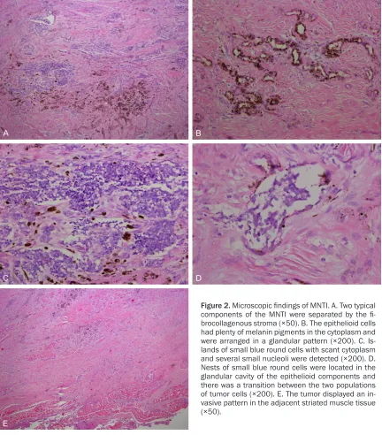
Microscopically, the tumor was biphasic in
cyto-plasm. They were arranged in a glandular pat-tern (Figure 2B). Islands of primitive-looking small blue round cells with scant cytoplasm and small round nucleoli were seen focally
(Figure 2C). And these two components were
separated from each other by the collagen. Some small blue round cells were located in the glandular cavity of the epithelioid components and there was a transition between these two populations of cells (Figure 2D). The tumor invaded the peripheral adipose and striated muscle tissue (Figure 2E). Occasionally mitotic
figures were found in the small cell component. Necrosis and calcification was not seen in the
tumor.
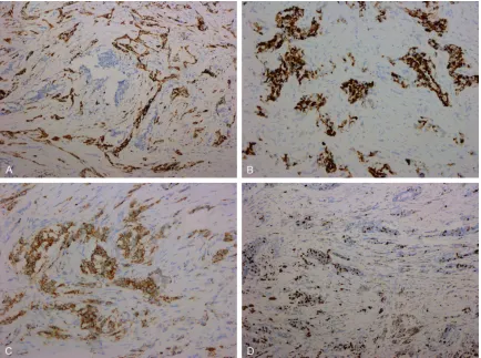
Immunohistochemically, the glandular compo-nent was positive for pan-cytokeratin (AE1/ AE3) (Figure 3A) and HMB45 (Figure 3B). The small blue cell component was positive for syn-aptophysin (SYN) (Figure 3C). All the compo-nents were negative for S-100, SMA, DES, GFAP, and CD99. The proliferation index Ki-67 was relatively high at 25% in the small blue cell component, however, negative in the glandular component (Figure 3D). The diagnosis was mel-anotic neuroectodermal tumor of infancy.
Discussion
Melanotic neuroectodermal tumor of infancy is a very rare benign tumor in infancy derived from the neural crest. A broad nomenclature
has been used for this tumor in the literature: Congenital melanocarcinoma, retinal anlage tumor, pigmented congenital epulis, melanotic progonoma, melanotic neuroectodermal tumor of infancy, pigmented ameloblastoma, pig-mented teratoma, atypical melanoblastoma, melanotic adamantinoma, retinal choristoma, melanotic epithelial odontoma, and benign melanotic tumor [13, 17, 18]. Most patients are
in the first year of life with a peak between the
second and the sixth month [19]. The mean age is 4.3 months [20]. MNTI has a male predilec-tion [19]. 92.8% of MNTIs occur in craniofacial region, mostly in maxilla (68-80%), skull (10.8%), mandible (5.8%), and brain (4.3%). Some other uncommon location includes epi-didymis, paratesticular region, testis, ovary, mediastinum, uterus and extremities [19]. Only 8 cases of MNTI were reported in the extremi-ties including our case. Half of the 8 cases were located in the soft tissue. Clinicopathologic fea-tures of all the 4 cases in the soft tissue of extremites were summarized in Table 1. They were all girls. The age interval was between 5 months and 10 months and all the patients were less than 1 year which was the common age range. Of the 4 cases, two were in the thigh, one in the upper arm and one in the forearm. Our case was the only one in the forearm. The tumors were 2.0-4.0 cm in maximum diameter.
Grossly, the tumors were firm, solid and well-defined. The cut surface was gray-black.
Histologically, all the 4 cases showed a bipha-sic feature and had two populations of cells. One population of cells was the epithelioid cells. They were cuboid and had plenty of eosin-ophilic cytoplasm and abundant intracellular melanin granules. These cells arranged in a glandular pattern. The other population of cells was the small blue round cells which just looked like the neuroblasts and had scanty cytoplasm. This kind of cells was aggregated in islands. These two components were embedded by a
dense fibrocollargenous stroma. Occasional mitotic figures were found in the small cell com
-ponent. 3 of the 4 tumors infiltrated the adja -cent soft tissue. Immunohistochemically, the
two kinds of cells had different immunoprofiles
(Table 2). The epithelioid cells were positive for cytokeratin (4/4) and HMB45 (3/4).
Neuron-specific enolase or synaptophysin was positive
in the small blue round cells in all the cases. Ultrastructual study was conducted in 2 of the
4 cases and confirmed the presence of a bipha
sic cell population [14, 16]. Primitive junctions
and premelanosomes were identified in the epi -thelioid components. There were intracytoplas-mic dense-core granules in the small blue round cells. The genetic study of MNTI was rarely reported. Gomes et al reported that MNTI might harbor the oncogenic BRAFV600E
muta-tion which first provided the insights to the tar -geted therapy of MNTI [4].
Our case shared the same clinicopathological features with the previous 3 cases: infants,
tumor size, the grey-black cut surface, classic biphasic pattern in morphology, and similar immunohistochemical phenotype. The
diagno-sis of MNTI was definitive. However, the loca -tion of our case was still uncommon for MNTI although 3 cases had been reported in the soft tissue of the extremities.
[image:3.612.91.523.74.566.2]The differential diagnosis for MNTI includes metastatic neuroblastoma, rhabdomyosarco-ma, Ewing’s sarcoma/peripheral primitive neu-roectodermal tumor, desmoplastic round cell
tumor (DSRCT), malignant melanoma, myeloid sarcoma and lymphoma. Metastatic neuroblas-toma does not have the biphasic feature in morphology and epithelial markers are nega-tive for this tumor. Rhabdomyosarcoma would have immunoreactivity for myogenic markers such as desmin, myogenin and myoD1. Ewing’s sarcoma/peripheral primitive neuroectodermal tumor usually has strong membranous CD99 expression and EWS-FLI1 fusion also can be detected. DSRCT does not have the epithelial component and melanin pigments are absent
in the tumor. Malignant melanoma is very rare in infants and does not have the small round cell component. Myeloid sarcoma and lympho-ma expresses a panel of helympho-matopoietic lympho- mark-ers which are negative for MNTI.
[image:4.612.90.524.71.394.2]The treatment guidelines have not been estab-lished for MNTI for their scarcity. Early diagno-sis, total resection and regular follow-up are optimal. Radical excision should be empha-sized and is the core of the treatment strategy. Chemotherapy may be effective for the cases
Figure 3. Immunohistochemical features of MNTI. A. Pancytokeratin was diffusely positive in the epithelioid compo-nents, however, negative in the small blue cell components (×100). B. The glandular components were positive for HMB45 (×200). C. The small blue cell components were positive for SYN (×200). D. Ki-67 index was about 25% in the small blue cell components (×100).
Table 1. Clinicopathologic features of MNTI in the soft tissue of the extremities
Case Sex/Age (Mo) Location complaintChief Maximum tumor diameter (cm) Management Follow-up
Scheck et al [14] F/6 Left thigh mass 2.0 Operation AWRM 15 m after surgery Al-Marzooq et al [15] F/6 Right upper arm mass 4.0 Operation Unknown
Lacy et al [16] F/5 Right thigh mass 2.5 Operation for twice AWRM 40 m after the initial surgery
[image:4.612.93.522.481.550.2]that have difficulties in surgery, tumor residue
or metastases. Reported chemotherapy was effectively used in 3 cases of facial MNTI [21]. Willi Woessmann presented a case of MNTI in the right maxilla who was primarily cured with chemotherapy without operation [22]. Radiation therapy was restrictedly used for it might cause severe late side effects in infants. All the data suggest the prognoses of MNTI are good. All the 4 cases just received extensive tumor resection without any adjuvant therapy includ-ing 1 case received surgery for twice for the ini-tial positive surgical margins. The follow-up was between 3 and 40 months except the follow-up of 1 case was unstated in the report. 3 cases were all uneventful in the reported follow-up time. Occasionally recurrences and metasta-ses can happen in some MNTI cametasta-ses. The local recurrence rate after resection was between 10% and 15% [19]. Kruse-Lösler et al made an analysis of 140 MNTI cases reported between 1990 and 2004 and found that the overall recurrence rate was 20% [19]. 5-10% of MNTI cases had metastases [15]. Metastases appeared in 23 MNTI cases which accounted for 6.5% of total 355 cases [19]. Some experts had suggested that aneuploidy in MNTI was a useful prognostic marker for predicting tumor recurrence or metastases, but it was still con-troversial [17, 20, 23]. Comprehensive therapy may be helpful for these intractable cases. In conclusion, we presented a rare case of MNTI in the soft tissue of the extremities. This
is the first case in the forearm. Although this tumor has specific histological,
immunohis-tochemical, and ultrastructural features, MNTI
still must be remembered to enter into the dif-ferential diagnosis of small round cell tumors or pigmented tumors in the soft tissue of the extremities. More data are needed to under-stand the behaviour of this kind of tumor in the extremities.
Disclosure of conflict of interest
None.
Address correspondence to: Dr. Lian Chen, De- partment of Pathology, Children’s Hospital of Fudan University, Shanghai, China. Tel: (86) 21-6493-1728; Fax: (86) 21-6493-1914; E-mail: doctchenlian@163. com
References
[1] Krompecher E. Zur Histogenese and Morpho- logie der Adamantinome und sonstiger Kiefergeschwulste. Beitr Path Anat 1918; 64: 165-197.
[2] Borello ED, Gorlin RJ. Melanotic neuroectoder-mal tumor of infancy-a neoplasm of neural crest origin. Report of a case associated with high urinary excretion of vanimandelic acid. Cancer 1966; 19: 196-206.
[3] Gochhait D, Dey P, Mitra S, Saikia UN. Fine-needle aspiration cytology of melanotic neuro-ectodermal tumor of infancy. Diagn Cytopathol 2015; 43: 770-772.
[4] Gomes CC, Diniz MG, de Menezes GH, Castro WH, Gomez RS. BRAFV600E Mutation in Melanotic Neuroectodermal Tumor of Infancy: Toward Personalized Medicine? Pediatrics 2015; 136: e267-269.
[image:5.612.93.523.83.254.2][5] Rachidi S, Sood AJ, Patel KG, Nguyen SA, Hamilton H, Neville BW, Day TA. Melanotic
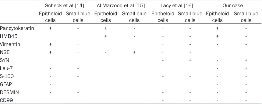
Table 2. Immunohistochemical features ofMNTI in the soft tissue of the extremities
Scheck et al [14] Al-Marzooq et al [15] Lacy et al [16] Our case Epitheloid
cells Small blue cells Epitheloid cells Small blue cells Epitheloid cells Small blue cells Epitheloid cells Small blue cells
Pancytokeratin + - + - + - +
-HMB45 + - + - +
-Vimentin + + + - -
-NSE + + - + + +
SYN - + - +
Leu-7 - - - +
S-100 - - -
-GFAP - -
-DESMIN - - -
-CD99 - - -
-+, positive; -, negative. Abbreviations: NSE, neuron-specific enolase; SYN, synaptophysin; GFAP,glial fibrillary acidic protein.
Neuroectodermal Tumor of Infancy: A Systematic Review. J Oral Maxillofac Surg 2015; 73: 1946-1956.
[6] Gupta R, Gupta R, Kumar S, Saxena S. Melanotic neuroectodermal tumor of infancy: review of literature, report of a case and follow up at 7 years. J Plast Reconstr Aesthet Surg 2015; 68: e53-54.
[7] Cui Y, Mao Z, Liao C. Melanotic neuroectoder-mal tumor of infancy: A case report and review of the surgical treatment. Oncol Lett 2015; 9: 29-34.
[8] Davis JM, DeBenedictis M, Frank DK, Lessin ME. Melanotic neuroectodermal tumor of in-fancy: a wolf in sheep’s clothing. Ann Otol Rhinol Laryngol 2015; 124: 97-101.
[9] Chaudhary S, Manuja N, Ravishankar CT, Sinha A, Vijayran M, Singh M. Oral melanotic neuroectodermal tumor of infancy. J Indian Soc Pedod Prev Dent 2014; 32: 71-73.
[10] Johnson RE, Scheithauer BW, Dahlin DC. Melanotic neuroectodermal tumor of infancy-a review of seven cases. Cancer 1983; 52: 661-666.
[11] Elli M, Aydin O, Pinarli FG, Dagdemir A, Dabak N, Selcuk MB, Acar S. Melanotic neuroectoder-mal tumor of infancy of the femur. Pediatr Hematol Oncol 2006; 23: 579-586.
[12] Choi IS, Kook H, Han DK, Baek HJ, Jung ST, Lee JH, Park JG, Hwang TJ. Melanotic neuroecto-dermal tumor of infancy in the femur: a case report and review of the literature. J Pediatr Hematol Oncol 2007; 29: 854-857.
[13] Rekhi B, Suryavanshi P, Desai S, Gulia A, Desai S, Juvekar SL, Puri A, Jambhekar NA. Melanotic neuroectodermal tumor of infancy in thigh of an infant--a rare case report with diagnostic implications. Skeletal Radiol 2011; 40: 1079-1084.
[14] Scheck O, Ruck P, Harms D, Kaiserling E. Melanotic neuroectodermal tumor of infancy occurring in the left thigh of a 6-month-old fe-male infant. Ultrastruct Pathol 1989; 13: 23-33.
[15] Al-Marzooq YM, Al-Bagshi MH, Chopra R, Hashsish H. Melanotic neuroectodermal tumor of infancy in the soft tissues of the arm: fine needle aspiration biopsy and histologic corre-lation-a case report. Diagn Cytopathol 2003; 29: 352-355.
[16] Lacy SR, Kuhar M. Melanotic neuroectodermal tumor of infancy presenting in the subcutane-ous soft tissue of the thigh. Am J Dermatopathol 2010; 32: 282-286.
[17] Pettinato G, Manivel JC, d’Amore ES, Jaszcz W, Gorlin RJ. Melanotic neuroectodermal tumor of infancy: a reexamination of a histogenetic problem based on immunohistochemical, flow cytometric, and ultrastructural study of 10 cas-es. Am J Surg Pathol 1991; 15: 233-245. [18] Kaya S, Unal OF, Saraç S, Gedikoğlu G.
Melanotic neuroectodermal tumor of infancy: report of two cases and review of literature. Int J Pediatr Otorhinolaryngol 2000; 52: 169-172. [19] Kruse-Lösler B, Gaertner C, Bürger H, Seper L,
Joos U, Kleinheinz J. Melanotic neuroectoder-mal tumor of infancy: a systematic review of the literature and presentation of a case. Oral Surg Oral Med Oral Pathol Oral Radiol Endod 2006; 102: 204-216.
[20] Yoo IH, Yum SK, Oh SJ, Kim KM, Jeong DC. Melanotic neuroectodermal tumor of infancy disseminated by a ventriculoperitoneal shunt and diagnosed from the inguinal sac. J Pediatr Hematol Oncol 2014; 36: e61-64.
[21] Kumari TP, Venugopal M, Mathews A, Kusumakumary P. Effectiveness of chemother-apy in melanotic neurectodermal tumor of in-fancy. Pediatr Hematol Oncol 2005; 22: 199-206.
[22] Woessmann W, Neugebauer M, Gossen R, Blütters-Sawatzki R, Reiter A. Successful che-motherapy for melanotic neuroectodermal tu-mor of infancy in a baby. Med Pediatr Oncol 2003; 40: 198-199.