Original Article
Expression of eag1 channel associated with the
aggressive clinicopathological features and
subtype of breast cancer
Guang-Xu Liu1,2,3*, Yun-Cui Yu1,2*, Xiang-Ping He4*, Sheng-Nan Ren5, Xue-Dong Fang5, Fen Liu1,2, Yan He1,2
1Department of Epidemiology and Health Statistics, School of Public Health, Capital Medical University, Beijing
100069, China; 2Beijing Municipal Key Laboratory of Clinical Epidemiology, Beijing 100069, China; 3Physical
Examination Center, 731 Hospital, China Aerospace Science and Industry Corporation, Beijing, China; 4Center
of Prevention and Cure of Breast Diseases, Haidian Maternal & Child Health Hospital, Beijing 100080, China;
5Department of General Surgery, China-Japan Union Hospital of Jilin University, Changchun 130021, Jilin, China. *Equal contributors.
Received September 18, 2015; Accepted October 24, 2015; Epub November 1, 2015; Published November 15, 2015
Abstract: Backgrounds: Expression of eag1 channel (Eag1) is associated with cell malignant transformation, tumor cell metastasis and poor prognosis of the patient. This study aimed at examining whether expression of the Eag1 associated with aggressive clinicopathological feature and the molecular subtype of breast cancer. Materials and Methods: 109 patients who received breast cancer operation during January 2009 to December 2010 in Chinese-Japanese Friendship Hospital of Jilin University were recruited. We investigated the association of the Eag1 with clinicopathological features and molecular subtype of in triple negative breast cancer (TNBC) by univariate or mul-tivariate analysis in a cross-section study.Results: The positive rate of Eag1 was 18.5% higher in TNBC compared with non-triple negative breast cancer (Non-TNBC) (P = 0.012, OR = 2.83, 95% CI = 2.16-3.47). Compared with the Eag1 negative group, the expression of Eag1 was linked to the larger tumor size (P = 0.002), advanced TNM stage (P = 0.029), high proportion of positive lymph node (87.6% vs. 65%, P = 0.014) and invasive ductal carcinoma (91% vs. 75%, P = 0.046). Conclusions: The expression of Eag1 may be partially explained the aggressive behavior of TNBC in the breast cancer tissue.
Keywords: Eag1 channel, clinicopathological features, triple negative breast cancer, association
Introduction
Breast cancer is the most frequently diagnosed cancer and becomes the leading cause of can-cer death in female worldwide [1]. Furthermore, breast cancer will increase to 85 per 10,000 women by 2021 [2]. In China, the crude inci-dence rate and mortality rate of female breast cancer during 2003-2007 were 41.64 per 100,000, which was ranked as the top among female cancers, respectively [3], leading to a serious threat to women’s physical and mental health.
For the heterogeneity and complexity of the dis-ease, single factor assessment is difficult to make the most suitable strategy for breast can-cer treatment and predict the prognosis [4].
Table 1. Clinicopathological features and expression of biomarkers in breast cancer patients
Clinicopathological features N % (range) Mean age (M ± S) 51.2 ± 10.4 (26-85) Tumor size (cm) 3.4 ± 1.9 (2.3-5.9) < 2 14 12.8 2-5 80 73.4 > 5 15 13.8 Pathologic types
Invasive ductal 96 88.1 Others 13 11.9 TNM stage
I 14 12.8
II 51 46.8
III 44 40.4
Histologic grade
I 20 18.3
II 45 41.3
III 44 40.4
Lymph node metastasis
Positive 91 83.5 Negative 18 16.5 ER
Positive 73 66.9 Negative 36 33.1 PR
Positive 65 59.6 Negative 44 40.4 HER-2
Positive 32 29.4 Negative 77 70.6 Eag1
Positive 89 81.7 Negative 20 18.3
further analyze the new breast cancer target associated with clinicopathological features in breast cancer patients.
Eag1 channel (ether a-go-go potassium chan-nel member 1, also known as Kv10.1), in physi-ological conditions, is restricted expression in the central nervous system, placenta, testes and prostate. However, overexpression of Eag1 has been found in many cancer, including cervi-cal, breast, melanoma, colon, lung, and ovarian cancers [7-11].Notably, Eag1 has been demon-strated to participate the processes of transfor-mation, migration and metastasis of the tumor
cell [12, 13]. It was reported that the proportion of Eag1 positive expression in breast cancer patients was over 82% [14], so it is worthwhile to examine the association of Eag1 with the clinical progression of the tumor.
In this study, we analyze the association of Eag1 expression with clinicopathological fea-tures in breast cancer, and further evaluate the difference of eag1 channel expression between TNBC and non-TNBC, aim at investigated the role of eag1 channel in the breast cancer progression.
Materials and methods
Patients and diagnostic criteria
In this study, 109 patients who received breast cancer operation during January 2009 to December 2010 in Chinese-Japanese Frie- ndship Hospital of Jilin University were recruit-ed. All patients were Chinese Han population. The TNM stage was determined based on the American Joint Committee on Cancer (AJCC) criteria and the histological grade was assessed according to the modified Bloom-Richardson classification. The pathological types were assessed in accordance with the criterion of the world health organization (WHO) breast cancer pathology. Lymph node metastasis was defined as any metastasis in local region of ipsilateral axillary, supraclavicular, or internal mammary lymph nodes, or distant region of other contralateral axillary lymph nodes as well as supraclavicular lymph nodes.
Immunohistochemical staining and judgments of results
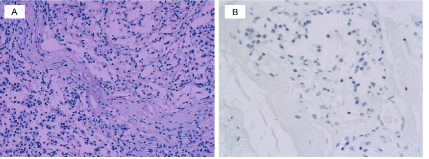
Figure 1. Representative Immunostaining of Eag1 in breast cancer carcinoma tissue. A. Positive staining for Eag1 in breast carcinoma tissues. B. Negative staining in breast carcinoma tissues (× 400).
dilution in 3% bovine serum albumin/PBS. Slides were incubated overnight in a humidified chamber at 4°C with anti-Eag1 rabbit antise-rum (Abcam, ab86204) at a 1:1200 dilution, followed by incubation with the EnVision Peroxidase System and DAB (Zhongshanjinqiao, Beijing, China).
Tumors with 1% or more positively nuclear-stained cells were considered positive for estrogen receptor (ER) and progesterone recep-tor (PR) expression; human epidermal growth factor receptor-2 (HER2) staining was scored by counting the number of cells positively stained on the membrane and expressed as a percent-age of total tumor cells, according to the report of Hemmerlein et al [14]. Eag1 stains were con-sidered positive if any (weak or strong) mem-branous invasive carcinoma cell staining was observed. The diagnosis was made by two pathologists who specialized in breast patholo-gy through assessed each archival hematoxylin and eosin (H&E) stained slides.
Statistical analysis
The different distribution of the continuous variable of age between eag1 channel positive and negative groups as well as between TNBC and non-TNBC were examined by the indepen-dent t-test or one-way ANOVA. The differences of clinicopathological features, including cate-gory variables of age, histological type, and sta-tus of lymph node metastasis were assessed by chi-square test when compared the eag1 positive group with negative group. The differ-ences clinicopathological features of
histologi-cal grade, ranked tumor size and TNM stage between eag1 channel positive and negative groups were evaluated by the Wilcoxon rank sum test. Chi-square test was also used to compare the difference of eag1 channel posi-tive rate between TNBC and non-TNBC. The associations of eag1 channel with clinicopatho-logical features of breast cancer was assessed by the unconditional logistic regression. All sta-tistical tests were two-sided, and P < 0.05 was considered significant. The statistical software SPSS version 19.0 (IBM Corp., Armonk, USA) was used for all statistical analyses.
Results
Clinicopathological features of the patients
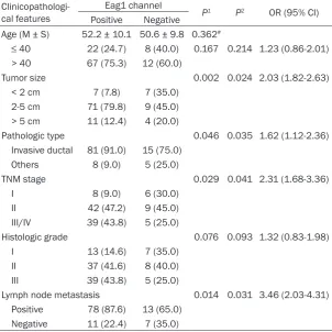
Table 3. Eag1 expression associated with Clinicopathological features
Clinicopathologi-cal features
Eag1 channel
P1 P2 OR (95% CI)
Positive Negative
Age (M ± S) 52.2 ± 10.1 50.6 ± 9.8 0.362#
≤ 40 22 (24.7) 8 (40.0) 0.167 0.214 1.23 (0.86-2.01) > 40 67 (75.3) 12 (60.0)
Tumor size 0.002 0.024 2.03 (1.82-2.63) < 2 cm 7 (7.8) 7 (35.0)
2-5 cm 71 (79.8) 9 (45.0) > 5 cm 11 (12.4) 4 (20.0)
Pathologic type 0.046 0.035 1.62 (1.12-2.36) Invasive ductal 81 (91.0) 15 (75.0)
Others 8 (9.0) 5 (25.0)
TNM stage 0.029 0.041 2.31 (1.68-3.36) I 8 (9.0) 6 (30.0)
II 42 (47.2) 9 (45.0) III/IV 39 (43.8) 5 (25.0)
Histologic grade 0.076 0.093 1.32 (0.83-1.98) I 13 (14.6) 7 (35.0)
II 37 (41.6) 8 (40.0) III 39 (43.8) 5 (25.0)
Lymph node metastasis 0.014 0.031 3.46 (2.03-4.31) Positive 78 (87.6) 13 (65.0)
Negative 11 (22.4) 7 (35.0)
#: P value from Student’s t test. P1: P-value from Chi-square test or Wilcoxon rank sum
test. P2: P-value from unconditional logistic regression analysis. CI:confidence interval.
the expression of Eag1 in TNBC was still significant-ly higher than that in non-TNBC (P = 0.012, OR = 2.83, 95% CI = 2.14-3.47), indicated that there was a correlation between Eag1 and TNBC (Table 2).
Expression of eag1 asso-ciated with clinicopatho-logical features of breast cancer
The result demonstrated that the cases of eag1 channel positive expres-sion showed a significant difference in Tumor size (P = 0.002) and TNM stage (P = 0.029) compared with the cases of eag1 channel negative expres-sion. The proportion of 2-5 cm of tumor size in the cases of eag1 chan-nel positive expression was the highest (79.8%), which was higher 34.8% than in the cases of eag1 channel negative expres-sion. The proportion of TNM stage II and III in the cases of eag1 channel positive expression was higher 13.8%, 7.2% res- pectively than in the cases of eag1 channel negative expression. The most cases of eag1
chan-Table 2. Expression of Eag1 between TNBC and non-TNBC
Eag1 TNBC Non-TNBC P1 P2 OR (95% CI)
Positive (N, %) 31 (93.8) 58 (75.3) 0.008 0.012 2.83 (2.16-3.47) Negative (N, %) 1 (6.2) 19 (24.7)
Total (N, %) 32 (100%) 77 (100%)
P1: P-value fromChi-square test. P2: P-value from unconditional logistic regression
analy-sis adjusting for age. CI:confidence interval.
HER-2 and eag1 (Figure 1) channel were 66.9%, 59.6%, 29.4% and 81.7%, respectively.
Eag1 was highly expressed in TNBC
All cases of breast cancer was classified into two subgroup: TNBC and non-TNBC, based on IHC stain of ER, PR and HER-2. The results revealed that the proportion of TNBC and non-TNBC accounted 29.4% and 70.6%, respective-ly. The results indicated that the positive expression of Eag1 in TNBC was 18.5% higher than that in non-TNBC (P = 0.008). After adjust-ing for age in the unconditional logistic regres-sion analysis, the results demonstrated that
[image:4.629.100.403.207.509.2]posi-tive group was significantly difference in tumor size (P = 0.024, OR = 2.03, 95% CI = 1.82-2.63), pathologic types (P = 0.035, OR = 1.62, 95% CI = 1.12-2.36), TNM stage (P = 0.041, OR = 2.31, 95% CI = 1.68-3.36) and lymph node metastasis (P = 0.031, OR = 3.46, 95% CI = 2.03-4.31) (Table 3).
Discussion
To our knowledge, this study is the first to dem-onstrate that the positive rate of eag1 in TNBC was significantly higher than that in the non-TNBC, furthermore, Eag1 expression was asso-ciated with aggressive clinicopathological fea-tures, such as larger tumor size, advanced TNM stage, high proportion of positive lymph node and invasive ductal carcinoma.
Breast cancer molecular subtypes based on immunohistochemical markers have been con-firmed to provide the usefulness of therapeutic and prognostic [16-19]. However, TNBC is a special subtype of breast cancer, which exhib-ited a poor prognosis due to its aggressive bio-logical behavior and lack of effective treatment method, as this type of cancer is insensitive to targeted and endocrine therapy [20]. In order to explore the reason for the poor outcomes of TNBC, ongoing studies on TNBC are currently conducted clinically and experimentally. Me- anwhile, many reports showed that Eag1 was overexpressed in distinct malignancies, includ-ing cervical, breast, lung, liver, prostate, colon, ovarian and gastric cancers [14, 21-23]. Hemmerlein et al. have reported that Eag1 was overexpressed in breast cancer. What’s more, Eag1 has been demonstrated to participate the processes of transformation, migration and metastasis of the tumor cell [12, 13]. In this study, Eag1 was expressed in 81.7% of breast cancer, which was consistent with Hemmerlein study [14].
Eag1 expression was associated with some clinical parameters in different categories of cancer, such as tumor size in head and neck cancer and lymph node metastasis in colorec-tal cancer [24, 25]. Furthermore, Eag1 overex-pression has been confirmed to correlate with the poorer prognosis, shorter overall survival in acute myelocytic leukemia (AML), ovarian, colorectal and oesophageal cancer [11, 26, 27]. However, no evidence was reported that eag1 expression associated with
clinicopatho-gical features in breast cancer. Our study dem-onstrated that eag1 positive expression was associated with larger tumor size, advanced histological grade, more positive lymph node and invasive ductal carcinoma compared with Non-Eag1 expression, which indicated that eag1 overexpression was correlated with more aggressive clinicopathological features, which also indicated that high expression rate of Eag1 in TNBC may at least partially account for its more aggressive biological behavior.
Despite the relative small sample size and lack of the follow-up data of the patient, this study still provided the evidence that Eag1 may play an important role in the pathological process of breast cancer, indicated its potential role in the therapy for breast cancer, especially for TNBC.
Acknowledgements
This work was supported by grants of key proj-ects in the National Science & Technology Pillar Program (No.SQ2015BA1300692) and Natural Science Foundation of Beijing Municipality to Yan He (7132027).
Disclosure of conflict of interest
None.
Address correspondence to: Dr. Xue-Dong Fang, Department of General Surgery, China-Japan Union Hospital of Jilin University, Changchun 130021, Jilin, China. Tel: +86-431-88796888; E-mail: fangxue-dong@medmail.com.cn; Drs. Fen Liu and Yan He, Department of Epidemiology and Health Statistics, School of Public Health, Capital Medical University, Xitoutiao 10, Youanmen, Beijing 100069, China. Tel: +86-10-83911778; Fax: +86-10-83911778; E-mail: liufen05@ccmu.edu.cn (FL); Tel: +86-10-83911779; Fax: +86-10-83911779; E-mail: yanhe1220@126. com (YH)
References
[1] Kamangar F, Dores GM, Anderson WF. Patterns of cancer incidence, mortality, and prevalence across five continents: defining priorities to re-duce cancer disparities in different geographic regions of the world. J Clin Oncol 2006; 24: 2137-50.
[3] Yang L, Sun TT, Wang N. [The incidence and mortality trends of female breast cancer in Beijing, China: between 2004 and 2008]. Zhonghua Yu Fang Yi Xue Za Zhi 2012; 46: 1009-14.
[4] DiGiovanna MP, Stern DF, Edgerton SM, Whalen SG, Moore D, Thor AD. Relationship of epidermal growth factor receptor expression to ErbB-2 signaling activity and prognosis in breast cancer patients. J Clin Oncol 2005; 23: 1152-60.
[5] Cheang MC, Chia SK, Voduc D, Gao D, Leung S, Snider J, Watson M, Davies S, Bernard PS, Parker JS, Perou CM, Ellis MJ, Nielsen TO. Ki67 index, HER2 status, and prognosis of patients with luminal B breast cancer. J Natl Cancer Inst 2009; 101: 736-50.
[6] Oakman C, Pestrin M, Zafarana E, Cantisani E, Leo A Di. Role of lapatinib in the first-line treat-ment of patients with metastatic breast can-cer. Cancer Manag Res 2010; 2: 13-25. [7] Meyer R, Schönherr R, Gavrilova-Ruch O,
Wohlrab W, Heinemann SH. Identification of ether à go-go and calcium-activated potassium channels in human melanoma cells. J Membr Biol 1999; 171: 107-15.
[8] Pardo LA, Camino D del, Sánchez A, Alves F, Brüggemann A, Beckh S, Stühmer W. Oncogenic potential of EAG K(+) channels. EMBO J 1999; 18: 5540-7.
[9] Ding XW, Yan JJ, An P, Lü P, Luo HS. Aberrant expression of ether à go-go potassium channel in colorectal cancer patients and cell lines. World J. Gastroenterol 2007; 13: 1257-61. [10] Díaz L, Ceja-Ochoa I, Restrepo-Angulo I, Larrea
F, Avila-Chávez E, García-Becerra R, Borja-Cacho E, Barrera D, Ahumada E, Gariglio P, Alvarez-Rios E, Ocadiz-Delgado R, Garcia-Villa E, Hernández-Gallegos E, Camacho-Arroyo I, Morales A, Ordaz-Rosado D, García-Latorre E, Escamilla J, Sánchez-Peña LC, Saqui-Salces M, Gamboa-Dominguez A, Vera E, Uribe-Ramírez M, Murbartián J, Ortiz CS, Rivera-Guevara C, De Vizcaya-Ruiz A, Camacho J. Estrogens and human papilloma virus oncogenes regulate human ether-à-go-go-1 potassium channel ex-pression. Cancer Res 2009; 69: 3300-7. [11] Asher V, Khan R, Warren A, Shaw R, Schalkwyk
GV, Bali A, Sowter HM. The Eag potassium channel as a new prognostic marker in ovarian cancer. Diagn Pathol 2010; 5: 78.
[12] Hegle AP, Marble DD, Wilson GF. A voltage-driv-en switch for ion-indepvoltage-driv-endvoltage-driv-ent signaling by ether-à-go-go K+ channels. Proc Natl Acad Sci U S A 2006; 103: 2886-91.
[13] Downie BR, Sánchez A, Knötgen H, Contreras-Jurado C, Gymnopoulos M, Weber C, Stühmer W, Pardo LA. Eag1 expression interferes with hypoxia homeostasis and induces
angiogene-sis in tumors. J Biol Chem 2008; 283: 36234-40.
[14] Hemmerlein B, Weseloh RM, Mello de Queiroz F, Knötgen H, Sánchez A, Rubio ME, Martin S, Schliephacke T, Jenke M, Heinz-Joachim-Radzun, Stühmer W, Pardo LA. Overexpression of Eag1 potassium channels in clinical tu-mours. Mol Cancer 2006; 5: 41.
[15] Park S, Koo JS, Kim MS, Park HS, Lee JS, Lee JS, Lee JS, Kim SI, Park BW. Characteristics and outcomes according to molecular sub-types of breast cancer as classified by a panel of four biomarkers using immunohistochemis-try. Breast 2012; 21: 50-7.
[16] Hugh J, Hanson J, Cheang MC, Nielsen TO, Perou CM, Dumontet C, Reed J, Krajewska M, Treilleux I, Rupin M, Magherini E, Mackey J, Martin M, Vogel C. Breast cancer subtypes and response to docetaxel in node-positive breast cancer: use of an immunohistochemical defini-tion in the BCIRG 001 trial. J Clin Oncol 2009; 27: 1168-76.
[17] Cancello G, Maisonneuve P, Rotmensz N, Viale G, Mastropasqua MG, Pruneri G, Veronesi P, Torrisi R, Montagna E, Luini A, Intra M, Gen- tilini O, Ghisini R,Goldhirsch A, Colleoni M. Prognosis and adjuvant treatment effects in selected breast cancer subtypes of very young women (< 35 years) with operable breast can-cer. Ann Oncol 2010; 21: 1974-81.
[18] Nielsen TO, Parker JS, Leung S, Voduc D, Ebbert M, Vickery T, Davies SR, Snider J, Stijleman IJ, Reed J, Cheang MC, Mardis ER, Perou CM, Bernard PS, Ellis MJ. A comparison of PAM50 intrinsic subtyping with immunohis-tochemistry and clinical prognostic factors in tamoxifen-treated estrogen receptor-positive breast cancer. Clin Cancer Res 2010; 16: 5222-32.
[19] Cuzick J, Dowsett M, Pineda S, Wale C, Salter J, Quinn E, Zabaglo L, Mallon E, Green AR, Ellis IO, Howell A, Buzdar AU, Forbes JF. Prognostic value of a combined estrogen receptor, pro-gesterone receptor, Ki-67, and human epider-mal growth factor receptor 2 immunohisto-chemical score and comparison with the Genomic Health recurrence score in early breast cancer. J Clin Oncol 2011; 29: 4273-8. [20] Yuan N, Meng M, Liu C, Feng L, Hou L, Ning Q,
Xin G, Pei L, Gu S, Li X, Zhao X. Clinical charac-teristics and prognostic analysis of triple-nega-tive breast cancer patients. Mol Clin Oncol 2014; 2: 245-251.
Camacho J. Ether a go-go potassium channels as human cervical cancer markers. Cancer Res 2004; 64: 6996-7001.
[22] Patt S, Preussat K, Beetz C, Kraft R, Schrey M, Kalff R, Schönherr K, Heinemann SH. Expression of ether à go-go potassium chan-nels in human gliomas. Neurosci Lett 2004; 368: 249-53.
[23] Mello de Queiroz F, Suarez-Kurtz G, Stühmer W, Pardo LA. Ether à go-go potassium channel expression in soft tissue sarcoma patients. Mol Cancer 2006; 5: 42.
[24] Menéndez ST, Villaronga MA, Rodrigo JP, Alvarez-Teijeiro S, García-Carracedo D, Urdinguio RG, Fraga MF, Pardo LA, Viloria CG, Suárez C, García-Pedrero JM. Frequent aber-rant expression of the human ether à go-go (hEAG1) potassium channel in head and neck cancer: pathobiological mechanisms and clini-cal implications. J Mol Med 2012; 90: 1173-84.
[25] Ding XW, Luo HS, Jin X, Yan JJ, Ai YW. Aberrant expression of Eag1 potassium channels in gastric cancer patients and cell lines. Med. Oncol 2007; 24: 345-50.
[26] Agarwal JR, Griesinger F, Stühmer W, Pardo LA. The potassium channel Ether à go-go is a novel prognostic factor with functional relevance in acute myeloid leukemia. Mol Cancer 2010; 9: 18.