Original Article
Expression of livin, survivin and caspase-3 in prostatic
cancer and their clinical significance
Junfei Gu*, Lixin Ren*, Xiaolu Wang, Changbao Qu, Yong Zhang
Department of Urology, The Second Hospital of Hebei Medical University, Shijiazhuang 050000, Hebei, China.
*Equal contributors.
Received August 31, 2015; Accepted October 19, 2015; Epub November 1, 2015; Published November 15, 2015
Abstract: To explore the expressions level of Livin, Survivin and Caspase-3 in prostatic cancer and the relationship among the 3 proteins and the clinicopathological features as well as the correlation among them. Totally, 43
paraf-fin-embedded prostate cancer tissues obtained from patients who were performed with rectal prostate biopsy or ex
-cision and 17 paraffin-embedded prostatic hyperplasia tissues were collected. All the specimens were confirmed by
pathology. Immunohistochemistry SP method was used to detect the expressions of Livin, Survivin and Caspase-3 in prostatic cancer compared to hyperplasia tissues. The positive expression rates of both Livin and Survivin in pros-tatic cancer tissue were higher than those in prospros-tatic hyperplasia tissue (93.02% vs. 64.70%, P < 0.05; 83.72% vs. 35.29%, P < 0.01). However, the positive expression rate of Caspase-3 in prostatic cancer tissue was obviously lower than that in prostatic hyperplasia tissue (25.58% vs. 58.82%, P < 0.01). Both Livin and Survivin expressions in prostatic cancer tissue were related to pathological grading (Gleason scores) (X2 = 14.000, P = 0.001), but not related to preoperative PSA, clinical stages and distant metastasis (P > 0.05). Capsase-3 expression in prostatic cancer tissue was related to pathological grading (Gleason scores) (X2 = 14.000, P = 0.001) and clinical stages (X2 = 4.896, P= 0.027), but not related to preoperative PSA and distant metastasis (P > 0.05). In prostatic cancer tissue, Livin expression had no correlation with Survivin expression (r = 0.127, P = 0.419 > 0.05), but negatively correlated with Caspase-3 expression (r = -0.497, P = 0.001). Survivin expression was negatively correlated with Caspase-3 expression (r = -0.354, P = 0.020). Livin, Survivin and Caspase-3 are closely related to the occurrence and devel-opment of prostatic cancer and which are expected to become new targets for diagnosis and treatment in future.
Keywords: Prostatic cancer, livin, survivin, caspase-3, correlation
Introduction
Prostatic cancer is a disease of serious impact-ing the life equality of the elderly and ranks the second of case fatality rate in males in Europe and the United States, even all over the world, second only to lung cancer [1, 2]. In recent years, with the changes of environment, high fat diet and population aging, the morbidity of prostatic cancer is increasing year by year in China and ranks the third of male urogenital system tumors, which is gradually close to the European and American. A study in the United State shows that the morbidity of prostatic can-cer is 12%~46%, which is found in a study of general autopsy of the male elderly over 50 years old that the morbidity has been increas-ing with the patients get older. Patients with prostatic cancer have no obvious symptoms in early stage, but when cancer tissue increases
to a certain extent which leads to suppression of the urethra, and the abnormal urination. For example, dysuria and hematuresis occur in a small number of patients, while the distant metastasis such as mostly bone metastasis is found in a large number of patients. It is in advanced stage that patients feel lower back pain, which with poor therapeutic effect and unfavorable prognosis.
Inhibitory of apoptosis proteins (IAPs) family is presently the only endogenous Caspases inhib-itory factors which can directly inhibit end effectors including Caspases-3 and Caspases -7. The family consists of 8 members, namely, HIAP-1, HIAP-2, NIAP, XIAP, ILP-2, Bruce, Survivin and Livin [3, 4]. Liven is often over-expression in some malignant tumors tissues and makes cell apoptosis being restrained, so it is closely related to the occurrence of malignant tumors [5]. Survivin has function of antiapoptosis, which can regulate cell cycle and participate in angiogenesis. Survivin is seldom expression in normal differentiated mature tissues except from thymus gland and gonad, but is highly expressed in most malignant tumors, so it can be regarded as tumor associated factors. Prostatic cancer belongs to a genetic disease. At present, studies on the regulatory methods and new targets for it were to explore in gene level at home and abroad. Therefore, in this study, Immunohistochemical staining method was used to detect the expressions of Livin, Survivin and Caspase-3 and explore the rela-tionship among the 3 proteins, furthermore, the clinicopathological features and the corre-lation between Livin and Survivin, Livin and Caspase-3, Survivin and Caspase-3 were also investigated. In hoping to provide the optimal markers for the diagnosis and prognosis of prostatic cancer and the scientific experimen-tal basis were applied for the effective targets in the treatment of prostatic cancer.
Materials and methods
Specimens collection
Totally 43 paraffin-embedded prostate cancer specimens obtaining from patients performed with rectal prostate biopsy or excision were col-lected from the Second Hospital of Hebei Medical University from Jan., 2013 to Sept, 2014. All specimens were confirmed by histo-pathology and have complete clinical and path-ological data. All patients were not treated with radiotherapy, chemotherapy and endocrino-therapy. Patients aged 55 to 81years old, with a mean age of 72.6 years. The average volume of prostate was 51.24 cm3. The average value of prostate-specific antigen (PSA) was 20.55 ng/ mL, including 4 cases with PSA < 4 ng/mL, 16 with 4~10 ng/mL and 19 with PSA > 10 ng/mL. Pathological grading was judged by Gleason
points-scoring system, including 4 cases in G1 (Gleason 2~4 points, well differentiated), 10 cases in G2 (Gleason 5~6 points, medium dif-ferentiated), 29 cases in G3 (Gleason 7~10 points, poorly differentiated or undifferentiat-ed). According to TNM staging system of American Joint Committee on Cancer (AJCC) in 2002, there were 2 cases in stage I, 17 cases in stage II, 12 cases in stage III and 12 cases in stage IV.
A total of 17 paraffin-embedded prostatic hyperplasia specimens obtaining from patients performed with excision and confirmed by pathology were selected as control. Patients aged 52 to 81 years old with the mean age of 71.9 years. The average volume of prostate was 59.06 cm3 and the average value of PSA was 4.59 ng/mL.
Key reagents
Rabbit anti-human Livin polyclonal (aa264-280) antibody and rabbit anti-human Caspase- 3 polyclonal antibody were purchased from Beijing Bo Orson Biological Technology Co., LTD and Santa Cruz Biotechnology, Inc. Rabbit anti-human Survivin polyclonal antibody, SP kit and DAB chromogenic reagent kit were bought from Wuhan Boster Biological Technology, Ltd. Immunohistochemistry
buffer for time periods determined by the response of antigen with antibody. The sec-tions were counterstained with hematoxylin for 4-5 min, washed, dehydrated in ethanol and xylene and then mounted on slides.
Evaluation criterion
Double blind was used for reading sections by two pathologists. Cells in which Livin, Survivin and Caspase-3 showed yellow-brown in cyto-plasm or cell nucleus were considered to be positive. Staining intensity and number of posi-tive cells were used to conduct semi-quantita-tive analysis. The expression was graded according to following staining intensity scores: 0 (no expression), 1+ (weak), 2+ (moderate) and 3+ (strong) scores stand for colorless, light yel-low, clay bank and brown, respectively; and per-centage of positive stained tumor cells: 0 <
10%, 1 (10%~50%), 2 (51%-75%), 3 (> 75%). Five visual fields in each slice were selected and the proportion of positive cells in 100 cells under microscope (×400) was deemed as the percentage of positive cells. The product of integral of staining intensity and positive cells proportion was used for evaluating the positive degree: 0~1, 2~4, 5~8 and 9~12 points refers to negative (-), weak positive (+), positive (++) and strong positive (+++).
Statistical data analysis
[image:3.612.89.289.72.220.2]SPSS15.0 software package was applied for data analysis. Comparison of sample rate was analyzed by chi-square test. Correlation between Livin and Survivin, Livin and Caspase-3, Survivin and Caspase-3 was ana-lyzed by Spearman rank correlation. A value of P < 0.05 was considered to be statistically significant.
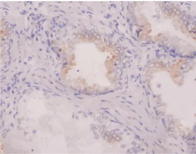
Figure 1. Positive expression of livin in prostatic can-cer tissue (×400).
[image:3.612.323.522.73.224.2]Figure 2. Positive expression of livin in prostatic hy-perplasia tissue (×400).
Figure 3. Positive expression of survivin in prostatic cancer tissue (×400).
[image:3.612.89.290.274.436.2] [image:3.612.324.522.275.431.2]Results
Expressions of livin, survivin and caspase-3 in prostatic hyperplasia and prostatic cancer tissues
The positive expression rates of Livin, Survivin and Caspase-3 in prostatic cancer and pros-tatic hyperplasia tissues were 93.02% (40/43) and 64.70% (11/17); 83.72% (36/43) and 35.29% (6/17), respectively, the both differ-ences were significant (P < 0.05 and P < 0.01). The positive expression rate of Caspase-3 in prostatic cancer tissue was 25.58% (10/43), which obviously lower than that [58.82% (10/17)] in prostatic hyperplasia tissue (P < 0.01).
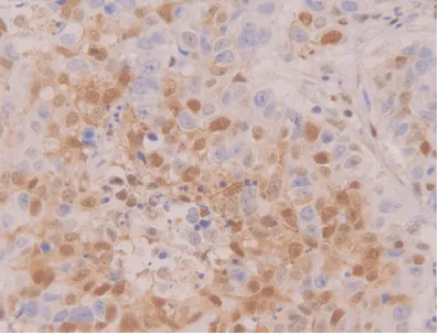
Positive staining of Livin, Survivin and Caspase-3 presented yellow-brown color. Livin, Survivin and Caspase-3 were positively expressed in cytoplasm and nucleus (Figures 1-6).
Relationship among livin, survivin, caspase-3 expressions and clinicopathological features of prostatic cancer
The expression of both Livin and Survivin in prostatic cancer tissue was related to patho-logical grading (Gleason scores) (X2 = 14.000,
P = 0.001), but not related to preoperative PSA, clinical stages and distant metastasis is (P > 0.05). Capsase-3 expression in prostatic can-cer tissue was related to pathological grading (Gleason scores) (X2 = 14.000, P = 0.001) and
clinical stages (X2 = 4.896, P = 0.027), but not
related to preoperative PSA and whether dis-tant metastasis is or not (P > 0.05, Table 1).
Pairwise comparison between livin and sur-vivin, livin and caspase-3, survivin and cas-pase-3 in prostatic cancer
In prostatic cancer tissue, Livin expression had no correlation with Survivin expression (r = 0.127, P = 0.419 > 0.05), but negatively corre-lated with Caspase-3 expression (r = -0.497, P = 0.001). Survivin expression was negatively correlated with Caspase-3 expression (r = -0.354, P = 0.020).
Discussion
[image:4.612.325.524.72.223.2]It is believed that the occurrence and develop-ment of tumors are molecular events, which are involving in stage process at the multi-gene and multi-molecular levels. The occur-rence of tumors, on one hand, is caused by the indefinite cell proliferation which is due to the disorder of cell cycle and out of control in cell division, on the other hand, it is the result of inadequate apoptosis. When the induction of cell apoptosis happens, apoptotic genes are activated by signal transduction pathway, which leading to programmed cell death. Once the apoptotic process is suppressed, cells of gene mutation survive and proliferate, which thus result in tumor genesis. In recent years, IAPs, as a kind of important anti-apoptosis factor, increasingly catch the attention of the scholars and researchers. IAPs family is a kind of highly conservative anti-apoptotic adjustment fac-tors, with amino terminal containing baculovi-rus IAP repeat (BIR) domain which consists of one or up to three highly conservative amino acid residues. IAPs play a role of anti-apoptosis by directly and indirectly inhibiting members of caspases family [6, 7].
Figure 5. Positive expression of caspase-3 in
[image:4.612.90.289.73.225.2]pros-Livin gene is located on chromosome 20q13.3, with the length of 4.6 kb and includes 7 exons and 6 intron. At present, relevant studies were found in the following anti-apoptosis pathways of Livin: (1) It bindings to Caspase-3, Caspase-7 and Caspase-9 directly/indirectly by regulating Caspase signal pathway to inhibit the apoptosis [8]; (2) The activation of TAK1/JNK1 signal transduction pathway inhibits the apoptosis [9]; (3) The interaction of Livin with Smac/ DLABLO regulates apoptosis [10]. Livin is not expressed or low expressed in most of terminal differentiated tissues of normal adults, but highly specific overly expressed in certain malignant tumors, such as esophageal cancer, gastric cancer, liver cancer, intestinal cancer, prostatic cancer, bladder cancer, renal carci-noma, lymphadecarci-noma, neuroblastoma and leu-kemia [11-17]. Survivin was firstly separated in hybridization of effector cell protease recep-tor-1 (EPR-1) cDNA in human genome by Ambrosini G et al in 1997. Survivin gene is located on chromosome 20q13.3, which con-tains 4 exons and 3 introns and a BIR domain without RING. Survivin plays a role of apoptosis by directly and indirectly binding to Caspase-3 and Caspase-7, and participates in regulating effect of cell proliferation, division and cycle and angiogenesis effect. Survivin is expressed during embryonic development, but not in ter-minal differentiated tissues. However, it is high-ly expressed in most transformed cells and human tumors. Another study was found that Caspase-3 showed a trend of decrease gradu-ally in non-hyperplasia tissue, adenoma and
cancer tissues, which indicated that the abnor-mal expression of Caspase-3 was closely asso-ciated with the occurrence and development of tumors [18].
In this study, the positive expression rate of Livin in prostatic cancer and prostatic hyperpla-sia tissues was 93.02% (40/43) and 64.70% (11/17), respectively, and the difference between them was significant. The positive expression rate of Survivin in prostatic cancer and prostatic hyperplasia tissues was 83.72% (36/43) and 35.29% (6/17), respectively, and there was significant difference between them. The positive expression rate of Caspase-3 in prostatic cancer tissue was 25.58% (10/43), obviously lower than that 58.82% (10/17) in prostatic hyperplasia tissue. Moreover, Livin expression had no correlation with Survivin expression, but negatively correlated with Caspase-3 expression. Survivin expression was negatively correlated with Caspase-3 expression. Those results indicate that Livin and Survivin which are highly expressed in prostatic cancer tissue play an important role in tumorgenesis of prostatic cancer probably by inhibition of apoptosis. Moreover, low-expresse-Caspase-3 in prostatic cancer may inhibit apoptosis and promote the tumorgenesis and development of prostatic cancer.
[image:5.612.90.522.95.284.2]Livin and Survivin belong to the members of IAPs family and they are resistant to apoptosis by directly acting on Caspase-3 and Capspase-7. However, they are not completely the same in
Table 1. Relationship among livin, survivin, caspase-3 expressions and clinicopathological features [n (%)]
n Livin P Survivin P Caspase-3 P
Positive Negative Positive Negative Positive Negative
PSA
< 4 ng/mL 8 (18.60) 8 (100.00) 0 (0.00) 0.536 7 (87.50) 1 (12.50) 0.477 1 (12.50) 7 (87.50) 0.317 4~10 ng/mL 16 (37.21) 14 (87.50) 2 (12.50) 12 (75.00) 4 (25.00) 5 (31.25) 11 (68.75) > 10 ng/mL 19 (44.19) 18 (94.74) 1 (5.26) 17 (89.47) 2 (10.53) 4 (21.05) 15 (78.95) Clinical staging
I+II 19 (44.19) 18 (94.74) 1 (2.33) 1.000 15 (78.95) 4 (21.05) 0.451 7 (36.84) 12 (63.16) 0.027 III+IV 24 (55.81) 22 (91.67) 2 (8.33) 21 (87.50) 3 (12.50) 3 (12.5) 21 (87.50) Pathological grading
G1 (2~4) 4 (9.30) 4 (100.00) 0 (0.00) 0.001 4 (100.00) 0 (0.00) 0.001 0 (0.00) 4 (100.00) 0.001 G2 (5~6) 10 (23.26) 9 (90.00) 1 (10.00) 9 (90.00) 1 (10.00) 4 (40.00) 6 (60.00) G3 (7~10) 29 (67.44) 27 (93.10) 2 (6.90) 23 (79.31) 6 (20.69) 6 (20.69) 23 (79.31) Distant metastasis
such function. Whether the mechanisms of both are correlated, it is still unclear. Livin and surviving are highly expressed in prostatic can-cer, more obviously than those in prostatic hyperplasia, which indicates there may be a dif-ferent mechanism for roles of Livin and Survivin in tumorgenesis and development of prostatic cancer. In conclusion, Livin, Survivin and Caspase-3 are closely related to the occur-rence and development of prostatic cancer and are expected to become new targets for diag-nosis and treatment of prostatic cancer.
Disclosure of conflict of interest
None.
Address correspondence to: Dr. Yong Zhang, De- partment of Urology, The Second Hospital of Hebei Medical University, 215 Hepingxi Road, Shijiazhuang 050000, Hebei, China. Tel: +86-311-66002999; Fax: +86-311-66002999; E-mail: yongzhang258@ sina.cn
References
[1] Jemal A, Tiwari RC, Murray T, Ghafoor A, Samuels A, Ward E, Feuer EJ, Thun MJ; American Cancer Society. Cancer statistics 2004. CA Cancer J Clin 2004; 54: 8-29. [2] Hoffman KE, Chen MH, Moran BJ, Braccioforte
MH, Dosoretz D, Salenius S, Katin MJ, Ross R,
D’Amico AV. Prostate cancer-specific mortality
and the extent of therapy in healthy elderly men with high-risk prostate cancer. Cancer 2010; 116: 2590-2595.
[3] Pan H, Yin C, Van Dyke T. Apoptosis and cancer mechanisms. Cancer Surv 1997; 29: 305-327. [4] Vaux DL, Korsmeyer SJ. Cell death in
develop-ment. Cell 1999; 96: 245-254.
[5] Chang H, Schimmer AD. Livin/melanoma in-hibitor of apoptosis protein as a potential ther-apeutic target for the treatment of malignancy. Mol Cancer Ther 2007; 6: 24-30.
[6] Chen X, Li YS. Expression and significance of
Livin in prostate cancer tissue. Fujian Med J 2013; 35: 80-83.
[7] Chao XJ, Zhao L, Wang ZM, Song XX, Du LY. Expression of Livin and Survivin in gastric
car-cinoma and its clinical significance. Hebei Med
J 2013; 35: 327-329.
[8] Huerta S, Heinzerling JH, Anguiano-Hernandez YM, Huerta-Yepez S, Lin J, Chen D, Bonavida B,
Livingston EH. Modification of gene products
involved in resistance to apoptosis in meta-static colon cancer cells: roles of Fas, Apaf-1, NFkappaB, IAPs, Smac/DIABLO, and AIF. J Surg Res 2007; 142: 184-194.
[9] Maas C, Verbrugge I, de Vries E, Savich G, van de Kooij LW, Tait SW, Borst J. Smac/DIABLO re-lease from mitochondria and XIAP inhibition are essential to limit clonogenicity of Type I tu-mor cells after TRAIL receptor stimulation. Cell Death Differ 2010; 17: 1613-1623.
[10] Chen YS, Li HR, Lin M, Chen G, Xie BS, Xu NL, Lin LF. Livin abrogates apoptosis of SPC-A1 cell by regulating JNKI signaling pathway. Mol Biol Rep 2010; 37: 2241-2247.
[11] Dubrez-Daloz L, Dupoux A, Cartier J. IAPs: more than just inhibitors of apoptosis proteins. Cell Cycle 2008; 7: 1036-1046.
[12] Chen L, Ren GS, Li F, Sun SQ. Expression of livin and vascular endothelial growth factor in different clinical stages of human esophageal carcinoma. World J Gastroenterol 2008; 14: 5749-5754.
[13] Augello C, Caruso L, Maggioni M, Donadon M, Montorsi M, Santambrogio R, Torzilli G, Vaira V, Pellegrini C, Roncalli M, Coggi G, Bosari S. Inhibitors of apoptosis proteins (IAPs)
expres-sion and their prognostic significance in hepa -tocellular carcinoma. BMC Cancer 2009; 9: 125.
[14] Yang YL, Lin SR, Chen JS, Lin SW, Yu SL, Chen HY, Yen CT, Lin CY, Lin JF, Lin KH, Jou ST, Hu CY, Chang SK, Lu MY, Chang HH, Chang WH, Lin
KS, Lin DT. Expression and prognostic signifi -cance of the apoptotic genes BCL2L13, Livin, and CASP8AP2 in childhood acute lymphoblas-tic leukemia. Leuk Res 2010; 34: 18-23. [15] Ye L, Song X, Li S, Yang D, Zhang J, Che X, Chen
X, Wang J, Zhang Z. Livin-alpha promotes cell proliferation by regulating G1-S cell cycle tran-sition in prostate cancer. Prostate 2011; 71: 42-51.
[16] Cheng T, Zhang JG, Cheng YH, Gao ZW, Ren XQ. Relationship between PTEN and Livin expres-sion and malignancy of renal cell carcinomas. Asian Pac J Cancer Prev 2012; 13: 2681-2685.
[17] Ibrahim L, Aladle D, Mansour A, Hammad A, Al Wakeel AA, Abd El-Hameed SA. Expression and
prognostic significance of livin/BIRC7 in child -hood acute lymphoblastic leukemia. Med Oncol 2014; 31: 941.
![Table 1. Relationship among livin, survivin, caspase-3 expressions and clinicopathological features [n (%)]](https://thumb-us.123doks.com/thumbv2/123dok_us/859221.597147/5.612.90.522.95.284/table-relationship-livin-survivin-caspase-expressions-clinicopathological-features.webp)
