Original Article
Frequency and spectrum of metachronous
malignancies in heart transplant recipients:
a 11-year-experience at a German heart center
Thomas Strecker1, Johannes Rösch1, Michael Weyand1, Abbas Agaimy2
1Center of Cardiac Surgery and 2Institute of Pathology, Friedrich-Alexander-University Erlangen-Nuremberg,
Ger-many
Received December 23, 2012; Accepted January 21, 2013; Epub February 15, 2013; Published March 1, 2013 Abstract: Background and aim: Heart transplantation (HTX) has become an established therapy for patients with end-stage heart failure. However, cancer incidence has been shown to be increased in the context of transplant-associated immunosuppression. The objective of this study is to analyze the incidence, histological spectrum, treat-ment and survival of various cancer types in HTX patients. Methods: We evaluated retrospectively all patients who underwent orthotopic HTX between 2000 and 2011 at our hospital including those patients who underwent HTX in other centers, but did their routine follow-up examinations at our department because of changing residence. Results: 142 patients had HTX performed at our center in the last 11 years and another 9 patients visited our department for monitoring after HTX performed at an external center (total: 151). Ten patients (6.6%) developed a metachronous malignancy (3 non-melanoma skin cancer, 2 lung cancer and 1 each parotid gland cancer, prostate cancer, renal cancer, urinary bladder cancer and ductal pancreatic cancer). The latency between HTX and the di-agnosis of the secondary neoplasm ranged from 33 to 152 months (median 76 months; mean 88 months). In all cases, surgery with or without chemoradiation was the treatment for the metachronous cancer. While most cases followed a favorable course after appropriate surgical and/or oncological treatment, four tumors (1 salivary duct carcinoma, 1 urinary bladder carcinoma, 1 ductal pancreatic cancer and 1 skin cancer) revealed a remarkable ag-gressiveness with wide-spread metastatic disease at the time of diagnosis or shortly thereafter. Conclusions: Inci-dence of various cancer types among HTX patients in this survey was consistent with previous studies, with lung and skin cancer as the commonest malignancies encountered. Regular cancer screening may be of benefit in reducing morbidity and mortality in these patients.
Keywords: Heart transplantation, secondary malignancy, immunosuppression, lung cancer, skin cancer, parotid gland cancer, urinary tract cancer, pancreatic cancer
Introduction
Heart transplantation (HTX) has become the gold standard for patients of all ages with end-stage heart failure resistant to medical or con-ventional surgical therapy [1, 2]. While the advances in immunosuppressive therapies have led to a concomitant decrease in cardiac allograft rejection, one uncommon but serious complication is the occurrence of malignancy after transplantation [3, 4].
Development of metachronous neoplasms is a well recognized complication in solid organ transplant recipients. The incidence and types
lymphoprolifera-tive diseases (PTLD) [9] and EBV-associated smooth muscle neoplasms [10].
In the study by Collett et al who analyzed a total of 37617 organ transplant recipients at a large transplantation center [11], 15% developed cancer at a median follow-up of 16 years; non-melanoma skin cancer was most common type encountered (57%). Of the group who devel-oped non-melanoma skin cancer, 13% were reported to have developed another de novo cancer other than skin cancer. In that study, the adjusted incidence of cancer among transplant recipients in England was more than double the incidence in the general population during the same period (90/1000 vs. 36/1000). The authors noted that the overall risk of cancer among transplant recipients is almost constant from two years after transplantation. Among the different cancer types listed, lung cancer and bladder cancer showed a significant increase among transplant recipients com-pared to the expected frequency in the general population. Thus close monitoring of organ transplant recipients to timely diagnose malig-nancies after transplantation is a major chal-lenge for all medical professions of all disci-plines involved in the management of affected patients.
The aim of this study was to analyze the fre-quency and spectrum of metachronous neo-plasms in a cohort of heart transplant
recipi-ents treated and/or followed-up at our center during the last 11 years.
Patients and methods
[image:2.612.90.375.73.263.2]All patients who underwent an HTX from 2000 to 2011 at the Center for Cardiac Surgery, University Hospital of Erlangen, Germany have been included in this retrospective analysis. These were 142 cases (1.3%) of all 10693 consecutive open heart procedures performed during the same period at our department (Figure 1). Additi- onally, we included 9 patients who underwent HTX in other hospitals, but visited our cen-ter for routine follow-up exami-nations because of changing residence.
Figure 1. Cardiac Surgery at the University of Erlangen between 2000 and 2011. CABG = Coronary Artery Bypass Grafting; VP = Valve Procedures; TAVI = Transcatheter Aortic Valve Implantation; AS = Aortic Surgery; VAD = Ven-tricular Assist Device; HTX = Heart Transplantation; AOCS = Any Other Car-diac Surgery.
The majority of our recovered patients were men (n=125; 82.8%) with a mean age of 53.2 ± 10.0 years (range 16.4 – 68.2 years) at the time of HTX. The mean age for the 26 females was 47.4 ± 17.4 years (range 3.7 – 69.1 years). Patients characteristic are summarized in Table 1. Mean serum creatinine of all transplant patients was 1.4 ± 0.7 mg/dl, mean ischemic time duringtransplantation was 176.3 ± 51.8 min. Mean donor age for all patients was 35.7 ± 11.4 years. Among donors there were 123 male (81.5% of total). For further statistical analysis, all 151 transplant patients were divid-ed into two groups: patients without metachro-nous malignancies (n=141) and patients with post-transplant malignancies (n=10, Table 1). HTX was performed in most of the cases due to dilated (n=71, 47.0%, DCM) and ischemic (n=70, 46.4%, ICM) cardiomyopathy. Further underly-ing causes for HTX were hypertrophic non-obstructive cardiomyopathy (n=3, HNOCM), aortic valve disease (n=1, AVD), congenital heart defect (n=2, CHD), non-compaction car-diomyopathy (n=1, NCCM), sarcoidosis (n=1), myocarditis (n=1), and sarcoma (n=1) (Figure 2).
Immunosuppressive regimen
aza-Table 1. HTX-Patients Characteristics
Patient Characteristics All Patients (n = 151) Patients Without Malignancies (n = 141) Patients With Malignancies (n = 10)
Recipient age (years±SD) 52.2±11.8 52.0±12.0 54.9±8.8
Recipient gender (n=male/% of subgroup) 125/82.8 115/81.5 10/100
No. of treated rejection episodes, grade 2R and 3R
(n/% of all biopsies of subgroup) 162/8.5 149/8.6 13/7.8
Blood group (n=0/A/B/AB) 44/69/25/13 43/62/23/13 1/7/2/0
Ischemic time (min±SD) 176.3±51.8 176.8±52.1 168.3±48.5
Serum creatinine (mg/dl±SD) 1.4±0.7 1.4±0.7 1.9±0.9
Donor age (years±SD) 35.7±11.4 35.8±11.4 33.5±10.7
[image:3.792.89.702.88.207.2]Donor Gender (n=male/% of subgroup) 123/81.5 117/83.0 8/80.0
Table 2. Clinicopathological features of cardiac transplant recipients who developed metachronous malignancies
Case (years)/genderAge at HTX Age at neoplasm (years) Duration after HTX (months) Site of neoplasm Histological type TNM stage Outcome
1 47/M 60 152 Skin SCC pT1 22 months alive and well
2 66/M 68 33 Skin SCC pT1 60 months alive and well
3 56/M 62 74 Lung SCC G2, pT2 pN0(0/28), R0 37 months alive and well
4 57/M 64 77 Lung SCC G3, pT2, pN1, R0 Died after 8 months
5 39/M 51 143 Parotid Salivary duct carcinoma G3 pT3 pN2b(26/30), R0 Recent case, alive
6 65/M 69 47 Skin SCC G3 pN2b(14/23), R0 Recent case, alive
7 45/M 50 65 Urinary bladder Urothelial carcinoma G3 pT2a pM1, R1 Died after 16 months
8 61/M 72 129 Prostate Acinar carcinoma (3+4) Right lobe, Gleason-score 7 18 months alive and well
9 52/M 60 100 Right kidney 2 papillary carcinomas G1, pT1a (m=2), R0 38 months alive and well
10 56/M 61 57 Pancreas Mucinous ductal carcinoma Miliary lung metastases Died shortly after diagnosis
[image:3.792.91.706.243.385.2]thioprine and steroids. In 2002/2003 this regi-men was changed to CsA, mycophenolate mofetil (MMF) and steroids. If repeatedly severe allograft rejections appeared, CsA was substi-tuted with tacrolimus. Mammalian target of rapamycin (mTOR) inhibitiors (everolimus) were used since 2004/2005, either as calcineurin inhibitor (CNI)-free immunosuppression or com-bined with CNI after progressive renal insuffi -ciency or repeated allograft rejection.
Concomitant oral prednisone was given 10 mg for the first three months, 7.5 mg for the next three months and 5 mg lifelong. In the first six months after HTX, CsA levels in combination with azathioprine or MMF were kept between 250-350 ng/ml. Thereafter CsA levels were sequentially reduced (200-250 ng/ml months 6-12, and 150-200 as of one year post trans-plantation. Azathioprine doses were adjusted on white blood cell count (total WBC ranging from 4000-10.000 cells/mm3). Tacrolimus
lev-els were maintained between 12-15 ng/ml within the first 6 months after HTX. Since the next six months, tacrolimus levels were kept between 10-12 ng/ml, followed by levels of 7-10 ng/ml as of one year post HTX. Everolimus was titrated to maintain a level between 4-7 ng/ml lifelong. In combination with everolimus, tacrolimus levels were a little bit lower (8-10 ng/ml in the first year, and thereafter between 6-8 ng/ml, everolimus levels were allays between 4-7 ng/ml). Particular notably, is the individual adjustment of medication for each HTX-Patient.
Follow-up
[image:4.612.94.518.71.405.2]in cases with clinically suspected allograft dys-function or rejection. The normal biopsy sched-ule was: weekly for the first month, every 2 weeks for the next month, once for the next 4 weeks, once for the next 6 weeks, then every 3 months for the next two years, and afterwards every 6 months for the next years. Allograft rejection was diagnosed according to the International Society for Heart and Lung Transplantation (ISHLT) guidelines [12, 13]. After all EMB procedures, a transthoracic echo-cardiogram (TTE), a conventional chest x-ray in two planes and a complete blood screening were performed. Additionally, all cardiac trans-plant recipients underwent right and left heart catheterization once every year after HTX. Tumor screening included a dermatological, urological, gynaecological, gastroenterological or any other medical examination whenever clinically indicated. From all 151 patients who underwent HTX in the last 11 years, a total fol-low-up of 607.8 patient years was created (mean 4.0 years). The follow-up varied from one day to 11 years. Two patients from this study have been published previously as case reports [14, 15] and were summarized in a recently published paper [16], respectively.
Results
General frequency of malignant neoplasms in HTX patients
Ten of the 151 patients (6.6%) developed a metachronous malignant neoplasm during the follow-up period (Table 2). The latency between HTX and the diagnosis of the secondary neo-plasm ranged from 33 to 152 months (median 76 months; mean, 88 months). Taken by histo-logical type, the mean time to diagnosis of the neoplasm was 77 and 109 months for squa-mous cell carcinoma (SCC) and the non-squa-mous neoplasms, respectively. Taken by site, SCC of the lung developed earlier than SCC of the skin (75.5 versus 92.5 months respective-ly). However, the frequency of neoplasms would be higher if recent cases with <2 years follow-up are excluded (10.6%) and would even rise to 21.2% if only patients who have reached the mean follow-up (91 months, i.e. 7.6 years) for those who developed malignancy were taken into consideration.
All of the 10 patients were males with a mean age at the time of HTX of 54.9 ± 8.8 years
(range 39.2 – 66.0 years). Their age at the time of diagnosis of the malignant neoplasm ranged from 51.1 – 72.6 years (mean 63.2 years) (Table 2). In comparison to patients without malignancies, patients with neoplasms were not significant older at time of transplantation (52.0 years versus 54.9 years respectively, Table 1). However, the percentage of male recipients was significantly higher among patients with malignancies (n=10, 100% of subgroup) than in the group without neoplasms (115 men = 81.5% of subgroup). Concerning underlying reasons for HTX, five of the ten patients suffered from DCM and 5 from ICM, respectively.
Effects of immunosuppression
Interestingly, the number of treated rejection episodes, grade 2 and 3, scored by the International Society for Heart and Lung Transplantation (ISHLT) guidelines [12], and revised in 2005 [13], were not significantly dif -ferent between patients with and those without metachronous malignancies (8.6% versus 7.8% respectively, Table 1). Repeated moderate or severe allograft rejection required modification of immunosuppressive therapy in 44 of the 151 cardiac transplant patients (29.1%), in the malignancy group, 4 of the 9 patients (44,4%) received a switch of immunosuppression proto-col. Furthermore, no statistically significant cor -relation between development of metachro-nous malignancies and the use of different immunosuppressive drugs as cyclosporine A (CsA), azathioprine, mycophenolate mofetil (MMF), tacrolimus or mTOR inhibitior (everolim-us) could be established during the follow-up period (data not shown).
The mortality rate after diagnosis of malignan-cy during follow-up was 33.3% (3 of 10 patients with malignancies). Clinicopathological fea-tures and outcome of these 10 cardiac trans-plant recipients who developed metachronous malignancies are summarized in Table 2. Additionally, no statistically significant correla -tion between diagnosis of malignancy and serum creatinine pre-HTX, ischemic time during HTX or donor age as well as donor gender was seen (Table 1).
summarized in Figure 3. There were 3 non-melanocytic skin cancers, 2 lung cancers, 1 parotid gland cancer, 1 prostate cancer, 1 renal cancer, 1 ductal pancreatic cancer and 1 uri-nary bladder cancer. Well differentiated SCC was diagnosed in the head and neck skin (eye, cheek and ear) in two patients. One patient had two distinct SCC of the skin and one Bowen dis-ease. This patient developed extensive cervical lymph node metastasis and parotid gland metastasis 3 months after diagnosis of skin cancer. Two patients developed SCC of the lung; both had involved the right lower lobe of the lung (in addition to the right middle lobe in
one) (Figure 4). Lymph node involvement was seen in one of the two patients, but there was no histological or clinical evidence of increased tumor aggressiveness. One patient was alive 37 months after diagnosis of lung cancer, the other died 8 months later due to progressive cardiopulmonary failure.
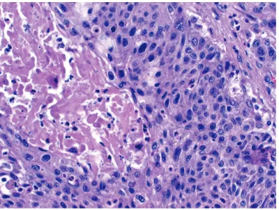
[image:6.612.98.518.139.301.2]Salivary gland cancer: One patient had poorly differentiated (high-grade) salivary duct carci-noma. The tumor in this patient (recent case) Figure 3. Distribution of second malignancies in heart transplant recipients.
[image:6.612.90.290.345.496.2]Figure 4. Example of keratinizing squamous cell car-cinoma of the lung after HTX (original magnification x400).
[image:6.612.324.525.345.495.2]showed extensive permeation of the lymphat-ics and venous channels in the soft tissue of the head and neck with more than 20 regional lymph node metastases (Figure 5). A few months later he developed liver metastasis and received radiation and chemotherapy. At last follow-up (currently), he is alive with disease under palliative treatment. As mentioned above, the parotid gland was also involved by metastatic disease from SCC of skin in another patient.
Other miscellaneous neoplasms: One patient developed acinar carcinoma of the prostate that was diagnosed by core needle biopsy because of elevated PSA (PSA: 15.8 ng/ml; Gleason score: 3+4=7) [17]. The tumor was lim-ited to the right lobe of the gland. Another patient was diagnosed with high-grade widely invasive sarcomatoid transitional cell carcino-ma of the urinary bladder, shortly followed by metastatic disease (Figure 6). Another patient who developed end-stage vascular renal dis-ease that necessitated renal transplantation was diagnosed with two small well differentiat-ed papillary renal cell carcinomas containdifferentiat-ed within the explanted right kidney. The last patient developed a widely metastatic pancre-atic ductal adenocarcinoma that presented with miliary bilateral pulmonary metastasis. This patient was a recent case diagnosed dur-ing preparation of this manuscript. He died short after diagnosis due to cancer-related pro-gressive cardiopulmonary failure.
Discussion
In this study, we have analyzed the frequency, histological spectrum, treatment and outcome of metachronous malignant neoplasms in patients with a history of HTX. We found 10 patients (6.6%) with a malignant neoplasm that developed after HTX. This rate of metachro-nous malignancies in cardiac transplant recipi-ents is consistent with previously published series with a range of 4.1% to 46.1% up to 15 years after HTX [18-21]. However, it is well rec-ognized that the incidence and types of immune suppression-associated neoplasia usually vary with the extent and completeness of follow-up. Our patients had a mean follow-up period of 4 years. The metachronous malignancy devel-oped at a range of 33 to 152 months (median, 76; mean, 88 months). However, the frequency of neoplasms would be higher if recent cases
with < 2 years follow-up are excluded (10.6%) and would even rise to 21.2% if only those who reached the mean follow-up for metachronous cancer were considered. In the study by Collett et al, 15% of solid organ transplant recipients developed cancer at a median follow-up of 16 years. The authors noted that the overall risk of cancer among transplant recipients is almost constant from two years after transplantation. However, 13% of those who initially developed a skin cancer suffered later from another non-cutaneous malignancy.
[image:7.612.324.525.71.223.2]that this patient had incidental papillary renal carcinoma associated with his end-stage renal disease but unrelated to his immunosuppr- ession.
Three neoplasms in our cases showed remark-able tumor aggressiveness with wide-spread metastasis and/or extensive permeation of lymphatic and blood vessels. Although both neoplasms were of histological types well known to display a highly aggressive clinical course, it is not completely excluded that the long-term immunosuppression might have influenced the tumor behaviour. Remarkably, all three neoplasms have developed after more than 5 years from HTX. Of note, two of these neoplasms were histogentically related; both were ductal adenocarcinoma of salivary-type tissue (parotid gland and pancreas). Both developed more than 10 years after immuno-suppression and presented initially with exten-sive local or widely metastatic disease. These features in both cases suggest a possible com-mon pathogenesis and enhanced aggressive-ness but this remains currently unclear. The remaining patients did not follow an aggressive course and both have survived well after appro-priate surgical and/or oncological treatment. Of note, we did not observe a higher frequency of non-melanocytic skin cancer (SCC and Bowen disease) in our cases, thus contrasting with some previous studies [4, 6]. This may be due to limited extent of follow-up after HTX in some of our patients, variation in regimens used for immunosuppression by different cen-ters, age of the cohort analyzed in different studies, presence or absence of additional risk factors predisposing to skin cancer (actinic skin damage, HPV infection, etc.), missing diagno-ses of skin lesions treated at external hospital and denied by the patients or various combina-tions of the above. Likewise, post-transplant lymphoproliferative disorder (PTLD) has not been recorded in our cases. This might be due to the rarity of early-onset PTLD as most cases developed after long-term immunosuppressive therapy at a mean of 128 months [9]. Interestingly, the incidence of different types of metachronous malignant neoplasms varied with the organ transplanted [11]. Moreover, fur-ther prospective clinical studies are presently underway to test the concept that different immunosuppressive drugs reduce cancer while
simultaneously inhibiting allograft rejection [23, 24].
In summary, we reported our experience with metachronous malignancies in HTX patients, illustrating the histological heterogeneity of recorded neoplasms, variation of anatomic sites affected and the increasing frequency with the extent of follow-up after HTX. Overrepresentation of some organ systems (urinary tract, lung and salivary glands) in our study and some previous reports underlines the possibility of and the need for establishing a screening program for early detection of these aggressive malignancies in HTX patients. The unusually aggressive course of some of these neoplasms underscores the need for studying their biological distinctness from their sporadic counterparts in non-transplant patients to allow for conceptualizing more appropriate therapeutic regimens for them. Acknowledgement
We thank all colleagues at our heart failure and transplantation ambulance, University Hospital Erlangen, for their untiring assistance and supervision of all heart transplant recipients. Address correspondence to: Dr. Thomas Strecker, Center of Cardiac Surgery, Friedrich-Alexander-University Erlangen-Nuremberg, Krankenhaus-strasse 12, 91054 Erlangen, Germany. Phone: +49-9131-85-33985; Fax: +49-9131-85-36088; E-mail: thomas.strecker@uk-erlangen.de
References
[1] Stehlik J, Edwards LB, Kucheryavaya AY, Ben-den C, Christie JD, Dobbels F, Kirk R, Rahmel AO, Hertz MI. The Registry of the International Society for Heart and Lung Transplantation: Twenty-eighth Adult Heart Transplant Re-port--2011. J Heart Lung Transplant 2011; 30: 1078-94.
[2] Gambino A, Cerutti A, Feltrin G, Toscano G, Tar-antini G, Milanesi O, Angelini A, Gerosa G. Out-come after pediatric heart transplantation: two decades of a single center experience. Eur J Cardiothorac Surg 2007; 32: 220-4.
[4] Doesch AO, Müller S, Konstandin M, Celik S,
Kristen A, Frankenstein L, Ehlermann P, Sack FU, Katus HA, Dengler TJ. Malignancies after heart transplantation: incidence, risk factors, and effects of calcineurin inhibitor withdrawal. Transplant Proc 2010; 42: 3694-9.
[5] Yildirim Y, Ozyilkan O, Emiroglu R, Bemirhan B, Karakayali H, Haberal M. Early diagnosis of cancer in renal transplant patients: A single center experience. Asian Pacific J Cancer Prev 2006; 7: 336-339.
[6] Alam M, Brown RN, Silber DH, Mullen GM,
Feldman DS, Oren RM, Yancy CW. Cardiac Transplant Research Database Group: In-creased incidence and mortality associated with skin cancers after cardiac transplant. Am J Transplant 2011; 11: 1488-97.
[7] Boeckle E, Boesmueller C, Wiesmayr S, Mark W, Rieger M, Tabarelli D, Graziadei I, Hoefer D, Antretter H, Stelzmueller I, Krugmann J, Zangerle R, Huemer H, Poelzl G, Margreiter R, Bonatti H. Kaposi sarcoma in solid organ trans-plant recipients: a single center report. Trans-plant Proc 2005; 37: 1905-9.
[8] Reuschenbach M, Tran T, Faulstich F, Hartschuh W, Vinokurova S, Kloor M, Kraut-krämer E, Zeier M, von Knebel Doeberitz M, Sommerer C. High-risk human papillomavirus in non-melanoma skin lesions from renal al-lograft recipients and immunocompetent pa-tients. Br J Cancer 2011; 104: 1334-41. [9] Chan TS, Hwang YY, Gill H, Au WY, Leung AY,
Tse E, Chim CS, Loong F, Kwong YL. Post-trans-plant lymphoproliferative diseases in Asian solid organ transplant recipients: late onset and favorable response to treatment. Clin Transplant 2012 Sep-Oct; 26: 679-83.
[10] Deyrup AT, Lee VK, Hill CE, Cheuk W, Toh HC,
Kesavan S, Chan EW, Weiss SW. Epstein-Barr-Virus-associated smooth muscle tumors are distinctive mesenchymal tumors reflecting multiple infection events. A clinicopathologic and molecular analysis of 29 tumors from 19 patients. Am J Surg Pathol 2006; 30: 75-82. [11] Collett D, Mumford L, Banner NR, Neuberger J,
Watson C. Comparison of the incidence of ma-lignancy in recipients of different types of or-gan: a UK Registry audit. Am J Transplant 2010; 10: 1889-96.
[12] Billingham ME, Cary NR, Hammond ME, Kem-nitz J, Marboe C, McCallister HA, Snovar DC, Winters GL, Zerbe A. A working formulation for the standardization of nomenclature in the di-agnosis of heart and lung rejection: Heart Re-jection Study Group. The International Society for Heart Transplantation. J Heart Transplant 1990; 9: 587-93.
[13] Stewart S, Winters GL, Fishbein MC, Tazelaar HD, Kobashigawa J, Abrams J, Andersen CB,
Angelini A, Berry GJ, Burke MM, Demetris AJ, Hammond E, Itescu S, Marboe CC, McManus B, Reed EF, Reinsmoen NL, Rodriguez ER, Rose AG, Rose M, Suciu-Focia N, Zeevi A, Bill-ingham ME. Revision of the 1990 working for-mulation for the standardization of nomencla-ture in the diagnosis of heart rejection. J Heart Lung Transplant 2005; 24: 1710-20.
[14] Strecker T, Zimmermann I, Wiest GH. Pulmo-nary and cardiac recurrence of sarcoidosis in a heart transplant recipient. Dtsch Med Wochen-schr 2007; 132: 1159-62.
[15] Strecker T, Schmid A, Agaimy A, Hugo C, Wey-and M, Zielezinski T. Giant metastatic alveolar soft part sarcoma in the left ventricle: appear-ance in echocardiography, magnetic reso-nance imaging, and histopathology. Clin Cardi-ol 2011; 34: E6-8.
[16] Strecker T, Rösch J, Weyand M, Agaimy A. Pri-mary and metastatic cardiac tumors: imaging characteristics, surgical treatment and histo-pathological spectrum: a 10-year-experience at a German Heart Center. Cardiovasc Pathol 2012 Sep-Oct; 21: 436-43.
[17] Humphrey PA. Gleason grading and prognostic factors in carcinoma of the prostate. Mod Pathol 2004; 17: 292-306.
[18] Garlicki M, Wierzbicki K, Przybyłowski P, Drop D, Biernat M, Rudziński P, Olszewska B, Dziat-kowiak A. The incidence of malignancy in heart transplant recipients. Ann Transplant 1998; 3: 41-7.
[19] Crespo-Leiro MG, Alonso-Pulpón LA, Villa-Ar-ranz A, Brossa-Loidi V, Almenar-Bonet L,
González-Vilchez F, Delgado-Jiménez JF, Mani-to-Lorite N, Díaz-Molina B, Rábago G, Arizón-del Prado JM, Romero-Rodríguez N, Brossa V,
Blasco-Peiró T, Pascual-Figal D, de la Fuente-Galán L, Muñiz-García J, The prognosis of non-cutaneous, nonlymphomatous malignancy af-ter heart transplantation: data from the Spanish Post-Heart Transplant Tumour Regis-try. Transplant Proc 2010; 42: 3011-3.
[20] Crespo-Leiro MG, Villa-Arranz A, Manito-Lorite N, Paniagua-Martin MJ, Rábago G, Almenar-Bonet L, Alonso-Pulpón L, Mirabet-Pérez S, Di-az-Molina B, González-Vilchez F, Arizón de Pra-do JM, Romero-Rodriguez N, Delgado-Jimenez J, Roig E, Blasco-Peiró T, Pascual-Figal D, De la Fuente Galán L, Muñiz J. Lung cancer after heart transplantation: results from a large mul-ticenter registry. Am J Transplant 2011; 11: 1035-40.
[21] Brewer JD, Colegio OR, Phillips PK, Roenigk RK, Jacobs MA, Van de Beek D, Dierkhising RA,
[22] Hurst FP, Jindal RM, Fletcher JJ, Dharnidharka V, Gorman G, Lechner B, Nee R, Agodoa LY, Ab-bott KC. Incidence, predictors and associated outcomes of renal cell carcinoma in long-term dialysis patients. Urology 2011; 77: 1271-6.
[23] Geissler EK. The impact of mTOR inhibitors on the development of malignancy. Transplant Proc 2008; 40: S32-5.