Functional Analysis of Third Ventriculostomy
Patency by Quantification of CSF Stroke Volume
by Using Cine Phase-Contrast MR Imaging
Nu´ria Bargallo´, Lourdes Olondo, Ana I. Garcia, Sebastian Capurro, Luis Caral, and Jordi Rumia
OBJECTIVE: Endoscopic third ventriculostomy (ETV) is increasingly used as alternative treatment for obstructive hydrocephalus. The aim of this study was to determine the utility of quantitative and qualitative examinations with cine phase-contrast MR imaging to determine the efficacy of ventriculostomy across time and whether CSF pulsation is restored after ETV. METHODS:Thirty-eight patients treated with ETV were evaluated with cine phase-contrast MR within 1 month after surgery. Follow-up studies were performed after 1 year in 25 patients and after 2 years in 12. We evaluated flow void changes in the floor of the third ventricle and quantified the stroke volume at the site of the ventriculostomy. We also recorded changes in ventricular size and clinical outcome. To determine the restoration of CSF pulsation, we compared the CSF waveform at the ventriculostomy with the CSF waveform at the aqueduct in a healthy control group.
RESULTS: After ventriculostomy, restoration of pulsate motion characteristics of CSF circulation was observed. The stroke volume registered at ventriculostomy was maintained with time. There was a statistically significant relationship between clinical outcome and stroke volume. Overall flow magnitude was the most effective variable to determine which patients would improve after surgery. Values >75 mm3showed a sensitivity of 76.7% and a specificity of 87.5% There was no relationship between ventricular size changes and clinical outcome. Patients with primary aqueduct stenosis had the best response to surgery, whereas patients with Arnold Chiari malformation or communicating hydrocephalus had the worst response.
CONCLUSION:Quantitative analysis with phase-contrast MR imaging indicates that ETV is an efficient technique for restoring CSF pulsation, with efficacy being maintained during the follow-up controls. Quantification of stroke volume at ventriculostomy is a good indicator of the functional status of ETV, and a high stroke volume in the ventriculostomy appears to be a positive predictor of favorable clinical outcome.
Neuroendoscopic third ventriculostomy has become a first-line treatment for obstructive hydrocephalus in several centers. The morbidity associated with this technique is low, and the success rates are high. It is essential, however, to ensure that this new internal shunt is effective. The outcome of the patients is obviously a key factor. Some reviews of the current literature have identified parameters that can be used to determine malfunction of the ventriculostomy. In addition to clinical outcome, particular attention has
been paid to ventricular size reduction and the pres-ence of a flow void in cine phase-contrast MR imag-ing (1–10). The aim of this study was to assess the usefulness of a cine phase-contrast MR measure-ment—stroke volume—as a parameter for predicting the functional evolution and state of ventriculostomy. We correlated quantitative volume data with clinical follow-up, ventricular size, and flow void signal inten-sity in cine phase-contrast MR. We were also inter-ested in whether CSF circulation is restored after endoscopic third ventriculostomy (ETV) and whether the CSF flow pattern in the ventriculostomy is similar to that in the aqueduct.
Materials and Methods
All patients treated with neuroendoscopic ventriculostomy in the neurosurgery department between 1997 and 2000 were included in the study. The decision to perform this technique Received October 29, 2004; accepted after revision February 11,
2005.
From the Departments of Radiology (N.B., L.O., A.I.G., S.C.) and Neurosurgery (L.C., J.R.), Hospital Clı´nic i Provincial de Barcelona, Barcelona, Spain.
Address correspondence to Nu´ria Bargallo´, CDIC, Servicio de Radiodiagno´stico, Hospital Clinico y Provincial de Barcelona, C/ Villarroel 170, Barcelona 08003, Spain.
©American Society of Neuroradiology
rather than ventriculoperitoneal shunt was made by the neurosurgeons.
Thirty-eight patients (25 men and 13 women; mean age, 39.5 years; age range, 11–74 years) with hydrocephalus were treated by neuroendoscopic ventriculostomy at the floor of the third ventricle anterior to the mammillary bodies during this period of time. The study was approved by our institutional review board, but patient informed consent was not provided because phase-contrast MR imaging was included during the standard follow-up MR studies after ventriculostomy.
Occlusive hydrocephalus was diagnosed in all but 3 patients. In 11 patients, the cause was primary aqueduct stenosis; in 18 patients, the cause was secondary stenosis due to intraventric-ular or extraventricintraventric-ular cystic or tumor lesions compromising CSF circulation at the third ventricle or aqueduct—9 tectum tumors, 6 pineal tumors, and 3 cystic lesions, including colloid cyst (Fig 1); and in 6 patients the cause was choroid plexus papilloma and neuroepithelial cysts) and Arnold Chiari mal-formation. We also included 3 patients with communicating hydrocephalus (2 with previous history of meningitis and the other with previous history of subarachnoid hemorrhage). No other diseases were associated.
Previous clinical symptoms in all patients (headaches, nau-sea and vomiting, vertigo, and, in some cases, absence seizures) were associated with increased intracranial pressure.
After the ETV, CSF flow MR studies were performed in all patients within the first postoperative month. Follow-up CSF flow MR studies were undertaken 1 year after the procedure in 25 patients and 2 years after the procedure in 12.
MR Imaging Techniques. All initial CSF flow MR studies were performed with a 1.5T Siemens Magnetom 63SP system. Two series of cine phase-contrast MR imaging techniques were applied after performing spin-echo coronal T1-weighted im-ages to locate the third ventriculostomy and evaluating ventric-ular size after surgery: one in the axial plane, with through-plane velocity encoding in the craniocaudal direction for flow quantification, and one in the sagittal plane, with in-plane velocity encoding in the craniocaudal direction for qualitative assessment.
Axial Technique. CSF flow dynamics were quantitatively studied with the use of a prospective cardiac-gated high-reso-lution axial phase-contrast protocol with an imaging plane perpendicular to the ventriculostomy. The direction of flow encoding was craniocaudal. The imaging parameters were as follows: TR, 100 msec; TE, 16 msec; flip angle, 15°; number of acquisitions, 2; field of view, 160 mm; matrix, 512⫻512; scan thickness, 4 mm. Velocity encoding at 20 cm/s was initially selected and increased to 30 cm/s if aliasing was encountered. Depending on the patient’s heart rate, the measurement time lasted from 10 to 15 minutes, and 19 phase images were calculated.
Once the imaging data had been acquired, we transferred the images to a workstation and processed the images by using software provided by Siemens (Numaris version 2.5). A
spher-ical region of interest was placed in the ventriculostomy shown on a magnified image and a CSF flow waveform was generated. During CSF diastole, CSF moves in the caudocranial direc-tion (positive velocity), whereas, during CSF systole, the flow is craniocaudal (negative velocity). The CSF velocity data finally used to calculate the third ventriculostomy CSF stroke volume (net CSF volume inflow and net CSF volume outflow during the cardiac cycle) were obtained from these images. We also calculated the overall flow amplitude (OFA⫽systolic stroke volume [SSV] plus net diastolic stroke volume [DSV]) and the ratio of the absolute value of these 2 measurements (ratio⫽ DSV/SSV).
To evaluate the reproducibility of the quantification values, some regions of interest were performed twice by the same radiologist, on different occasions, and we determined that no significant differences were observed between the 2 measurements.
Sagittal Technique.For qualitative assessment of CSF flow, midsagittal contrast images were displayed in closed-loop cine format (Fig 2). The direction of flow encoding was craniocau-dal and velocity encoding 10 cm/s. The imaging parameters were as follows: TR, 70 msec; TE, 13 msec; flip angle, 15°; field of view, 250 mm; matrix, 192 ⫻256; scan thickness, 4 mm. Measurement time was approximately 7 minutes. We evaluated the presence of signal intensity void in the ventriculostomy (at the level of mammillary bodies.)
Some of the follow-up studies were performed in other 1.5T MR scanning equipment (Signa GE, Milwaukee, WI) by using similar parameters and the same velocity encoding. Previous validation between the 2 scans was performed.
Ventricular size was measured in millimeters from MR stud-ies conducted preoperatively and compared with the last post-operative MR studies. The third ventricle diameter was mea-sured at the widest coronal section, and the lateral ventricle was measured at the maximum bifrontal distance obtained, also in the coronal plane.
Clinical data after treatment were collected from chart re-view and discussion with the neurosurgeons. The patients were evaluated at least 3 months after surgery. Outcome was graded as I (complete resolution of hydrocephalus symptoms), II (par-tial resolution of the symptoms), III (no improvement), or and IV (worsening of the symptoms).
To evaluate the pattern of CSF circulation a healthy control group, 22 individuals were studied (12 men and 10 women; mean age, 49.36 years) with cine phase-contrast MR imaging. To evaluate aqueductal CSF flow, the MR protocol was the same as described above, but the section thickness of 4 mm was positioned perpendicular to the aqueduct. DSVs and SSVs in the aqueduct were quantified.
Pearson correlation coefficient was used to evaluate the corre-lation between stroke volume values obtained in the ventricu-lostomy in patients and those obtained in the aqueduct in the control group. The Spearman correlation coefficient was used to evaluate the stroke volume values obtained in the ventricu-lostomy with clinical outcome and ventricular changes. The2
test was used to evaluate the relation between clinical outcome and ventricular size. To determine a baseline stroke volume value useful to predict the clinical outcome, the receiver oper-ating characteristic analysis (ROC) curve was performed.
Results
Thirty patients showed improvement after third ventriculostomy, 17 patients presented partial im-provement, and 13 had complete resolution of the symptoms. Two patients presented initial improve-ment but progressive worsening of the symptoms, and 6 patients did not improve at all.
By using cine phase-contrast MR imaging of the sagittal plane, in the initial follow-up studies in 36 patients we detected flow void in the ventriculostomy hole (from the floor of the third ventricle body toward basal cisterns). Two patients with communicating hy-drocephalus never showed flow void in the follow-up studies, and neither of them improved after surgery. Two patients who initially had flow void in the low-up studies and clinical improvement in later fol-low-up studies manifested lack of flow void in the ventriculostomy. One of these patients was an 11-year-old boy with primary aqueduct stenosis who had clinical deterioration, and the ventriculostomy was
re-examined, which demonstrated arachnoid septa (Fig 2). The other patient who did not present flow in the ventriculostomy in the later follow-up studies had a pineal tumor compromising the aqueduct. After treatment, the tumor size decreased, the aqueduct became permeable again, and the ventriculostomy flow disappeared (Fig 3).
In 6 patients, we observed reversed flow circulation at the ventriculostomy in the first follow-up study, but in subsequent controls the flow returned to being caudocranial in diastole and craniocaudal in systole.
Fifteen patients (39.5%) of all groups presented a reduction in ventricular size. The relation between clinical outcome and ventricular size changes is de-scribed in Table 1.
Five of the 13 patients whose symptoms resolved completely (38.5%) and 9 patients of the 17 (52.9%) who had partial improvement showed ventricular size reduction. Patients with secondary aqueduct stenosis showed more ventricular size reduction than the other groups; 66.7% of these patients presented re-duction in ventricular size, whereas only 26.7% of patients with primary aqueduct stenosis had ventric-ular size reduction.
The relation between clinical outcome and hydro-cephalus etiology is represented in Table 2. Patients with aqueduct stenosis demonstrated better clinical out-come than patients with Arnold Chiari malformation or communicating hydrocephalus. In fact, 2 of the 6 pa-tients with Arnold Chiari malformation and 2 of the 3 FIG 2. An 11-year-old boy with
patients with communicating hydrocephalus partially improved after surgery, whereas 10 of the 11 patients
with primary aqueduct stenosis (90.9%) showed res-olution of symptoms after ventriculostomy.
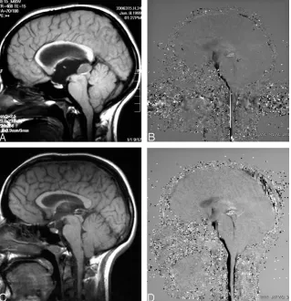
[image:4.585.55.374.59.389.2]FIG 3. Patients with secondary aque-duct stenosis induced by pineal lesion. A,Sagittal T1-weighted image demon-strated a cystic lesion in the pineal re-gion blocking the aqueduct and produc-ing hydrocephalus. B, Sagittal cine phase-contrast in the ventriculostomy site performed shortly after surgery and treatment of pineal lesion demonstrates a filiform flow signal intensity in the ven-triculostomy and subtle flow signal in-tensity in the aqueduct (arrows).C, Sag-ittal T1-weighted image obtained long after treatment demonstrates reduction of pineal lesion and aqueduct decom-pression. D, Sagittal cine phase-con-trast performed in the same examination shows absence of flow in the ventricu-lostomy and aqueduct permeability.
TABLE 1: Clinical outcome related to ventricular size changes
Clinical Outcome
Ventricular Size Changes
No Improvement (n⫽6; 15.8%)
Partial Improvement (n⫽17; 44.7%)
Good Outcome (n⫽13; 34.2%)
Clinical Worsening (n⫽2; 5.3%)
No reduction (n⫽23; 60.5%)
5 (13.2%) 8 (21.1%) 8 (21.1%) 2 (5.3%)
Reduction (n⫽15; 39.5%)
1 (2.6%) 9 (23.7%) 5 (13.2)
TABLE 2: Relationship between hydrocephalus etiology and clinical outcome
Hydrocephalus
Clinical Outcome
PAS (n⫽11; 28.9%)
SAS (n⫽18; 47.4%)
AC (n⫽6; 15.8%)
CH (n⫽3; 7.9%)
No improvement (n⫽6; 15.8%)
1 (2.6%) 4 (10.5%) 1 (2.6%)
Partial Improvement (n⫽17; 44.7%)
3 (7.9%) 12 (31.6%) 1 (2.6%) 1 (2.6%)
Good outcome (n⫽13; 34.2%)
7 (18.4%) 5 (13.2%) 1 (2.6%)
Clinical worsening (n⫽2; 5.3%)
1 (2.6%) 1 (2.6%)
[image:4.585.50.541.421.518.2] [image:4.585.55.533.545.674.2]Stroke volume values obtained in the ventriculos-tomy in the patient group were significantly higher than the stroke volume values registered in the aque-duct in the control group (P ⬎ .01). In the control group the median of OFA was 80 mm3, whereas in the patient group the median obtained in the ventriculos-tomy was 137 mm3.
These results confirm the higher flow of CSF dur-ing the cardiac cycle passdur-ing through the ventriculos-tomy than passing through the aqueduct. This result was to be expected, because the diameter of the ventriculostomy was greater than the aqueduct size. We also observed that the stroke volume values were maintained in the follow-up studies.
When we compared the stroke volume values be-tween patients with clinical improvement and patients without any clinical changes or worsening of the symptoms, we found that the patients with bad out-come had lower stroke volume values (Fig 4). This finding is statistically significant if we use SSV and DSV and OFA (P⬎.01). We did not, however, find any differences between patients with total or partial improvement.
Comparative ROC curves by using the stroke vol-ume values demonstrated that OFA is a better test than the other variables to predict the response to surgery in the postoperative studies. When OFA value is⬎75 mm3, the sensitivity and specificity of this technique to determine which patient will improve were 76.7% and 87.5%, respectively, with a positive likelihood ratio of 6.13 and a negative likelihood ratio of 0.27.
Discussion
The indications for ETV are still a matter of con-troversy. Although the technique has proved to be highly successful in treating occlusive hydrocephalus caused by primary or secondary aqueductal stenosis
and space-occupying lesions of the midbrain, the pi-neal region or the posterior fossa (11–15), it is thought to be less effective in patients with hydro-cephalus caused by intraventricular or subarachnoid hemorrhage, in patients with meningitis, in pediatric patients with associated spinal dysraphism and in nor-mal pressure hydrocephalus (1, 2, 5, 12, 16 –18). In Chiari type I malformations, the results with ETV reported in the literature vary widely. Although hy-drocephalus associated with Chiari type I malforma-tions may be considered obstructive, the reason for the disparity in the results is unknown (5, 18). In our series the only 2 cases with Chiari type I malformation presenting clinical improvement were patients in whom the main cause of hydrocephalus was aqueduc-tal stenosis.
A range of image parameters have been assessed to evaluate the permeability of the ETV, including ven-tricular size changes, flow void signal intensity, and MR patency, by using cine phase-contrast MR (2–5, 7, 12, 19, 20). MR imaging velocity measurements in the third ventriculostomy have also been evaluated by Lev et al (13).
Ventricular size is not always reduced after third ventriculostomy. The proportion of subjects who did not present ventricular size reduction ranges between 11% and 38% in the series reported to date. In our series, we found a reduction in only 39.5% of the patients, and, on correlating the ventricular size changes with clinical improvement and hydrocepha-lus etiology, we found that even among patients in whom the symptoms improved completely only 38% had ventricular size reduction. We also found that the patients with secondary aqueductal stenosis caused by tumor compression had the greatest ventricular size reduction. This finding suggests that ventricular size reduction is easier to identify in patients with acute hydrocephalus, whereas in chronic hydrocephalus the ventricular size changes are subtler and in some cases are only perceived when detailed measurements or volumetric studies are performed (2, 20 –22). Cur-rently, the consensus is that ventricular size reduction does not seem to correlate with clinical outcome, and, thus, anatomic neuroimaging during follow-up is only useful for ruling out increasing ventricular size.
Numerous investigators have studied flow void as an indicator of ETV patency. Flow void signal inten-sity in the floor of the third ventricle assessed by MR flow studies is observed in most patients with clinical improvement and indicates evidence of flow through the ETV (3, 16, 23). Some patency of the ETV site, however, has been demonstrated, even in cases deemed clinical failures with an incidence of as high as 50% in some series (24). In our series, 4 patients did not show flow void signal intensity in the floor of the third ventricle in the follow-up sagittal cine phase-contrast MR imaging studies, but the quantitative studies still indicated low values in mean velocity and stroke volume. In one of these patients with primary aqueduct stenosis, the ventriculostomy was examined and arachnoid septa were observed. The other 3 pa-tients were also reevaluated—2 who had communi-FIG 4. Box plot of the OFA of stroke volume related to clinical
cating hydrocephalus were treated with ventriculo-peritoneal shunt, and in the other patient who presented reduction of secondary aqueductal stenosis after radiation therapy we considered that the ven-triculostomy was not functioning but the CSF flow in the aqueduct had been restored.
Phase-contrast flow-sensitive MR imaging techniques offer more physiologic data than structural MR images and qualitative assessment of the patency of ventricu-lostomy. The sagittal acquisition tends to underestimate CSF flow because of intravoxel phase dispersion from in-plane flow, but, by contrast, it produces an
easy-to-read cine display. The angle-axial acquisitions perpen-dicular to the ventriculostomy takes advantage of through-plane flow and is more accurate for quantita-tive analysis, because the partial volume effects are min-imized. Velocity encoding has to be similar or a little higher than 20 cm/s to avoid aliasing artifacts. Although in this study the MR acquisitions were frankly larger, new MR images provide faster hardware that allows performing these studies in⬍15 minutes. Only patients with severe bradycardia and cardiac arrhythmia have to be excluded, because cardiac cycle registration will be difficult.
Lev et al (13) reported the utility of cine phase-contrast MR velocity measurements in determining the functional status of third ventriculostomy. They examined 6 patients with third ventriculostomy and 12 normal subjects by phase-contrast MR and corre-lated the quantitative velocity data with clinical fol-low-up. They concluded that phase-contrast MR ve-locity measurements, specifically the veve-locity ratio between the high pontine cistern and the space ante-rior to the spinal cord, may help to determine the functional status of third ventriculostomy.
In our study, we compared stroke volume measure-ment in the third ventriculostomy and the aqueduct in a healthy control group and observed that the pattern of the stroke volume was similar, indicating that the cardiac cycle-related pulsated bidirectional CSF mo-tion through the aqueduct in the healthy volunteers is also present in the ventriculostomy orifice. The low resistance provided by the ventriculostomy allows in-creased CSF pulsation during the cardiac cycle. Stroke volume measurements in the ventriculostomy are expected to be higher than in the aqueduct be-cause the diameter of the ventriculostomy is greater than the aqueduct size. Our results demonstrated these differences between stroke volume in the ven-triculostomy and the aqueduct and also the perma-nency of these high stroke volume values across time in the follow-up studies. We also observed that in some patients the caudocranial flow was higher than the craniocaudal in the first postoperative control but this pattern was reversed in the follow-up studies. After surgery there may be a turbulent flow in the ventriculostomy that progressively returns to normal and this inverted flow pattern in the first postoperative control does not necessarily indicate failure of the ven-triculostomy. None of the patients in our series showed worsening or lack of improvement of symptoms.
Of the 6 patients who did not improve after ven-triculostomy, 4 had Arnold Chiari malformation and another had communicating hydrocephalus. The lack of success of ventriculostomy in these conditions may not have been due to inefficient surgery technique but rather to erroneous indication. In each case, the stoke volume was low, and, thus, ventriculostomy function was poor because of an insufficient pressure gradient between the third ventricle floor and the basal cis-terns. An obstructive intraventricular condition leads to the appearance of a pressure gradient between the ventricular system and the subarachnoid space and the opening between these compartments. This pres-sure gradient allows the CSF to move forward into the subarachnoid space. Quantitative analysis of the CSF flow in the ventriculostomy in these cases with lack of success indicated that the amount of CSF flow across the fenestration was insufficient to resolve the CSF circulation problem, possibly because of inap-propriate treatment in most patients with nonintra-ventricular obstruction (Fig 5).
An important finding in this study is that, if the OFA of the stroke volume in the initial follow-up study is high (⬎75 mm3), ventriculostomy is effective and the patient improves. If the value of stroke
vol-ume is⬍75 mm3, further radiologic follow-up is nec-essary to determine the usefulness of ventriculos-tomy. We also found that a decrease in stroke volume during the follow-up control was associated with ven-triculostomy failure and clinical deterioration.
Conclusion
Endoscopic third ventriculostomy restores the pul-satile bidirectional CSF motion. In addition, the mea-surement of stroke volume in ventriculostomy by us-ing cine phase-contrast MR imagus-ing provides functional information about the third ventriculos-tomy. Our results show a good correlation between clinical improvement and stroke volume. When the stroke volume obtained in the ventriculostomy is high, the clinical outcome is usually good and further quantification radiologic study is only needed in the appearance of clinical deterioration. Ventricular size is not a good indicator of ventriculostomy patency. Similar to other authors, we consider ETV to be the best technique for primary and secondary aqueductal stenosis, but not for communicating hydrocephalus.
References
1. Buxton N, Ho KJ, Macarthur D, et al. Neuroendoscopic third ventriculostomy for hydrocephalus in adults: report of a single unit’s experience with 63 cases.Surg Neurol2001;55:74 –78 2. Buxton N, Turner B, Ramli N, Vloeberghs M.Changes in third
ventricular size with neuroendoscopic third ventriculostomy: a blinded study.J Neurol Neurosurg Psychiatry2002;72:385–387 3. Cinalli G, Sainte-Rose C, Chumas P, et al.Failure of third
ventric-ulostomy in the treatment of aqueductal stenosis in children.
J Neurosurg1999;90:448 – 454
4. Fukuhara T, Vorster SJ, Ruggieri P, Luciano MG.Third ventric-ulostomy patency: comparison of findings at cine phase-contrast MR imaging and at direct exploration.AJNR Am J Neuroradiol
1999;20:1560 –1566
5. Fukuhara T, Vorster SJ, Luciano MG.Risk Factors for failure of endoscopic third ventriculostomy for obstructive hydrocephalus.
Neurosurgery2000;46:1100 –1111
6. Hayashi N, Hamada H, Hirashima Y, et al.Clinical features in patients requiring reoperation after failed endoscopic procedures for hydrocephalus.Minim Invas Neurosurg2000;43:181–186 7. Kulkarni AV, Drake JM, Amstrong DC, Dirks PB.Imaging
corre-lates of successful endoscopic third ventriculostomy.J Neurosurg
2000;92:915–919
8. Meier U, Zeilinger FS, Schonherr B.Endoscopic ventriculostomy versus shunt operation in normal pressure hydrocephalus: diag-nostics and indication.Minim Invas Neurosurg2000;43:87–90 9. Murshid WR.Endoscopic third ventriculostomy: toward more
in-dications for the treatment of non-communicating hydrocephalus.
Minim Invas Neurosurg2000;43:75– 82
10. Schroeder HW, Schweim C, Schweim HH, Gaab MR.Analysis of aqueductal cerebrospinal fluid flow after endoscopic aqueducto-plasty by using cine phase-contrast magnetic resonance imaging.
J Neurosurg2000;93:238 –244
11. Fukuhara T, Luciano MG.Clinical features of late-onset idiopathic aqueductal stenosis.Surg Neurol2001;55:132–137
12. Hopf NJ, Grunert P, Fries G, et al.Endoscopic third ventriculos-tomy: outcome analysis of 100 consecutive procedures. Neurosur-gery1999;44:795– 806
13. Lev S, Bhadelia RA, Estin D, et al.Functional analysis of third ventriculostomy patency with phase contrast MRI velocity mea-surements.Neuroradiology1997;39:175–179
14. Rieger A, Rainov G, Brucke M, et al.Endoscopic third ventricu-lostomy is the treatment of choice for obstructive hydrocephalus due to pediatric pineal tumors. Minim Invas Neurosurg 2000; 43:83– 86
caused by primary aqueductal stenosis. Neurosurgery 2000;46: 104 –111
16. Buxton N, Macarthur D, Mallucci C, et al.Neuroendoscopic third ventriculostomy in patients less than 1 year old.Pediatr Neurosurg
1998;29:73–76
17. Jones RF, Stening WA, Brydon M.Endoscopic third ventriculos-tomy.Neurosurgery1990;26:86 –91
18. Suehiro T, Inamura T, Natori Y, et al.Successful neuroendoscopic third ventriculostomy for hydrocephalus and syringomyelia asso-ciated with fourth ventricle outlet obstruction. J Neurosurg
2000;93:326 –329
19. Rovira A, Capellades J, Grive E, et al.Spontaneous ventriculos-tomy: report of three cases revealed by flow-sensitive phase-con-trast cine MR imaging.AJNR Am J Neuroradiol1999;20:1647–1652
20. Schwartz TH, Ho B, Prestigiacomo CJ, et al.Ventricular volume following third ventriculostomy.J Neurosurg1999;91:21–25 21. Oka K, Go Y, Kin, et al. The radiographic restoration of the
ventricular system after third ventriculostomy.Minim Invas Neu-rosurg1995;38:158 –162
22. Schwartz TH, Yoon SS, Cutruzzola FW, Goodman RR. Third ventriculostomy: post-operative ventricular size and outcome.
Minim Invas Neurosurg1996;39:122–129
23. Goumnerova LC, Frim DM. Treatment of hydrocephalus with third vetriculocisternostomy: outcome and CSF flow patterns. Pe-diatr Neurosurg1997;27:149 –152