SHORT COMMUNICATION
Activity dependence of spreading depression in the locust CNS
Kristin E. Spong*, Tom R. Mazzetti and R. Meldrum RobertsonABSTRACT
Spreading depression (SD) is associated with large changes in extracellular ion concentrations and can be induced by impairing mechanisms of K+ion homeostasis. We tested activity dependence of SD in the locust model of ouabain-induced SD in the metathoracic ganglion. Wind activation of thoracic circuitry resulted in small increases of K+concentration that took 5–10 s to be cleared from the extracellular space. In the presence of the Na+/K+-ATPase inhibitor ouabain, wind stimulation every 30 s halved the latency to the first SD event and increased its duration. Wind stimulation was able to trigger the first event, suggesting that local activity could determine the origin of successive SD events. Perfusion with calcium-free saline blocked neural activity in the ganglion and prevented the occurrence of ouabain-induced SD. We conclude that ouabain-induced SD in the locust CNS is strongly dependent on the existing level of neural activity.
KEY WORDS: Potassium,Locusta migratoria, Metathoracic ganglion, Ouabain
INTRODUCTION
Spreading depression (SD) is a neural phenomenon first discovered by Leao (1944) in the cortex of the rabbit but has since been demonstrated to occur in many vertebrate and invertebrate systems. It is characterized by rapid cellular depolarization and an associated arrest in neural activity that slowly propagates throughout neural tissue (Leao, 1944; Somjen, 2001). The depolarization and disruption in electrical activity is reflective of the massive disturbance in ionic homeostasis that occurs at the onset of SD. Waves of SD are associated with abrupt increases in the extracellular potassium concentration ([K+]
o) and drop in extracellular sodium ([Na+]o),
calcium ([Ca2+]
o) and chloride ([Cl−]o) concentrations (Somjen,
2001). Restoration of ionic gradients occurs within minutes of an SD episode and ultimately allows for the recovery of neural activity (Rodgers et al., 2007; Somjen, 2001). Cortical spreading depression (CSD) in humans is thought to underlie the aura that accompanies migraine and has also been implicated in more severe pathologies such as stroke and traumatic brain injury (Somjen, 2001). Invertebrate SD has been best described in the CNS ofLocusta migratoriaand is associated with neural shutdown induced by environmental stress. For example, locusts enter a reversible coma when exposed to stimuli such as hyperthermia, hypothermia and anoxia during which SD-like events can be monitored within the metathoracic ganglion (MTG) (Rodgers et al., 2007, 2010). Interestingly, both the propagation rate (2.4 mm min−1) and magnitude of [K+]
odisturbance (∼50 mmol l−1)
during locust SD (Rodgers et al., 2007) are strikingly similar to measurements during CSD in mammals, which is associated with
[K+]
o increases of ∼50–60 mmol l−1 traveling at velocities of
2-5 mm min−1(Somjen, 2001).
In healthy neural tissue SD can be experimentally induced by disrupting mechanisms of K+homeostasis. For example, inhibition
of the Na+/K+-ATPase with ouabain reliably induces SD in both the
vertebrate and invertebrate CNS (Balestrino et al., 1999; Rodgers et al., 2009). In semi-intact locust preparations bath application of ouabain elicits repetitive waves of SD within the MTG, characterized by abrupt increases in [K+]
owhere the rise and fall
coincide with the arrest and recovery of electrical activity (Rodgers et al., 2009). Because of the robustness and repetitive nature of ouabain-induced SD, it has become a commonly used control procedure for investigations into the cellular mechanisms underlying SD in the locust CNS. Moreover, ouabain-induced SD in the locust has been proposed as a model for peri-infarct depolarizations (PIDs) which are detrimental depolarizations that spontaneously arise in ischemic brain regions following stroke (Rodgers et al., 2010).
Although the precise mechanisms underlying SD initiation are still not clearly understood, mechanistic models of locust SD suggest that onset occurs once K+levels exceed a critical threshold
triggering a positive-feedback cycle that ultimately leads to the characteristic all-or-none increase in [K+]
o(Armstrong et al., 2009;
Rodgers et al., 2010). It is predicted from these models that increased neural activity would predispose towards the generation of SD; however, direct evidence demonstrating the activity dependence of SD is lacking. Here, we test how increased neural activity, through wind activation of thoracic circuitry, affects ouabain-induced SD in the locust. Additionally, by manipulating the [Ca2+] in the bathing solution we reduce neural activity and
examine how this alters the response of the tissue to ouabain. Our results are consistent with the conclusion that ouabain-induced SD in the locust MTG is strongly influenced by existing levels of neural activity.
MATERIALS AND METHODS Animals
Locusts (Locusta migratoria Linnaeus 1758) were housed in a crowded colony located in the Queen’s University Animal Facility. The colony was maintained under 12 h light:12 h dark cycles at temperatures of approximately 25°C. Animals were fed once daily with wheat grass and a mixture of milk powder, yeast and bran. All experiments were performed using adult males at 2–5 weeks after imaginal ecdysis. Locusts were randomly chosen from the colony and held in ventilated plastic containers prior to experimentation. Animals used in the wind stimulation experiments were deprived of food for 1 day prior to experimentation to increase the probability of reliably activating locomotor circuitry (Davenport and Evans, 1984). The thoracic and abdominal cavities of semi-intact preparations (Robertson and Pearson, 1982) were continuously bathed in a saline solution to prevent desiccation. Standard locust saline contained (in mmol l−1): 147 NaCl, 10 KCl, 4 CaCl
2, 3 NaOH
and 10 HEPES buffer ( pH 7.2; all chemicals were obtained from
Received 21 September 2015; Accepted 17 December 2015
Department of Biology, Queen’s University, Bioscience complex, Room 3404, Kingston, Ontario, Canada K7L 3N6.
*Author for correspondence ([email protected])
Journal
of
Experimental
Sigma). Preparations were grounded by inserting a wire coated with silver chloride into either the abdomen or the upper thorax. To induce SD, semi-intact preparations were exposed to either 1×10⁻⁴mol l−1or 5×10⁻⁴mol l−1ouabain (Sigma) for 40 min.
Measuring extracellular K+
The [K+]
owas continuously monitored within the MTG using K+
-sensitive microelectrodes prepared using 1-mm-diameter unfilamented glass capillary tubes. Capillary tubes were rinsed with methanol (99.9%) and dried on a hotplate prior to being pulled. The microelectrodes were then silanized on a hotplate for 1 h by exposure to dichlorodimethylsilane (99%; Sigma) vapor. The microelectrode tips were filled with Potassium Ionophore I-Cocktail B (5% valinomycin; Sigma), back-filled with 500 mmol l−1KCl and stored in the dark with tips suspended in
distilled water until needed for experimentation. Reference microelectrodes were prepared prior to experimentation using 1-mm-diameter filamented glass capillary tubes and filled with 500 mmol l−1 KCl. Tip resistance of both the reference and K+
-sensitive microelectrodes was approximately 5–7 mΩonce filled. Just prior to each experiment, the K+-sensitive and reference
microelectrode pair were connected to a DUO773 two-channel intracellular/extracellular amplifier (World Precision Instruments, Sarasota, FL, USA) and calibrated using 15 mmol l−1 KCl+
135 mmol l−1NaCl and 150 mmol l−1KCl solutions to obtain the
voltage difference from a 10-fold change in K+ concentration.
Following calibration, the microelectrodes were inserted through the sheath of the MTG adjacent to one another and the extracellular K+
voltage within the neuropil was recorded. Voltage recordings were subsequently converted to [K+]
o (mmol l−1) using the Nernst
equation (for details, see Rodgers et al., 2007).
Measuring direct current (DC) potential
Abrupt negative deflections in DC field potential are indicative of SD, which reflects the massive cellular depolarization that takes place during the events (Somjen, 2001). Microelectrodes were prepared using 1-mm-diameter filamented glass capillary tubes. Once pulled, microelectrodes were filled with 500 mmol l−1KCl,
forming low-resistance tips (∼5–7 mΩ) and connected to a DUO773 two-channel intracellular/extracellular amplifier (World Precision Instruments). A single microelectrode was inserted through the sheath of the MTG and DC potential was continuously monitored throughout the experiment.
Wind stimulation protocol
To enhance neural activity, wind stimuli were applied to the head of semi-intact preparations. Wind stimulation was applied for 5 s and was administered through a 5-mm-diameter plastic tube situated at a distance∼2 cm from the locust’s head. In experiments measuring the extracellular K+ activity, wind stimulation was administered
every 30 s until the onset of ouabain-induced SD. In DC potential recordings, wind stimulation began following a 5 min baseline period and was administered every 30 s or 1 min for a total of 10 min. The flight central pattern generator (CPG) is one circuit likely to be activated by wind stimulation (Robertson and Pearson, 1982) and thus we monitored flight activity electromyographically by inserting a copper wire, insulated except at the tip, into a dorsal longitudinal wing depressor muscle located in the thorax. To test how wind activation of thoracic circuitry affects the susceptibility to SD, we compared preparations subjected to the wind stimulation protocol to control preparations that received no wind stimulation. In all experiments, repetitive SD was induced by bath application of ouabain (5×10⁻⁴ mol l−1), which was administered for 40 min
following a 5 min baseline period.
Calcium manipulations
Extracellular Ca2+levels were manipulated by altering the [Ca2+]
in the bathing saline solution. Preparations were treated with standard locust saline (as described above), high-Ca2+saline
or Ca2+-free saline. In the high-Ca2+ condition, the [Ca2+]
increased from 4 mmol l−1(standard locust saline) to 8 mmol l−1.
In Ca2+-free condition, zero-Ca2+ saline solution was used,
which also contained the Ca2+ chelator, ethylene glycol-bis
(β-aminoethylether)-N,N,N′,N′-tetraacetic acid (EGTA; Sigma) at a concentration of 10−3 mol l−1. Ouabain (10−4 mol l−1) was
dissolved in standard, high-Ca2+ or Ca2+-free saline and
administered for 40 min to semi-intact preparations following a 20 min treatment period with the corresponding saline type. Ouabain-induced SD was monitored by measuring the DC potential within the MTG.
Analyses of spreading depression
The latency to onset was measured from the time of ouabain application to the downward inflection point of the first SD event. The duration of the first event was calculated by taking time measurements at half maximum amplitude of the negative shift in DC potential. Total number of events represents the number of SD episodes that occurred within the 40 min treatment period with ouabain.
Statistical analyses
All data were plotted and analysed using SigmaPlot 12.5 (Systat Software). Parametric data are displayed using bar charts with columns representing the mean and s.e.m. Non-parametric data are plotted as box plots representing the 25th and 75th percentiles (interquartile range; IQR) with a line indicating median and whiskers extending to the 10th and 90th percentiles (individual points represent outliers). To determine significant differences between two groups eithert-tests or Mann–Whitney rank-sum tests were used for parametric and non-parametric data, respectively (P<0.05). A one-way ANOVA was used to determine significant differences between more than two groups and post hoc comparisons were performed using Holm–Šidák multiple comparisons (P<0.05).
RESULTS AND DISCUSSION
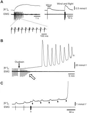
To increase neural activity, we applied repetitive wind stimuli to the head of semi-intact locust preparations activating thoracic circuitry including the flight CPG. Flight motor patterns were recorded electromyographically from a dorsal longitudinal wing depressor muscle and the [K+]
owas continuously monitored from within the
MTG (Fig. 1A). Wind activation of thoracic circuitry was associated List of symbols and abbreviations
[Ca2+] calcium concentration CNS central nervous system CPG central pattern generator CSD cortical spreading depression [K+]o extracellular potassium concentration MTG metathoracic ganglion
[Na+]o extracellular sodium concentration PID peri-infarct depolarization SD spreading depression
Journal
of
Experimental
with 0.2±0.03 mmol l−1(N=10) increases in the [K+]
owhich took
9.8±0.3 s (N=8) to return to baseline values (Fig. 1A) and fit nicely with an exponential [K+]
o clearance time constant of 3.1±0.4 s
(N=10). Wind alone was able to produce increases in [K+]
o;
however, the magnitude of increase was greater when flight was induced. The [K+]
o disturbances reported here are of similar
magnitude to the activity-dependent increases in [K+]
othat occur in
the CNS of the cockroach following electrical stimulation to the connectives (Grossman and Gutnick, 1981). Such modest increases in [K+]
oare sufficient to mediate interactions between neurons in
close proximity (Yarom and Spira, 1982). Furthermore, given the extensive yet restricted volume of the extracellular space, such K+
-mediated interactions are likely to spread, affecting more distant neurons (Spira et al., 1984).
Here, we show that wind stimulation could trigger the first ouabain-induced SD event. Repetitive SD was ouabain-induced within the MTG by bath application of ouabain and monitored by recording the characteristic all-or-none increases in the [K+]
o (Fig. 1B). Upon
ouabain wash-in, impairment in flight activity and a reduction in electromyography (EMG) amplitude was observed. Wind stimulation was applied every 30 s until the first ouabain-induced surge and in 5/9 preparations the first surge was clearly triggered by the 5 s wind stimulus (Fig. 1C). These results suggest that increases in local activity can determine the origin of successive SD events. This is consistent with recent reports investigating the origins of PIDs in mammalian cortex. Until recently the triggering factors leading to the
eruption of PIDs were unknown. However, it now has been demonstrated that somato-sensory activation of ischemic cortex reproducibly triggers PIDs and is associated with increased oxygen utilization in the stimulated region (von Bornstädt et al., 2015).
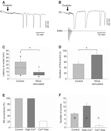
To further investigate the activity dependence of SD, we tested how wind activation of thoracic circuitry affects susceptibility to SD. Bath application of ouabain induced repetitive SD in 100% of control (Fig. 2A) and wind-stimulated preparations (Fig. 2B); however, wind-stimulated preparations were associated with shorter latencies to onset compared with control preparations (Fig. 2C; wind stimulated, median=6.8 min, IQR=5.4, 9.5; control, median=15.8 min, IQR=6.3, 24.1), suggesting that wind stimulation increased the susceptibility to ouabain-induced SD. Additionally, wind stimulation significantly increased the duration of the first SD event from 53.1±8.3 s, without stimulation, to 85.3± 10.3 s (Fig. 2D). A longer duration suggests impairment in the ability to restore ionic gradients. The event duration reported here probably reflects the increased demand on the Na+/K+-ATPase in
preparations experiencing increased neural activity as a result of wind stimulation.
In addition to investigating how increases in neural activity affect ouabain-induced SD, we also tested the effects of reducing neural activity by manipulating the [Ca2+] within the bathing medium. We
compared ouabain-induced SD under control, Ca2+-free and high-Ca2+
conditions. Reducing Ca2+levels can be predicted to reduce activity
by depressing synaptic transmission. In the current experiments,
30 s
1 mmol l–1
[K+] o
EMG
*
10 s Wind Wind and flight
A
5 min
20 mmol l–1
Ouabain
[K+] o
EMG
B
C
0.5 mmol l–1
1 s [K+]
o
EMG
[image:3.612.65.348.59.427.2]100 ms
Fig. 1. K+activity in response to wind stimulation, flight activity and ouabain exposure.(A-C) Simultaneous recordings of the [K+]owithin the MTG and flight activity (EMG) induced by wind stimulation. (A) Wind stimulation and flight activity transiently increase [K+]o. Inset shows an expansion of the flight motor pattern recorded electromyographically from a dorsal longitudinal wing depressor muscle. Note that wind stimulation alone increases [K+]oand that such wind-induced [K
+ ]o disturbances can be amplified when paired with flight activity. (B) Representative recording of ouabain-induced SD in a preparation subjected to wind stimulation (same preparation shown in A). Open arrow indicates impairment of flight and reduction of EMG amplitude as a result of ouabain wash-in. (C) Representative recording (different preparation from A and B) demonstrating that wind stimulation can trigger the first ouabain-induced event. The asterisk indicates a convulsive burst in DL motorneurons as flight CPG fails. Arrows indicate times of wind stimulation every 30 s after the EMG fails. Note that the final wind stimulation triggers the abrupt K+surge.
Journal
of
Experimental
a blockade of neural activity under Ca2+-free conditions was
evidenced by a cessation in ventalitory abdomen movements that occurred within 1–5 min of saline application. Bath application of ouabain reliably induced SD in 100% of preparations treated under control and high-Ca2+ conditions; however, under Ca2+-free
conditions, ouabain was found to induce SD in only 2/9 (∼22%) preparations (Fig. 2E). The two zero-Ca2+preparations were associated
with longer latencies to SD onset (17.61 min, 26.72 min) compared with the control (median=8.9 min, IQR=3.3, 15.1) and high-Ca2+
(median=5.2 min, IQR=4.4, 11.5) preparations. Additionally,
perfusion with zero-Ca2+saline significantly reduced the number of
individual events exhibited within the 40 min treatment period (Fig. 2F; Ca2+-free, 0.9±0.6; control, 6.7±0.7; high-Ca2+, 10.4±1.3).
These results demonstrate that a reduction in neural activity suppresses ouabain-induced SD. Reducing neural excitability has previously been shown to protect against stress-induced neural shutdown. For instance, blockade of Na+channels with tetrodotoxin (TTX) delays the onset of
hyperthermic-induced neural failure in the locust CNS (Rodgers et al., 2007). In the current experiments, preparations exposed to high-Ca2+
conditions were found to exhibit a significantly greater number of
Control High-Ca2+ Ca2+-free Control
Control Wind-stimulated
Control Wind-stimulated
*
*
EMG
10 min 10 min
20 mV 20 mV
Ouabain Ouabain
A
C
E
Latency to onset (min)
Duration of first event (s)
SD occurrence (%) Number of events
100
80
60
40
20
0
14 12 10 8 6 4 2 0 100
80
60
40
20
0 40
35 30 25 20 15 10 5 0
DC DC
B A C
B
D
F
[image:4.612.126.490.57.487.2]High-Ca2+ Ca2+-free
Fig. 2. Activity dependence of ouabain-induced SD.(A,B) Representative traces of the DC potential recorded from within the MTG during bath application of 5×10⁻⁴mol l−1ouabain under control conditions (A, no wind stimulation) and experimental conditions (B, wind stimulation applied). Note that following a delay, ouabain exposure induces repetitive negative deflections in DC potential indicative of SD. Simultaneous recordings of the DC potential and flight muscle activity (EMG) are shown during wind stimulation. (C) Wind-stimulated preparations (N=19) were associated with significantly shorter latencies to SD onset compared with control preparations (N=20). Data are plotted as the median and upper and lower quartiles. Asterisk indicates a significant difference between conditions (Mann– WhitneyU-statistic=96.000,P=0.009). (D) The duration of the first ouabain-induced event was significantly greater, as denoted by the asterisk, in preparations subjected to wind stimulation (N=19) compared with control preparations (N=20; two-tailedt-test,t37=−2.4,P=0.019). Data are plotted as means±s.e.m. (E) Percentage of preparations that exhibited ouabain-induced SD under control (N=9), high-Ca2+(N=10) and zero-Ca2+(N=9) conditions. Ca2+-free conditions could prevent SD occurrence. (F) Number of individual SD events recorded within the 40 min treatment period under control (N=9), high-Ca2+(N=10) and zero-Ca2+ (n=9) conditions. A one-way ANOVA revealed significant differences between groups (F2,25=25.8,P<0.001).Post hoccomparisons revealed that there were significantly fewer individual events under zero-Ca2+conditions compared with control and high-Ca2+conditions and a significantly greater number of events under high-Ca2+conditions compared with control conditions. Data are plotted as means±s.e.m. Columns assigned different letters are significantly different (Holm–Šidák,P<0.05).
Journal
of
Experimental
individual SD events compared with controls (Fig. 2F; data reported above). Interestingly, in the rat brain in vivo, increased influx of extracellular Ca2+ ions can trigger waves of CSD and facilitate
propagation rates, suggesting that Ca2+plays an important role in the
initiation of CSD (Torrente et al., 2014). Thus, the precise role that Ca2+
plays in locust SD merits further investigation.
To summarize, we tested the activity dependence of ouabain-induced SD in the locust CNS. Mechanistic models of locust SD speculate that increased neural activity would predispose towards occurrence of SD (Armstrong et al., 2009; Rodgers et al., 2010); however, this had seldom been tested. Here, we provide direct evidence that increases in neural activity heighten susceptibility to SD whereas reductions in activity attenuate SD. It is particularly interesting that wind stimulation could trigger the first SD event. Our findings demonstrate that ouabain-induced SD is strongly influenced by existing activity levels and help to substantiate previously proposed models of SD.
Acknowledgements
We would like to thank Madison Gunn for help collecting some of the data for the manuscript.
Competing interests
The authors declare no competing or financial interests.
Author contributions
K.E.S., T.R.M. and R.M.R. conceived and designed the research; K.E.S., T.R.M. and R.M.R. performed the experiments and analysed the data; K.E.S., T.R.M. and R.M.R. interpreted the results of the experiments; K.E.S. and R.M.R. prepared the figures; K.E.S. drafted the manuscript; K.E.S. and R.M.R. revised the manuscript; K.E.S., T.R.M. and R.M.R. approved the final version of the manuscript.
Funding
This work was supported by the Natural Sciences and Engineering Research Council of Canada [40930-2009].
References
Armstrong, G. A. B., Rodgers, C. I., Money, T. G. A. and Robertson, R. M.(2009). Suppression of spreading depression-like events in locusts by inhibition of the NO/ cGMP/PKG pathway.J. Neurosci.29, 8225-8235.
Balestrino, M., Young, J. and Aitken, P.(1999). Block of (Na+,K+)ATPase with ouabain induces spreading depression-like depolarization in hippocampal slices. Brain Res.838, 37-44.
Davenport, A. P. and Evans, P. D.(1984). Changes in haemolymph octopamine levels associated with food deprivation in the locust,Schistocerca gregaria. Physiol. Entomol.9, 269-274.
Grossman, Y. and Gutnick, M. J.(1981). Extracellular potassium activity during frequency-dependent conduction block of giant axons in the metathoracic ganglion of the cockroach.Brain Res.211, 196-201.
Leao, A. A. (1944). Spreading depression of activity in the cerebral cortex. J. Neurophysiol.7, 359-390.
Robertson, R. M. and Pearson, K. G.(1982). A preparation for the intracellular analysis of neuronal activity during flight in the locust.J. Comp. Physiol. A146, 311-320.
Rodgers, C. I., Armstrong, G. A. B., Shoemaker, K. L., LaBrie, J. D., Moyes, C. D. and Robertson, R. M.(2007). Stress preconditioning of spreading depression in the locust CNS.PLoS ONE2, e1366.
Rodgers, C. I., LaBrie, J. D. and Robertson, R. M.(2009). K+ homeostasis and central pattern generation in the metathoracic ganglion of the locust.J. Insect Physiol.55, 599-607.
Rodgers, C. I., Armstrong, G. A. and Robertson, R. M.(2010). Coma in response to environmental stress in the locust: a model for cortical spreading depression. J. Insect Physiol.56, 980-990.
Somjen, G. G.(2001). Mechanisms of spreading depression and hypoxic spreading depression-like depolarization.Physiol. Rev.81, 1065-1096.
Spira, M. E., Yarom, Y. and Zeldes, D.(1984). Neuronal interactions mediated by neurally evoked changes in the extracellular potassium concentration.J. Exp. Biol.112, 179-197.
Torrente, D., Mendes-da-Silva, R. F., Lopes, A. A. C., González, J., Barreto, G. E. and Guedes, R. C. A.(2014). Increased calcium influx triggers and accelerates cortical spreading depression in vivo in male adult rats.Neurosci. Lett.558, 87-90.
von Bornstädt, D., Houben, T., Seidel, J. L., Zheng, Y., Dilekoz, E., Qin, T., Sandow, N., Kura, S., Eikermann-Haerter, K., Endres, M. et al.(2015). Supply-demand mismatch transients in susceptible peri-infarct hot zones explain the origins of spreading injury depolarizations.Neuron85, 1117-1131.
Yarom, Y. and Spira, M. E.(1982). Extracellular potassium ions mediate specific neuronal interaction.Science216, 80-82.