Original Article
Enrichment of prostate cancer stem cells from primary
prostate cancer cultures of biopsy samples
Shunqi Wang1,3*, Shengsong Huang2*, Xin Zhao2, Qimin Zhang2, Min Wu2, Feng Sun3, Gang Han3, Denglong Wu2
1Institute of Life Science, College of Life Sciences and Food Engineering, Nanchang University, Nanchang, Jiangxi
330031, China; 2Department of Urology, Affiliated Tongji Hospital of Tongji University, Shanghai, Shanghai
200065, China; 3State Key Laboratory of Molecular Biology, Shanghai Key Laboratory of Andrology, Institute of
Biochemistry and Cell Biology, Shanghai Institutes for Biological Sciences, Chinese Academy of Sciences, Shang-hai 200031, China. *Equal contributors.
Received October 31, 2013; Accepted November 30, 2013; Epub December 15, 2013; Published January 1, 2014
Abstract: This study was to enrich prostate cancer stem cells (PrCSC) from primary prostate cancer cultures (PPrCC).
Primary prostate cancer cells were amplified in keratinocyte serum-free medium with epidermal growth factor (EGF) and bovine pituitary extract (BPE), supplemented with leukemia inhibitory factor (LIF), stem cell factor (SCF) and cholera toxin. After amplification, cells were transferred into ultra-low attachment dishes with serum-free DMEM/ F12 medium, supplemented with EGF, basic fibroblast growth factor (bFGF), bovine serum albumin (BSA), insu
-lin, and N2 nutrition. Expression of cell-type-specific markers was determined by RT-qPCR and immunostaining.
Tumorigenicity of enriched PrCSC was determined by soft agar assay and xenograft assay in NOD/SCID mice.
Bi-opsy samples from 19 confirmed prostate cancer patients were used for establishing PPrCC, and 18 cases (95%) succeeded. Both basal marker (CK5) and luminal markers (androgen receptor and CK8) strongly co-expressed in most of PPrCC, indicating their basal epithelial origin. After amplification under adherent culture condition in vitro,
transient amplifying cells were the dominant cells. Sphere formation efficiency (SFE) of passaged PPrCC was about 0.5%, which was 27 times lower than SFE of LNCaP (13.67%) in the same condition. Compared with adherent cells from PPrCC, prostasphere from PPrCC showed up regulated stem cell markers and increased tumorigenic potential
in soft-agar assay. However, spheroid cells from PPrCC prostasphere failed to initiate tumor in xenograft assay in 6
months. Thus, PPrCC can be established and amplified from prostate cancer biopsy samples. Our modified sphere
culture system can enrich PrCSC from PPrCC.
Keywords: Prostate cancer (PrCa), primary prostate cancer cultures (PPrCC), prostate cancer stem cells (PrCSC), sphere culture
Introduction
Prostate cancers are the most frequently diag-nosed cancers and the second most cause of cancer-related death in American men, accounting for 11% of all cancer related deaths [1]. In recent years, there has been a rapid increase in the incidence of prostate cancer in Chinese male [2], with ageing male population increasing briskly. Although there is a great progress in the diagnosis of localized prostate cancer and the disease can be treated early with surgery or radiation therapy, it recurs in approximately 20% to 30% of patients, which eventually progresses in most patients who receive further treatment [3]. Because most
prostate cancers are hormone dependent and respond to androgen deprivation therapy, androgen ablation remains the main treatment of the metastatic disease [4]. While initially effective, this treatment is followed by tumor recurrence in a few years, because the tumors eventually become androgen refractory and metastases, which give the strongest indica-tors of poor outcome [4, 5].
neurological cancer, cervical cancer, breast cancer, colon cancer, liver cancer, lung cancer, melanoma, ovarian cancer, testicular cancer and prostate cancer. Many prostate cancers relapse and metastasis in part due to the pres-ence of prostate cancer stem cells (PrCSC) [8], which do not express androgen receptor and do not directly respond to androgen deprivation therapy [9]. PrCSC may provide insight into the origin of prostate cancer and new therapeutics for prostate cancer. Cell-surface markers (also termed cancer stem cell markers) utilized in PrCSC research include CD133, CD44, integrin α2β1hi in prostate cancer tissue [10], CD44, CD133, integrin α2β1hi, CD24-, AlDH1 in human prostate cancer cell lines [11-16] and Lin -Sca-1+, CD49fhi, Trop2 in mouse model [17-19]. Single or different combinations of these can-cer stem cell markers are utilized to enrich PrCSC population.
Many human prostate cancer cell lines are used to isolate PrCSC, such as LNCaP [14, 16], PC3 [12, 13, 16, 20], DU145 [11-13, 21]. Few labs isolate PrCSC and do advanced research in PrCSC directly from prostate cancer tissues [10, 22], and definitive evidence for the exis -tence of PrCSC in prostate cancer is still lacking [23].
We adopted a sphere culture method to isolate and enrich PrCSC from primary prostate cancer cultures (PPrCC). Sphere culture has been used to isolate cancer stem cells from many types of cancers, and tumorsphere culture in serum-free growth factor defined medium was also used to isolate and propagate PrCSC from pros-tate cancer tissues [22], but has not been opti-mized to enrich PrCSC from PPrCC originating from prostate biopsy tissues. In this study, we amplify the PPrCC by optimized adherent cul-ture condition and enrich PrCSC by optimized suspension culture conditions. Our study dem-onstrates that this modified sphere culture sys -tem can be used to isolate and enrich PrCSC from prostate cancer biopsy samples.
Material and methods
Tissue collection and isolation of prostatic
cancer cells
Human prostatic tissue was obtained, with patient consent, from 28 patients (age range 50 to 86) undergoing radical prostatectomy for prostate cancer and/or prostatic needle biopsy
samples for cancer diagnosis. Initial PSA levels of these patients were from 5 μg/l to 370 μg/l, and Gleason score from 2+2 to 4+5. The condi-tion of prostatic cancer was confirmed by histo -logical examination of representative frag-ments. After pathological examination, primary cells cultured from prostate cancer were pre-pared for next step.
Tissues dissected from radical prostatectomy specimens were minced into small pieces (about 1 mm3), incubated with mixture of Trypsin/EDTA and collagenase I for 20 minutes at 37 °C, and passed through sterile 40-micron cell strainer (Falcon) to prepare single-cell sus-pension. The single cells were seeded into the 6-well plate coated by collagen I (10 μg/cm2). Needle biopsy samples were cut into small pieces (about 1 mm3), and the pieces seeded in the 6-well plate coated by collagen I (10 μg/ cm2) with 0.5 ml of culture medium for the first 12 hours and add 2 ml of culture medium, which increase the adhesion between the piec-es and the plate bottom.
Cell culture medium and cell cultures
The single cells from radical prostatectomy specimens or the small pieces from needle biopsy samples were maintained in complete keratinocyte growth medium (keratinocyte serum-free medium with epidermal growth fac-tor and bovine pituitary extract; Invitrogen), which were also supplemented with 2 ng/ml of leukemia inhibitory factor (LIF, Sigma), 2 ng/ml of stem cell factor (SCF, Sigma), and 100 ng/ml of cholera toxin (Sigma), referring to the meth-od described previously [10].
Cells were cultured in the plates or dishes coat-ed with collagen I in a humidificoat-ed incubator at 37 in an atmosphere of 95% air and 5% carbon dioxide. The medium was changed twice a week, and cells were passaged after detach -ment withTrypLE™ Express.
Immunocytochemistry
mono-clonal anti-cytokeratin 8 rAb (Abcam ab9023, 1:200), rabbit polyclonal anti-androgen recep-tor Ab (Santa Cruz Biotechnology sc-13062, 1:200), Goat anti-Rabbit-Cy3 Ab (Jackson 111-165-045, 1:1000), Goat anti-Mouse-Alexa488 Ab (Invitrogen A-11029, 1:1000). At last, cells were stained with 4’,6-diamidino-2-phenylin-dole dihydrochloride (DAPI) (Sigma D-8417, 1:8000) in PBS, were washed twice with PBS and were mounted with Vectashield (Vector Laboratories) before viewing under a fluores -cence microscope. Fluores-cence detection and imaging were carried out on Leica TCS SP5 con -focal microscope.
In vitro tumorsphere formation assay
Prostate cancer cells were counted and plant-ed in ultra-low dishes at a constant density of 50000 viable cells per milliliter. Cells were grown in serum-free DMEM/F12 medium (Gibco), which was supplemented with 20 ng/ ml epidermal growth factor (EGF, Sigma), 10 ng/ml basic fibroblast growth factor (bFGF, Sigma), 0.4% bovine serum albumin (BSA,
cutase (Innovative Cell Technologies) for 10 minutes at 37 °C, and mechanically dispersed by gently pipetting through a 23-gauge sterile needle. The single cells from prostasphere were used for soft agar assay in vitro and tumorigenesis assays in vivo.
Real-time PCR analysis
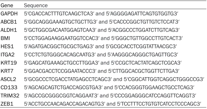
Total RNA from adherent cells and prosta-sphere was extracted using TRIzol Reagent (Invitrogen) according to the manufacturer’s instructions. 1 μg of total RNA was used to gen -erate cDNA using M-MLV reverse transcriptase (Invitrogen) according to the manufacturer’s instructions. 1 μl of cDNA was used for a single PCR reaction to determine the expression of self-renewal markers and stem cell markers. Part length cDNA fragments of the coding region for human genes were amplified by poly -merase chain reaction (PCR) using the primer sets as described in Table 2.
[image:3.612.91.520.83.203.2]Quantitative real-time PCR was performed on the Eppendorf MasterCycler RealPlex2 using Table 1. Antibodies used for immunocytochemical Staining (ICCS)
Antibodies Dilution Clone number Source and catalog number ICCS primary antibodies
Mouse monoclonal anti-CK8 1:200 M20 Abcam, ab9023
Rabbit monoclonal anti-CK5 1:400 EP1601Y Abcam, ab52635
Rabbit polyclonal Anti-AR 1:200 H-280 Santa Cruz Biotechnology, sc-13062 DAPI 1:8000 Sigma, D-8417, 1 mg/ml ICCS seconday antibodies
Goat anti-Rabbit-Cy3 1:1000 Jackson, 111-165-045
Goat anti-Mouse-Alexa488 1:1000 Invitrogen, A-11029
CK, Cytokeratin; AR, Androgen Receptor.
Table 2. Primer sequences for real-time PCR experiments
Gene Sequence
GAPDH 5′CGACCACTTTGTCAAGCTCA3′ and 5′AGGGGAGATTCAGTGTGGTG3′
ABCB1 5′GGCAGGGAAAGTGCTGCTTG3′ and 5′CACCCGGCTGTTGTCTCCAT3′
ALDH1 5′GCTGGCGACAATGGAGTCAA3′ and 5′ACGGCCCTGGATCTTGTCAG3′
BMI 5′CCTGGAGAAGGAATGGTCCAC3′ and 5′GGGCTGTTGGCCTTGTCACT3′
HES1 5′AGATGACGGCTGCGCTGAG3′ and 5′GCGCACCTCGGTATTAACGC3′
ITGA2 5′CCTCTGTGGGCACAGCAATG3′ and 5′AAGGGCAGGGCTGAGTTGC3′
KRT19 5′GAGCATGAAAGCTGCCTTGGA3′ and 5′CCGCTCACTATCAGCTCGCA3′
KRT7 5′GGACGACCTCCGGAATACCC3′ and 5′CTTGGCACGCTGGTTCTTGA3′
ASCL2 5′GCGCCCTCGACCTATGAGCCTCAGC3′ and 5′CGGCATTGGTCAGGCTGGGCCG3′
CD133 5′AGCAGCAGTCTGACCAGCGTGA3′ and 5′CCACGGGTGGAAGCTGCCTCAG3′
TRIM32 5′AGCCGCGGGCGGTCAGGAAT3′ and 5′CCCGGAGGGCATCCAGGTTCAGGT3′
ZEB1 5′ACCTGCCAACAGACCAGACAGTG3′ and 5′TCCTTTCCTGTGTCATCCTCCCAGC3′
Sigma), 5 μg/ml insulin (Sigma) and 1×N2 nutrition (St- emcell Technologi- es Inc.).
[image:3.612.92.423.247.422.2]SYBR Green PCR Master Mix (Toyobo). The rela-tive expression values were calculated relarela-tive to GAPDH by using the 2-ΔCT methods. Results
were calculated and normalized relative to the GAPDH control using the Microsoft Excel program.
Soft-agar assay
Prostate cancer adherent cells and/or single cells from prostasphere were resuspended in Dulbecco’s modified Eagle medium (DMEM; 4.5 g/L D-glucose, Invitrogen) supplemented with 10% FBS and 1% antibiotic containing 0.35% agarose prostasphere (5 000 cells per
35-millimeter well). Cells were grown on tissue culture plates containing a 1-millimeter layer of solidified 0.6% agarose in a DMEM supple -mented with 10% FBS and 1% antibiotic. After 3 weeks, the plates were then scanned and photographed, and number of colonies was quantified. For visualization, foci were metha -nol-fixed and stained with 0.1% trypan Blue. In vivo tumorigenesis assays
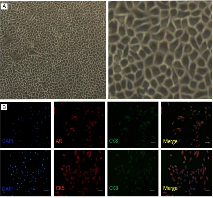
[image:4.612.91.522.73.473.2]Prostate cancer adherent cells and/or single cells from prostasphere were resuspended in 50 μl PBS and mixed with 50 μl Matrigel (Becton Dickinson) at a 1:1 ratio and held on ice. The Figure 1. Adherent cells from PPrCC. A: Adherent cells from PPrCC were observed under invert phase contrast
micro-scope. Cells are tight attachment to the bottom of the plate, which coated by collagen. Cells are belong to a kind of squamous epithelium cell, have polygonal, flat morphology and have compact structure with one another. B: Adher
entire 100 μl sample was injected into each flank of 6-8 weeks old Nude mice and/or NOD/ SCID mice anesthetized with isoflurane accord -ing to the animal protocol approved by the Shanghai Institute of Biology Sciences commit-tee for research in vertebrate animals. 8~12 weeks before that, the same confirmed pros -tate cancer tissues were injected into flank of NOD/SCID mice, xenograft tumors were observed and histological examination was underwent.
Results
Primary prostate cancer cultures (PPrCC)
To develop a method to culture primary pros-tate cancer cells in vitro, we optimized the cul-ture condition. Primary prostatic cancer cells grew well in keratinocyte serum-free medium with epidermal growth factor (EGF) and bovine pituitary extract (BPE), supplemented with leu-kemia inhibitory factor (LIF), stem cell factor (SCF) and cholera toxin. PPrCC tightly attached to the bottom of the plate coated by collagen I.
Like squamous epithelium cell, PPrCC have a polygonal flat morphology and compact struc -ture with one another (Figure 1A).
Biopsy samples from 28 patients were used for primary culture, 9 of them were not prostate cancer, and 18 cases (95%) were succeeded to culture prostate cancer cells in vitro.
Most prostate cancer cell lines were main-tained in regular medium (DMEM or RPMI1640 supplemented with FBS and antibiotic), such as LNCaP, DU145 and PC3. When PPrCC were cul -tured in regular medium containing FBS, FBS induced cell differentiation (data not shown). When PPrCC were grown in the medium includ-ing FBS, cells stop proliferatinclud-ing, the ratio between cytoplasm and cell nucleus increased seriously and cell flat size became much bigger than normal growing cells.
Cell origin of PPrCC
[image:5.612.95.520.73.339.2]Prostate epithelium consists of basal cells (including stem cells and transient amplifying Figure 2. Cells from passaged PPrCC form prostasphere in the suspension medium. A: Morphological examination of
prostasphere from passaged cells of PPrCC and/or prostate cell line (LNCaP) cultured in suspension medium. Pros -tasphere were maintained in the suspension medium (DMEM/F12 medium containing 10 ng/ml bFGF, 20 ng/ml
EGF, 0.4% BSA and 5 ìg/ml Insulin and N2 nutrition for 10 days. B: The number of prostasphere was counted which
were seeded into ultra-low attachment 6-well plates. The Y-axis shows prostasphere per one thousand seeded cells.
cells), luminal cells and neuroendocrine cells [17]. Prostate epithelial cells can be distin-guished based on expression of a variety of markers [18]. The majorities of basal cells express cytokeratin 5 (CK5), cytokeratin 14 (CK14), p63, CD44, Integrin alpha (ITGa), but no cytokeratin 8 (CK8), cytokeratin 18 (CK18), androgen receptor (AR) or prostate specific antigen (PSA) [17, 18]. In contrast, luminal cells express high levels of AR, PSA, CK8, CK18, but no p63 [17]. Transient amplifying (intermedi-ate) cells express basal marker, CK5 and often co-express the luminal marker, CK8 and pros -tate stem cell antigen (PSCA) [17, 18].
To determine the expression profile of PPrCC, adherent cultures of passaged PPrCC were immunostained using antibodies against both basal and luminal markers (Figure 1B). The basal marker CK5 was strongly expressed in the majority of primary culture cells. Luminal markers, including AR and CK8, were also
strongly expressed by most of primary culture cells. Co-expression of CK5 and CK8 in almost of all primary culture cells, suggested that pri-mary culture cells mainly were from basal epi-thelial cells, and after cultured in vitro, tran-sient amplifying cells were the dominant cells in the passaged adherent culture condition [24].
Tumorigenicity of cells from PPrCC in vitro
To determine the tumorigenicity of PPrCC in vitro, we performed soft-agar assay. Colony for-mation efficiency (CFE) of adherent cells from PPrCC (0.15%) is 23 times lower than CFE of LNCaP (3.5%).
Sphere formation of cells from PPrCC in vitro
[image:6.612.90.522.74.388.2]We attempted to employ stem cell suspension culture conditions to establish prostasphere-forming condition by using the cells from pas-saged PPrCC. By culturing cells in ultra-low Figure 3. Real-time PCR analysis was performed on prostasphere from adherent cells of PPrCC and adherent cells from PPrCC. The log2 (fold change) in gene expression between the prostasphere cells and adherent cells is shown.
attachment dishes, serum-free DMEM/F12 medium, supplemented with EGF, basic fibro -blast growth factor (bFGF), bovine serum albu-min (BSA), insulin and with/without N2 nutrition were used to enrich PrCSC. According to the prostasphere morphology and the prosta-sphere number, the optimized suspension medium is serum-free DMEM/F12 medium supplemented with EGF, bFGF, BSA, insulin and N2 nutrition in ultra-low attachment dishes. Ten days after initial plating, they formed increasingly larger multicellular spheroids. The prostasphere showed well-defined circular shape with evident marginal rims (Figure 2A), which has similar morphological appearance with the prostasphere from LNCaP cells. However, compared with the sphere forming efficiency (SFE) of LNCaP (13.67%), SFE of pas -saged cells from PPrCC (0.5%) in the same sup -plemented medium is 27 times lower than SFE of LNCaP (Figure 2B).
Prostasphere from PPrCC showed increased tumorigenic potential in vitro
To test whether prostasphere from PPrCC enriched for PrCSC, we first examined the
[image:7.612.92.518.73.312.2]expression profiles of putative stem cell mark -ers including HES1 (hairy and enhancer of split 1), ITGa2 (integrin alpha 2), CD133 (prominin 1), ASCL2 (achaete-scute complex homolog 2), ABCB1 (ATP-binding cassette, sub-family B, member 1), BMI (BMI1 polycomb ring finger oncogene), ALDH1 (aldehyde dehydrogenase 1) and differentiation markers including ZEB1 (zinc finger E-box binding homeobox 1), TRIM32 (tripartite motif containing 32), MUC1 (mucin 1, cell surface associated), CK19 (cytokeratin 19), CK7 (cytokeratin 7) in prostasphere derived from PPrCC. The log2 fold change of gene expression between the cells from prosta-sphere and adherent cells from PPrCC is shown in Figure 3. Quantitative real-time PCR results revealed that the expression levels of putative stem cell markers are significantly higher in cells from prostasphere than adherent cells, such as expression of ALDH1, ASCL2 and CD133 is 23-fold, 10-fold and 6-fold higher lev-els separately. Moreover, genes associated with differentiation are down regulated in cells form prostasphere compared to adherent cells, such as EMA and CK19. These gene expression profiles indicate that prostasphere enriched for PrCSC.
Figure 4.In vitro tumorigenicity of PPrCC in soft-agar assay. A: Soft-agar analysis of PPrCC and LNCaP cells. PPrCC
(1×10⁴ cells per 35-mm well) and LNCaP cells (1×10³ cells per 35-mm well) were re-suspended in DMEM medium
supplemented with 10% FBS and 1% antibiotic/antimycotic containing 0.35% agarose. B: Colonies derived from LNCaP and PPrCC were counting after 3 weeks separately. (PCP: Prostasphere cells from PPrCC; ACP: Adherent
To further test whether prostasphere from PPrCC increased tumorigenic potential in vitro, we performed soft-agar assay. As the size of prostasphere from PPrCC grew beyond 100μm in diameter, the prostasphere were enzymati-cally dissociated and then replanted as single-dissociated cells. Colonies were counted after 3 weeks. Though colony formation efficiency (CFE) of prostasphere from PPrCC (0.72%, Figure 4) was lower than CFE of LNCaP (3.47%, Figure 4), CFE of prostasphere from PPrCC was about 5-fold more than CFE of adherent cells from PPrCC (0.14%, Figure 4).
Prostasphere from PPrCC failed to give rise to form tumor in vivo
To further address whether prostasphere from PPrCC enriched for PrCSC, we tested whether prostasphere from PPrCC also showed enhanced tumor forming potential in vivo, by injection of cells from prostasphere and/or adherent cells from PPrCC into the flank region of Nude male mice and/or NOD/SCID male mice (4-6 weeks). LNCaP were able to form tumors (2/2) in 4 weeks when 500 thousand cells were injected into Nude male mice, but neither cells from prostasphere or adherent cells from PPrCC could form tumors even in 6 months at a high level cell dose (2.5 million per injection) in Nude male mice or NOD/SCID mice.
Discussion
Primary prostate culture cells
For decades, several different primary culture mediums with different supplementary had been used to maintain prostate cancer cells in vitro, such as PFMR-4A [25], KSFM [10] and PrEGM [26]. We found that KSFM medium with the supplementary is the best condition medi-um for us to culture primary prostate cancer cells in collagen-coated tissue culture dishes. And the cells growing from KSFM medium could not proliferate in PrEGM medium or in tissue culture dishes without collagen coating. In order to avoid variation in higher generation cells, we mainly used the 3rd and/or 4th pas-saged cells in our test assays, which could enlarge enough cells for different assay, though PPrCC could be subculture over 7 generation for most cancer prostate samples.
Prostate epithelium consists of CK5+/CK14+/ p63+/ basal cells and CK8+/CK18+ luminal cells [27] as well as neuroendocrine cells. Basal cells preferentially survive androgen ablation and give rise to luminal cell in vitro, which sup-port that basal layer is the origin of prostate stem cells. In vitro, undifferentiated cells with both basal marker and luminal marker could differentiate into luminal cells and basal cells [28]. In vitro, PPrCC express markers normally associated with both basal cells (CK5) and luminal cells (CK8 and AR). Potential uses of the PPrCC here were enrichment of PrCSC, understanding the properties of PrCSC from PPrCC, which was different from prostate can-cer cell line cultured in vitro for many generation.
The microenvironment is critical for PPrCC growth. Most prostate cell line is adherent in vitro, such as PC3, DU145, 22Rv1 and LNCaP, and certain amount of fetal bovine serum is essential for the cells proliferation. However, fetal bovine serum is a strong factor of differen-tiation for PPrCC growth, which does not only convert the phenotype from epithelium to fibro -blast, but also inhibit the proliferation with the result of cell apoptosis or necrosis. The other significant factor in our culture system is the collagen-coated tissue culture dish. PPrCC from collagen-coated dish could passaged for twice more in regular culture dish without coat-ing, but the morphology changed greatly with few typical epithelial cells, most cells grew big-ger than before and the cells were not close as before. Obviously, this is different from the cul-ture condition of prostate cancer cell lines, which grow very well in tissue culture dish with/ without coating treatment.
Prostate cancer stem cell
Many prostate cancer cell line have been used to separate PrCSC, such as LNCaP [12-14, 20], PC3 and Du145 [11, 13, 21]. Prostate cancer cell line have more mature culture system than PPrCC, and cells from the former proliferate much more quickly than cells from the latter, which also produce plenty of prostasphere by culture prostate cancer cell line in serum-free medium or harvest enough of PrCSC by flow cytometry assay basing on cancer stem cell markers, such as CD133/CD44.
sample: (1) prostate cancer cells limitation. About 10 000 cancer cells could be harvested from an 18-gauge-needle biopsy sample of prostate [29]. (2) Differentiation inducing by fetal bovine serum (FBS). Typical epithelial cells could be changed to fibroblast type if medium replaced by medium with FBS. Furthermore, the new fibroblast-like cells in our culture condi -tion have limit prolifera-tion in medium with FBS. (3) Slow-proliferation of PPrCC. Cell dou-bling time of adherent PPrCC in vitro is about 3 days, but the cancer cell death rate is close to that of its proliferation in vivo (xenograft to mice), net growth (i.e., tumor doubling time) is about one month [30]. Limitation is accompa -nied with the advantages of PPrCC, which give more variety (similar with in vivo) than prostate cancer cell line.
In this study, enriched PrCSC by sphere culture assay were evaluated by both soft agar assay and gene expression profiles of some putative cancer stem cell markers in vitro. Putative can-cer stem cells (with 50% Matri-gel) from sphere culture assay were subcutaneous into NOD/ SCID mice, and none tangible tumors could be detected in 6 months, which is similar to the results from the [30], though half of the biopsy sample could initiate tumor in 3 months by the same type of injection. The result indicates that our putative PrCSC from spheroids are quies-cent, further studies on xenograft system are needed to evaluate the property of PrCSC in vivo.
Acknowledgements
We wish to thank Ziqing Zhu, Hanqing Lin, Yanru Wang and other members in Chen’s lab (Shanghai Key Laboratory of Andrology, Institute of Biochemistry and Cell Biology, Shanghai Institutes for Biological Sciences, Chinese Academy of Sciences) for technical help. This work was supported by funds from “Strategic Priority Research Program” of the Chinese Academy of Sciences (XDA01040402) and National Natural Science Foundation of China (81172426).
Disclosure of conflict of interest
None.
Address correspondence to: Dr. Denglong Wu,
Department of Urology, Affiliated Tongji Hospital of
Tongji University, No. 389 Xincun Road, Shanghai 200065, China. E-mail: wuyy163@126.com; Dr.
Gang Han, State Key Laboratory of Molecular Biology, Shanghai Key Laboratory of Andrology,
Institute of Biochemistry and Cell Biology, Shanghai Institutes for Biological Sciences, Chinese Academy of Sciences, No. 320, Yueyang Road, Shanghai 200031, China. E-mail: ghan@sibs.ac.cn
References
[1] Siegel R, Ward E, Brawley O and Jemal A. Can-cer statistics, 2011: the impact of eliminating socioeconomic and racial disparities on pre-mature cancer deaths. CA Cancer J Clin 2011; 61: 212-236.
[2] Teng JF and Xu DF. Progress in Prostate Cancer Stem Cell. Journal of Clinical Urology 2011; 26: 309-311.
[3] Damber JE and Aus G. Prostate cancer. Lancet
2008; 371: 1710-1721.
[4] Debes JD and Tindall DJ. Mechanisms of an-drogen-refractory prostate cancer. N Engl J Med 2004; 351: 1488-1490.
[5] Feldman BJ and Feldman D. The development of androgen-independent prostate cancer. Nat Rev Cancer 2001; 1: 34-45.
[6] Lapidot T, Sirard C, Vormoor J, Murdoch B, Ho -ang T, Caceres-Cortes J, Minden M, Paterson
B, Caligiuri MA and Dick JE. A cell initiating hu
-man acute myeloid leukaemia after transplan -tation into SCID mice. Nature 1994; 367: 645-648.
[7] Al-Hajj M, Wicha MS, Benito-Hernandez A,
Mor-rison SJ and Clarke MF. Prospective identifica -tion of tumorigenic breast cancer cells. Proc Natl Acad Sci U S A 2003; 100: 3983-3988. [8] Singh S, Chitkara D, Mehrazin R, Behrman SW,
Wake RW and Mahato RI. Chemoresistance in
prostate cancer cells is regulated by miRNAs
and Hedgehog pathway. PLoS One 2012; 7:
e40021.
[9] Sharifi N, Kawasaki BT, Hurt EM and Farrar WL.
Stem cells in prostate cancer: resolving the castrate-resistant conundrum and implica-tions for hormonal therapy. Cancer Biol Ther 2006; 5: 901-906.
[10] Collins AT, Berry PA, Hyde C, Stower MJ and
Maitland NJ. Prospective identification of tu -morigenic prostate cancer stem cells. Cancer Res 2005; 65: 10946-10951.
[11] Wei C, Guomin W, Yujun L and Ruizhe Q. Can
-cer stem-like cells in human prostate carcino -ma cells DU145: the seeds of the cell line? Cancer Biol Ther 2007; 6: 763-768.
[12] Dubrovska A, Kim S, Salamone RJ, Walker JR,
Maira SM, Garcia-Echeverria C, Schultz PG and
Reddy VA. The role of PTEN/Akt/PI3K signaling
cancer stem-like cell populations. Proc Natl
Acad Sci U S A 2009; 106: 268-273.
[13] Dubrovska A, Elliott J, Salamone RJ, Telegeev GD, Stakhovsky AE, Schepotin IB, Yan F, Wang Y, Bouchez LC, Kularatne SA, Watson J, Trus -sell C, Reddy VA, Cho CY and Schultz PG. CXCR4 expression in prostate cancer
progeni-tor cells. PLoS One 2012; 7: e31226.
[14] Hurt EM, Kawasaki BT, Klarmann GJ, Thomas SB and Farrar WL. CD44+ CD24(-) prostate
cells are early cancer progenitor/stem cells that provide a model for patients with poor prognosis. Br J Cancer 2008; 98: 756-765. [15] Wu C, Wyatt AW, Lapuk AV, McPherson A, Mc
-Coneghy BJ, Bell RH, Anderson S, Haegert A,
Brahmbhatt S, Shukin R, Mo F, Li E, Fazli L, Hurtado-Coll A, Jones EC, Butterfield YS, Hach
F, Hormozdiari F, Hajirasouliha I, Boutros PC, Bristow RG, Jones SJ, Hirst M, Marra MA, Ma-her CA, Chinnaiyan AM, Sahinalp SC, Gleave
ME, Volik SV and Collins CC. Integrated ge
-nome and transcriptome sequencing identifies
a novel form of hybrid and aggressive prostate cancer. J Pathol 2012; 227: 53-61.
[16] Tang S, Mishra M, Frazier DP, Moore ML, Inoue K, Deora R, Sui G and Dubey P. Positive and
negative regulation of prostate stem cell anti-gen expression by Yin Yang 1 in prostate
epi-thelial cell lines. PLoS One 2012; 7: e35570.
[17] Mulholland DJ, Xin L, Morim A, Lawson D, Witte O and Wu H. Lin-Sca-1+CD49fhigh stem/pro -genitors are tumor-initiating cells in the Pten-null prostate cancer model. Cancer Res 2009; 69: 8555-8562.
[18] Liao CP, Adisetiyo H, Liang M and Roy-Burman P. Cancer-associated fibroblasts enhance the
gland-forming capability of prostate cancer stem cells. Cancer Res 2010; 70: 7294-7303. [19] Tang Y, Hamburger AW, Wang L, Khan MA and
Hussain A. Androgen deprivation and stem cell
markers in prostate cancers. Int J Clin Exp
Pathol 2009; 3: 128-138.
[20] Fan X, Liu S, Su F, Pan Q and Lin T. Effective
enrichment of prostate cancer stem cells from spheres in a suspension culture system. Urol Oncol 2012; 30: 314-318.
[21] Chen W and Wang GM. Gene expression profil -ing of cancer stem cells in the Du145 prostate
cancer cell line. Oncol Lett 2012; 3: 791-796.
[22] Guzman-Ramirez N, Voller M, Wetterwald A,
Germann M, Cross NA, Rentsch CA, Schalken
J, Thalmann GN and Cecchini MG. In vitro prop-agation and characterization of neoplastic
stem/progenitor-like cells from human pros -tate cancer tissue. Pros-tate 2009; 69: 1683-1693.
[23] Wang ZA and Shen MM. Revisiting the concept of cancer stem cells in prostate cancer. Onco-gene 2011; 30: 1261-1271.
[24] Buhler P, Wolf P, Katzenwadel A, Schultze-See -mann W, Wetterauer U, Freudenberg N and El-sasser-Beile U. Primary prostate cancer cul-tures are models for androgen-independent transit amplifying cells. Oncol Rep 2010; 23: 465-470.
[25] Peehl DM. Growth of prostatic epithelial and stromal cells in vitro. Methods Mol Med 2003; 81: 41-57.
[26] Goldstein AS, Drake JM, Burnes DL, Finley DS,
Zhang H, Reiter RE, Huang J and Witte ON.
Pu-rification and direct transformation of epitheli -al progenitor cells from primary human pros-tate. Nat Protoc 2011; 6: 656-667.
[27] Lee SO, Tian J, Huang CK, Ma Z, Lai KP, Hsiao
H, Jiang M, Yeh S and Chang C. Suppressor role of androgen receptor in proliferation of prostate basal epithelial and progenitor cells. J Endocrinol 2012; 213: 173-182.
[28] Barclay WW, Axanova LS, Chen W, Romero L, Maund SL, Soker S, Lees CJ and Cramer SD.
Characterization of adult prostatic progenitor/ stem cells exhibiting self-renewal and multilin-eage differentiation. Stem Cells 2008; 26: 600-610.
[29] Vander Griend DJ, Karthaus WL, Dalrymple S, Meeker A, DeMarzo AM and Isaacs JT. The role
of CD133 in normal human prostate stem cells and malignant cancer-initiating cells. Cancer Res 2008; 68: 9703-9711.
[30] Chen S, Principessa L and Isaacs JT. Human