Int J Clin Exp Pathol 2014;7(8):4720-4733 www.ijcep.com /ISSN:1936-2625/IJCEP0001204
Original Article
Role of biphasic changes in splenic dendritic cell
activity in a mouse model of multiple organ
dysfunction syndrome
Yi Lv*, Qian Liu*, Min Zhao, Yiduo Jin, Jiangyang Lu
Department of Pathology, The First Affiliated Hospital of General Hospital of PLA, Beijing 100048, China. *Equal contributors.
Received June 24, 2014; Accepted August 2, 2014; Epub July 15, 2014; Published August 1, 2014
Abstract: To analyze the changes in splenic dendritic cell (DC) activity and serum cytokine levels during the pro-gression of multiple organ dysfunction syndrome (MODS). A C57BL/6 mouse model of MODS was established by intraperitoneal injection of zymosan. Immunohistochemistry and flow cytometry were used to detect expression of I-Ab (MHC-II molecules of mice) as well as co-stimulatory and co-inhibitory molecules in spleen and DC surface. The levels of various cytokines in serum and spleen tissue were analyzed 6 h, 12 h, 24 h, 48 h, 5 d and 12 d after injury. Death occurred at 24-48 h and 10-12 d after injury. The expression of I-Ab and CD86 in spleen tissue and on DCs increased 6-12 h after injury, followed by gradual reduction and at 12 d. The inhibitory molecule, PD-L1, was expressed on normal DCs, but expression of PD-1 was undetectable. PD-L1 and PD-1 expression increased and remained high at 5 d and 12 d after injury. In addition, TNF and IL-1 levels increased 6-12 h after injury; HMGB1 and IL-10 levels increased 24 h and 5 d after injury, respectively. In contrast, IL-2 and IL-12 decreased with disease progression. At 12 d after injury, proinflammatory and anti-inflammatory cytokine levels remained high, while IL-2 and IL-12 were significantly reduced. IL-10 and IL-12 changes in spleen were consistent with those in serum. MODS progression was characterized by changes in splenic DC activity as well as altered serum pro-inflammatory and anti-inflammatory cytokine levels, suggesting early immune activation and predominant immune tolerance at the late stage.
Keywords: Splenic dendritic cell, MODS, immunohistochemical staining
Introduction
Multiple organ dysfunction syndrome (MODS) is an important cause of death in late-stage sepsis, and its mortality rate ranks first in the intensive care unit (ICU) [1, 2]. Interrupting the occurrence and development of sepsis as soon as possible is crucial for preventing the inci-dence of MODS in clinical practice. Although the effects of dozens of pro-inflammatory cyto -kine antagonists have been examined in clini-cal trials in the past 20 years, their efficacy in humans is far inferior to that observed in ani-mal studies [3-6], suggesting a deviation in the target or the timing of use [7-9].
In the early stages of sepsis, patients are char-acterized by an excessive inflammatory res-ponse or “cytokine cascade” [10, 11]. Advan- cements in clinical treatment enable the major-ity of patients to pass through this stage and
determining the appropriate measures for early excessive inflammatory response and late immune paralysis are critical for the prevention and treatment of sepsis. In addition, under-standing the mechanism by which immune acti-vation changes to immunosuppression in sep-sis and intervention prior to progression to immune paralysis will represent effective strat-egies for reducing the mortality of MODS. The spleen plays an important role in the innate and acquired immune responses [21, 22]; therefore, changes in splenic function will affect the systemic immune status. In a mouse model of cecal ligation and puncture (CLP) sep-sis, splenectomy reduced mortality and serum levels of high-mobility group protein B1 (HM- GB1) [23]. In addition, analysis of the choliner-gic anti-inflammatory pathway revealed that parasympathetic (vagal) activity attenuated systemic inflammation through regulating the activity of splenic immune cells [24], signifi -cantly reducing the death of septic animals [25, 26]. However, no anti-inflammatory effects were observed with vagus nerve excitation or cholinergic receptor activation in animals with splenectomy or when the splenic nerve was cut [26, 27], indicating that splenic function has an important impact on the prognosis of sepsis. Immunomodulation by the spleen requires the synergistic action of splenic dendritic cells (DCs) and various immune cells, including T cells, B cells, and Treg cells. DCs are the most important professional antigen-presenting cells (APCs), bridging the innate and adaptive immunity [28]. It was previously thought that DCs only present foreign antigens and activate T cells, thereby triggering immune responses. Recent studies have found that under certain conditions, DCs can produce immune tolerance and even negatively regulate immune function. Therefore, DCs have bidirectional immunomod-ulatory effects [29-31], which are achieved through interactions between co-stimulatory and co-inhibitory molecules located on the cell surface and the corresponding ligands on the lymphocytes [32, 33]. Understanding the func-tional changes of splenic DCs in MODS progres-sion and their impact may help develop new and more effective prevention and treatment strategies.
Our previous studies described an increase in the number and size of splenic DCs as well as
their activation in the early stage zymosan-induced MODS in mice [34]. Additionally, the activity of splenic DCs was reduced with increased DC and lymphocyte apoptosis and lytic necrosis in the late stage [34]. We specu-late that during the development of sepsis, changes in immune function of splenic DC could activity and therefore immune function mediate at least in part the early immune acti-vation and late immunosuppression through affacting regulating different T cell subsets. Thus, analyzing related changes in serological immune parameters (pro-inflammatory and anti-inflammatory cytokines and cytokines from different cell sources) may provide guid-ance for predicting disease progression and prognosis. This study used intraperitoneal injection of zymosan to replicate the MODS model and analyzed the changes in the splenic DC phenotype and various serum cytokines at different stages during MODS progression. We found that changes in splenic DC immune activ-ity and sequential changes in serum immuno-logical indicators were closely related to dis-ease progression.
Materials and methods
Establishment of an in vivo model of MODS
Male 6-8 week-old C57BL/6 mice (n = 165) weighing 20-25 g were purchased from the Experimental Animal Center of the Military Medical Academy of Sciences. Animals were acclimated for one week under 12 h light/12 h dark cycles. Mice were fed standard diet and had free access to drinking water. Prior to induction of MODS, animals were fasted for 12 h. Mice were randomly divided into a control group (n = 10) or experimental group (n = 155), and the experimental group was further divided into the following six subgroups based on time points post zymosan injury: 6 h group (n = 15), 12 h group (n = 15), 24 h group (n = 15), 48 h group (n = 30), 5 d group (n = 30), and 12 d group (n = 50).
injec-Analysis of immune parameters during MODS progression
tion of the zymosan suspension (800 mg/ kgBW). Then, the animals were fed convention-ally. In addition to monitoring for death, blood and spleen specimens were collected at corre-sponding time points for subsequent analysis.
Detection of cytokine levels in the serum and
spleen tissue
[image:3.612.94.526.73.570.2]for 30 min, the serum was isolated after cen-trifugation at 3000 rpm for 15 min and stored at -80°C. The spleen tissue was frozen in liquid nitrogen, and 100 mg was collected and homo- genized in 1 mL PBS in an ice bath. After cen-trifugation at 3000 rpm for 15 min at 4°C, the supernatant was collected. Enzyme-linked im- munosorbent assays (ELISAs) were used to detect serum concentrations of tumor necrosis factor (TNF) α, interleukin (IL)-1β, high-mobility group protein B1 (HMGB1), IL-10, IL-12 and IL-2 and levels of IL-10 and IL-12 in splenic tissue homogenate using commercial kits, following manufacturer’s instructions.
Immunohistochemical staining and immuno
-fluorescence labeling of spleen tissue
After the spleen tissues were collected, a por-tion of the spleen tissue was fixed with 10%
neutral formalin, paraffin embedded, and sliced into sections of 5-μm thickness. Conventional immunohistochemical staining methods were used, and horseradish peroxidase (HRP)-peroxide-diaminobenzidine (DAB) was used to label I-Ab, CD86, and TGFβ. The samples were
[image:4.612.93.518.72.392.2]counterstained with hematoxylin, and imaged using an Olympus BX40F microscope (Olympus, Melville, NY, USA). The other portion of spleen tissue was embedded in optimal cutting tem-perature (OCT) compound, and 4-µm frozen sections were prepared and attached onto the APES film. The samples were dried at room tem -perature and fixed in acetone at 4°C for 10 min. The immunofluorescence labeling of pro -grammed cell death ligand 1 (PD-L1) was per-formed, and samples were observed under a fluorescence microscope and photographed using a Nikon Eclipse 50i fluorescence micro-scope.
Analysis of immune parameters during MODS progression
Splenic DC isolation and culture and detection
of cytokines in cell culture supernatant
The spleen tissue was collected at correspond-ing time points and placed on a petri dish. The capsule was trimmed off, and the sample was immersed in 1.25 mL collagenase IV (1 mg/mL)
[image:5.612.92.526.73.578.2]The cell suspension was collected and isolated by centrifugation at room temperature and 2000 rpm for 10 min. After the supernatant was discarded, 5 mL pre-chilled PBS was added to the cells, which were dispersed evenly using a micropipette. The sample was slowly added into a sterile conical centrifuge tube with 10 mL lymphocyte separation solution (Ficoll-Papue) and centrifuged at room temperature at 3000 rpm for 15 min. The cells in the middle layer
[image:6.612.95.523.75.551.2]Analysis of immune parameters during MODS progression
The purified DCs were resuspended with RPMI-1640 (Hyclone, USA), and the cell concentra-tion was adjusted to 5×10⁶ cells/mL prior to
seeding onto 96-well culture plates (1.25×10⁶
cells/well). The cells were maintained in an incubator with 5% CO2 at 37°C. After 24 h, the cells were centrifuged at 1500 rpm for 5 min, and the supernatant was collected for analysis
of cytokine concentrations by ELISA as previ-ously described.
Detection of DC surface molecule expression using flow cytometry
[image:7.612.90.525.68.558.2](BSA), which were added into a flow cytometric tube (5×10⁴ cells/tube). After 2-3 µL FITC- or
PE-labeled fluorescent antibodies specific for target surface markers (or PE-IgG Isotype Control) were added, the samples were incu-bated at 4°C for 45 min and washed twice with PBS before centrifugation at 1500 rpm for 5 min. The supernatant was discarded, and the cells were resuspended in 400 µL PBS. Detection and analysis were carried out using a FACS calibur (Becton Dickinson, USA). The labeled antibodies included CD11c-PE, MHC-II (I-Ab)-FITC, PD-L1-PE, programmed death-1
(PD-1)-PE (all from BD Biosciences, USA), CD86- PE, and paired-immunoglobulin-like receptor (PIR)-B-PE (Biolegend, USA).
Statistical analysis
Data are shown as mean ± standard deviation (SD). SPSS13.0 software was used for single factor analysis of variance (ANOVA). Between-group comparisons were undertaken using the least significant difference (LSD) test, and the statistical significance level α was set at 0.05. Results
Symptoms, physical signs, and survival rate of MODS mice at different stages of disease
progression
The experimental mice showed reduced activi-ty and decreased dietary intake 3 h after injec-tion of zymosan. The symptoms were aggravat-ed at 12 h after injection as the mice were listless, shivering, refusing food intake, and had diarrhea, wet, dirty hair, closed eyes and increased secretions. The first peak of death occurred at 24-48 h post zymosan injection (mortality 30.1%). At 48 h, the status of experi -mental animals gradually improved, which was accompanied by gradual restoration of basic living habits. No deaths occurred during this period (mortality 0%). At 5-7 d after injection, animals showed systemic symptoms, which were aggravated at 10-12 d and were accom-panied by drowsiness, lack of food intake, and dyspnea. The second peak of death occurred during that period with a mortality rate of 21.9%.
Changes in the serum cytokine levels
In mice with zymosan-induced injury, serum cytokine levels were altered with disease
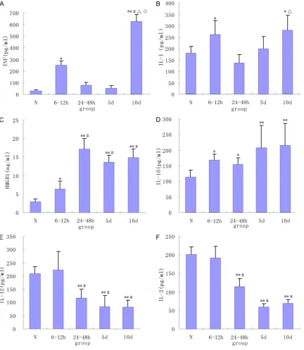
pro-gression (Figure 1). Specifically, serum concen -trations of proinflammatory cytokine, TNF-α, showed a bimodal increase that coincided with the biphasic changes in systemic symptoms and mortality (Figure 1A). In normal mice, serum TNF-α levels were low, rising sharply at 6 h-12 h after zymosan injection (P < 0.05) and returning to near normal levels at 24 h-5 d. TNF-α concentration increased again at 12 d to a greater extent than that observed at the 6-12 h time point (P < 0.01). A similar trend was observed with serum IL-1 levels, but to a lesser extent (Figure 1B; P < 0.05). In contrast, the anti-inflammatory cytokine, IL-10, increased slightly at 6 h-48 h after zymosan injection (P < 0.05) with greater levels observed at 5-12 d (Figure 1D; P < 0.01).
Serum concentrations of HMGB1, an inflamma -tory mediator in late-stage sepsis, increased gradually after injury, peaked at 24 h and remained at a high level for the duration of the study (Figure 1C; P < 0.01). Serum IL-12 secret-ed by mononuclear macrophages and antigen presenting DCs gradually decreased at 24-48 h after injury (Figure 1E). Its level was only 50% of that in the control group at 12 d after injury (P < 0.05), suggesting that the immune activity of antigen-presenting cells gradually decreased during MODS progression.
Secretion of IL-2 by activated T cells can pro-mote T cell proliferation of T cells; therefore, IL-2 levels are reflective of the degree of T cell activation. At the early stage of zymosan-induced injury (i.e., 6-12 h), no changes in IL-2 were noted (Figure 1F). However, its level was significantly reduced at 24-48 h after injury, which continued for the duration of the study (P < 0.01). Il-2 levels at 5 and 12 d were only one-third that of the control and 6-12 h groups, indi-cating that the T cell immune activity was sig-nificantly reduced at the later stages of MODS progression.
IL-12, IL-10 and TGF-β expression in the spleen and DC cultures during MODS progression
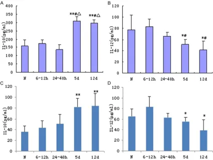
con-Analysis of immune parameters during MODS progression
trast, at 5 and 12 d after injury, IL-10 levels were significantly increased (P < 0.01) while IL-12 were reduced (P < 0.05) as compared to the control and 6-12 h groups. Similar changes in IL-10 and IL-12 secretion by splenic DCs were observed (Figure 2C and 2D, respectively; P < 0.05).
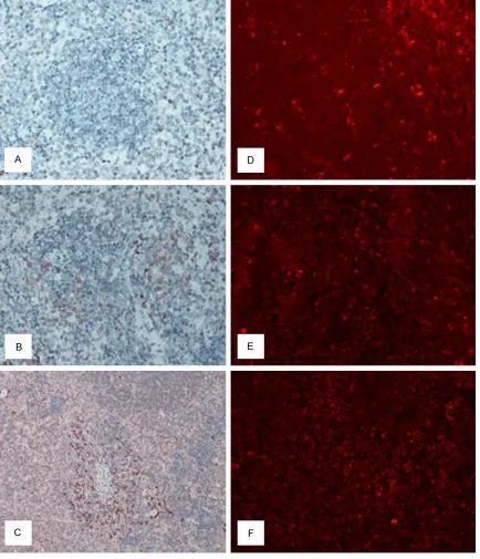
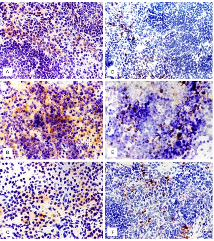
As shown in Figure 3A, scattered expression of TGF-β, an anti-inflammatory factor, was obser-ved in the spleen of mice in the normal control group; most of the TGF-β immunoreaction was detected in the marginal zone and red pulp area. At 6 h post induction of MODS injury, the number of TGF-β-positive cells increased slight -ly in the marginal zone of the white pulp (Figure 3A). At late-stage MODS, greater TGF-β expres -sion was observed with a patchy distribution in the red pulp area and the area close to the mar-ginal zone (Figure 3C). These results suggest that the immune activity of the spleen and splenic DCs was reduced and immunosuppres-sion enhanced with the disease progresimmunosuppres-sion.
Expression of co-stimulatory molecules in splenic tissue and DCs
DCs are the most powerful APC in the body. The expression of CD84 and I-Ab DC surface
mark-ers was next determined (Figure 4). Analysis of CD86 expression in a normal spleen revealed that splenic DCs were mainly distributed in the marginal zone with a small number of them located in the white pulp and splenic cord (Figure 4D). In the early stage of zymosan-induced injury (6-12 h), the number of CD86-positive cells increased (Figure 4E), which was subsequently reduced at 12 d (Figure 4F). During the MODS progression, changes in the expressions of the key signal molecule in the spleen which mediates antigen presentation, I-Ab, coincided with those observed for the
CD86 marker (Figure 4A-C).
The immunohistochemistry analysis of I-Ab and
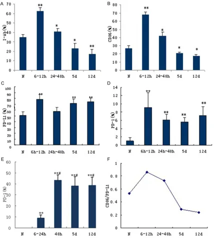
CD86 expression was also confirmed by FACS analysis of isolated splenic DCs. Specifically, the expression levels of I-Ab byisolated splenic
DCs significantly increased at 6 h-12 h after injury as compared with the normal group (Figure 5A; P < 0.01); its level dropped at 24-48 h and then continued to gradually decrease until the end of the study, resulting in a level that was only 50% of that in the control group (P < 0.01). Changes in the expression of CD86 on
the DCs were consistent with that of I-Ab,
sug-gesting that the immune activity of splenic APCs experienced a transformation from en- hancement to reduction during MODS progres-sion (Figure 5B).
Expression of PD-L1 and PD-1 in the spleen and on the surface of splenic DCs
PD-L1 and PD-1 transfer negative co-stimulato-ry signals. After double labeling of spleen tis-sue with CD11C-FITC and PD-L1-PE, a large number of splenic DCs were observed in nor-mal mice, with only a snor-mall proportion of DCs expressing PD-L1 (red-orange color) (Figure 3D). In late-stage MODS, the number of PD-L1-positive cells increased in the spleen (Figure 3F). In contrast, PD-1 expression was not detected in the spleen of normal mice; howev-er, its expression was increased at different time points during MODS progression.
Flow cytometry was used to also detect PD-L1 and PD-1 surface expression on splenic DCs (Figure 5C and 5D, respectively). Under normal conditions, the positive expression rate of PD-L1 was 50.31%, and a biphasic increase at 6-12 h and 5 d-12 d after injury was observed as compared with the normal group (Figure 5C; P < 0.05). In contrast, PD-1 expression was not detected in the normal control group; however, its expression increased at 6 h-24 h and contin-ued to be elevated for the duration of the study as compared with the normal group (P < 0.05). It is worth noting that while biphasic increases in PD-L1 expression were observed at 6-12 h and again at 5 d after injury, PD-1 expression was increased at 48 h after injury, which was before the second increase of PD-L1 expres-sion (Figure 5C and 5D). As shown in Figure 5F, ratio of co-stimulatory and co-inhibitory mole-cules on DCs at different stages during MODS progression revealed that the level of CD86 co-stimulatory molecule in the normal group was only half of that observed for the co-inhibitory molecule, PD-L1. In early MODS, the CD86/ PD-L1 ratio increased at 6 h -24 h, returned to normal levels at 48 h, and was half that of the control group at 12 d. Thus, DC function changed with MODS progression.
(over 50%). While its expression remained the same at the early stage after injury, PIR-B levels increased at 5 d and 12 d after injury as com-pared with the normal group (P < 0.05), which was the same profile observed for PD-L1/PD-1 at the late stage of MODS progression (Figure 5E).
Discussion
In this study, intraperitoneal injection of zymo-san was used to establish a mouse model of MODS [35]. The immune activation and disor-der of mice were consistent with that observed in MODS progression, which is in line with the inflammatory mediator theory of MODS [36, 37]. During the disease progression, changes in the immune phenotype of splenic DCs and levels of serum pro-inflammatory and anti-inflammatory cytokines were indicative of pre -dominant immune activation in the early stage and dominant immune tolerance in the late stage, which coincided with the two death peaks. During MODS progression, the sequen-tial changes in serum cytokine levels are impor-tant as a reference value for the timely detec-tion of immune status transidetec-tion and predicdetec-tion of disease progression.
The spleen is a core organ that regulates sys-temic immune balance. Its regulatory function is realized through interactions with a variety of immune cells, including macrophages, DCs, T and B lymphocytes and their subgroups [38]. DCs capture and process foreign antigens and present them to naive T cells, playing an impor-tant role in stimulating the adaptive immune response, inducing peripheral tolerance and determining the direction, intensity and dura-tion of the immune response [38-41]. DCs occupy a dominant position in spleen-mediated immunomodulation. The interactions between DCs and T cells are mediated through the bind-ing between the DC surface ligands and the receptors on the T cell surface (TCRs). DCs pro-cess the antigen and load it onto MHC-II mole-cules, forming a MHC-II-Ag complex prior to bind to TCRs and transferring the first signal. Co-stimulatory molecules, which are B7 family members and include CD86 and CD80, interact with CD28/CTLA to transfer the second signal. IL-12 released by APC is the third signal for T cell activation (Diebold 2008). Therefore, the expression levels of MHC-II, B7 family
mole-cules and IL-12 on DCs reflect the ability of DC to activate T cells, which determines the level of T cell activation. In this study, expression of co-stimulatory molecules, such as MHC-II and CD86 in the spleen and on the splenic DCs, at the early stage of MODS was higher than those in the normal control group, indicating en- hanced DC activity and activation of the immune response. In addition, increased ser- um concentrations of pro-inflammatory cyto -kines were observed at this stage. At the late stage, expression of MHC-II and CD86 on the DCs were significantly reduced, and IL-12 levels in the spleen were decreased. Also, IL-10 levels were greatly increased along with the number of TGF-β-positive cells. Isolated splenic DC cul -tures secreted less IL-12, but more IL-10. Thus, the results showed that the immune function of the spleen and splenic DCs was suppressed at the late stage of the disease. Moreover, the reduction in the T lymphocyte activity was relat-ed to the increasrelat-ed secretion of inhibitory cyto-kines by DCs [42].
DC immune regulation is also affected by the expression of inhibitory receptors and negative co-stimulatory molecules [43, 44]. Mouse PIR-B protein is a type of inhibitory receptor that is expressed on the surface of DCs. It binds to its natural ligand, MHC-I, affecting the activation of NF-κB by regulating IκB phosphor -ylation and dephosphor-ylation, leading to regulation of expression of its down-stream genes, including CD80, CD86, IL-12p70
Analysis of immune parameters during MODS progression
alterations in the antigen presentation second signaling pathway (CD86/CD28), which is the main pathway through which DCs mediate immune tolerance [48]. Our study found that in the late stage of the disease, expression of the co-inhibitory molecules, PD-L1 and PD-1, and the inhibitory receptor, PIR-B, on splenic DCs increased greatly, suggesting decreased DC activity and reduced antigen-presenting func-tion in the late stage of MODS. In addifunc-tion, immune phenotype characteristics of DC ance were shown [49]. In response to DC toler-ance, immune function is reduced, which can result in endogenous bacterial translocation or opportunistic pathogen infection. Upon induc-tion of immune cell synthesis and secreinduc-tion of a large number of pro-inflammatory cytokines, disease aggravation ensues. Thus, the chang-ing pattern of co-stimulatory and co-inhibitory molecules by splenic DCs indicates that their immune phenotype experienced a transition from activation to tolerance during MODS progression.
In mice of the normal control group, MHC-II and CD86 expression was low, and PD-L1 and PIR-B expression was high in DCs, indicating immune tolerance. It should be noted that normal DCs do not express PD-1; however, PD-1 expression increased gradually during the disease pro-gression. Therefore, we speculate that the co-expression of PD-L1 and PD-1 on DCs repre-sent a self-regulating mechanism by DCs. Under normal circumstances and in the early stage of disease, PD-L1 was increased, but PD-1 expression is low or absent. The mismatch between their expressions will not lead to the formation of immune tolerance. However, in the late stage, expression of both PD-L1 and PD-1 increased, inducing formation of immune toler-ance. It was also noted that PD-L1/PD-1 expressions on DCs increased at 6h after inju-ry, and IL-10 concentrations in the spleen and serum were also increased at this point, sug-gesting that the body has triggered a negative immunoregulatory mechanism at the early stage of the inflammatory response. If anti-inflammatory treatment is given to alleviate the excessive inflammatory response at that stage, immune suppression may be promoted, and the disease may be aggravated. This might par-tially explain the undesired effects of anti-inflammatory treatment given at the stage of excessive inflammatory response in early sep
-sis; however, it needs to be confirmed by fur -ther studies.
In the past, many treatments focusing on the inflammatory mediator “cascade” in sepsis and early MODS failed [3-5, 7-9], which suggests that selecting key targets according to the char-acteristics of immune disorder at different stages is the premise for identifying effective prevention and treatment of MODS. Therefore, we analyzed the expression of a variety of cyto-kines, including early-stage proinflammatory cytokines (TNF and IL-1), a late-stage inflamma -tory mediator (HMGB1) [50], an inhibi-tory cyto-kine (IL-10), a T-cell activator (IL-2) [51] and a cytokine related to APC activity (IL-12) [52]. During the progression of MODS, the serum concentrations of all of these cytokines were altered. For example, the early-stage inflamma -tory cytokines, TNF and IL-1, increased sharply in the acute injury phase at 6-12 h after the zymosan-induced injury and subsequently fell. Then, HMGB1 levels increased at 24 h, followed by a substantial increase in IL-10 levels at 5 d. The levels of IL-2 and IL-12 decreased gradually at 24-48 h after injury. Thus, changes in the serum inflammatory mediators characterized by an increase in pro-inflammatory cytokines coincided with the first death peak at the early stage of disease. During the second period of high mortality (10-12 d), the levels of serum pro-inflammatory cytokines (TNF and IL-1), a late-stage inflammatory cytokine (HMGB1), and an inhibitory cytokine (IL-10) all significantly increased, while the T cells- and APC-derived cytokines (IL-2 and IL-12) were significantly reduced. The changes in serum inflammatory mediators clearly reflect the transition of the immune function from an excessive inflamma -tory response (immune activation) to the coex-istence of immunosuppression and immune activation, which is consistent with the changes of splenic DC activity observed with disease progression.
IL-12 levels indicative of the typical serological characteristics of immunosuppression. Thus, the alleviation of the inflammatory response in remission did not result in disease alleviation, and may represent a potential signal of transi-tion to immunosuppression. Under the immu-nosuppressive conditions in the late stage of disease, the pro-inflammatory cytokines, TNF and IL-1, significantly increased again, suggest -ing the incidence of endogenous bacterial translocation or opportunistic pathogen infec-tion. This may be the reason that survivors of an early excessive inflammatory response ulti -mately died [54]. Therefore, paying close atten-tion to changes in the serum pro-inflammatory and anti-inflammatory cytokines may help detect the signs of immune suppression early during disease progression, permitting timely intervention to prevent disease progression. The late-stage serological markers indicated the coexistence of immune activation and immune tolerance, which suggests that strate-gies to induce immune suppression or immune activation should not be adopted. Instead, an immunomodulatory strategy should be taken. In summary, during the progression of MODS induced by intraperitoneal injection of zymo-san, changes in the stimulatory and co-inhibitory molecules on the splenic DCs and in serum concentrations of various pro-inflamma -tory and anti-inflamma-tory cytokines were characterized by early immune activation and predominant immune tolerance in the late stage, which is closely related to disease pro-gression. Dynamic monitoring of serum cyto-kine levels can provide early warning signs of the transition from excessive immune activa-tion to immune tolerance, which will provide an important basis for timely and effective inter- vention.
Acknowledgements
This study is funded by The Capital Medical Development Foundation (Project No. 2011- 5002-02).
Disclosure of conflict of interest
None.
Address correspondence to: Dr. Jiangyang Lu, De- partment of Pathology, The First Affiliated Hospital of General Hospital of PLA, 51 Fu-Cheng Road,
Beijing 100048, China. Tel: +86-10-68689159; Fax: +86-10-68689159; E-mail: [email protected]
References
[1] Deans KJ, Haley M, Natanson C, Eichacker PQ, Minneci PC. Novel therapies for sepsis: a re-view. J Trauma 2005; 58: 867-874.
[2] Riedemann NC, Guo RF, Ward PA. The enigma of sepsis. J Clin Invest2003; 112: 460-467. [3] Rittirsch D, Flierl MA, Ward PA. Harmful
mo-lecular mechanisms in sepsis. Nat Rev Immu-nol2008; 8: 776-787.
[4] Zingarelli B, Wheeler WJ, Wong HR. Novel phar-macologic approaches to the management of sepsis: targeting the host inflammatory re -sponse. Recent Pat Inflamm Allergy Drug Dis -cov2009; 3: 96-112.
[5] Sweeney DA, Danner RL, Eichacker PQ, Natan-son C. Once is not enough: Clinical trials in sep-sis. Intensive Care Med 2008; 34: 1955-1960. [6] Marshall JC. Such stuff as dreams are made
on: Mediator-directed therapy in sepsis. Nat Rev Drug Discov2003; 2: 391-405.
[7] Vincent JL, Sun Q, Dubois MJ. Clinical trials of immunomodulatorytherapies in severe sepsis and septic shock. Clin Infect Dis 2002; 34: 1084-1093.
[8] Marshall JC. Sepsis: Rethinking the approach to clinical research. J Leukoc Biol 2008; 83: 471-482.
[9] Carlet J, Cohen J, Calandra T, Opal SM, Masur H. Sepsis: time to reconsider the concept. Crit Care Med 2008; 36: 964-966.
[10] Hotchkiss RS, Karl IE. The pathophysiology and treatment of sepsis. N Engl J Med 2003; 348: 138-150.
[11] Rittirsch D, Flierl MA, Ward PA. Harmful mo-lecular mechanisms in sepsis.Nat Rev Immu-nol 2008; 8: 776-787.
[12] Kollef KE, Schramm GE, Wills AR, Reichley RM, Micek ST, Kollef MH. Predictors of 30-day mor-tality and hospital costs in patients with venti-lator-associated pneumonia attributed to po-tentially ntibiotic-resistant gram-negative bac- teria.Chest 2008; 134: 281-287.
[13] Levy MM, Dellinger RP, Townsend SR, Linde-Zwirble WT, Marshall JC, Bion J, Schorr C, Arti-gas A, Ramsay G, Beale R, Parker MM, Gerlach H, Reinhart K, Silva E, Harvey M, Regan S, An-gus DC. The Surviving Sepsis Campaign: re-sults of an international guideline-based per-formance improvement program targeting severe sepsis. Intensive Care Med 2010; 6: 222-231.
pro-Analysis of immune parameters during MODS progression
longed mechanical ventilation.Am J Respir Crit Care Med2007; 175: 935-942.
[15] Limaye AP, Kirby KA, Rubenfeld GD, Leisenring WM, Bulger EM, Neff MJ, Gibran NS, Huang ML, Santo Hayes TK, Corey L, Boeckh M. Cyto-megalovirus reactivation in critically ill immu-nocompetent patients. JAMA2008; 300: 413-422.
[16] Schefold JC, Hasper D, Reinke P, Monneret G, Volk HD. Consider delayed immunosuppres-sion into the concept of sepsis. Crit Care Med 2008; 36: 3118.
[17] Adib-Conquy M, Cavaillon JM. Compensatory anti-inflammatory response syndrome. Thromb Haemost 2009; 101: 36-47.
[18] Remick DG. Pathophysiology of sepsis. Am J Pathol 2007; 170: 1435-1444.
[19] Ward NS, Casserly B, Ayala A. The compensa-tory anti-inflammacompensa-tory response syndrome (CARS) in critically ill patients. Clin Chest Med 2008; 29: 617-625.
[20] Boomer JS, To K, Chang KC, Takasu O, Osborne DF, Walton AH, Bricker TL, Jarman SD 2nd, Kreisel D, Krupnick AS, Srivastava A, Swanson PE, Green JM, Hotchkiss RS. Immunosuppres-sion in patients eho die of sepsis and multiple organ failure.JAMA 2011; 306: 2594-2605. [21] Mebius RE, Kraal G. Structure and function of
the spleen. Nat Rev Immunol 2005; 5: 606-616.
[22] Wu L, Dakic A. Development of dendritic cell system. Cell Mol Immunol 2004; 1: 112-128. [23] Huston JM, Wang H, Ochani M, Ochani K,
Ro-sas-Ballina M, Gallowitsch-Puerta M, Ashok M, Yang L, Tracey KJ, Yang H. Splenectomy pro-tects against sepsis lethality and reduces se-rum HMGB1 levels. J Immunol 2008; 181: 3535-3539.
[24] Rosas-Ballina M, Ochani M, Parrish WR, Ochani K, Harris YT, Huston JM, Chavan S, Tracey KJ. Splenic nerve is required for cholinergic antiin-flammatory pathway control of TNF in endotox -emia. Proc Natl Acad Sci U S A 2008; 105: 11008-11013.
[25] Borovikova LV, Ivanova S, Zhang M, Yang H, Botchkina GI, Watkins LR, Wang H, Abumrad N, Eaton JW, Tracey KJ. Vagus nerve stimula-tion attenuates the systemic inflammatory re -sponse to endotoxin. Nature 2000; 405: 458-462.
[26] Peña G, Cai B, Ramos L, Vida G, Deitch EA, Ulloa L. Cholinergic regulatory lymphocytes re-establish neuromodulation of innate immune responses in sepsis. J Immunol 2011; 187: 718-725.
[27] Huston JM, Ochani M, Rosas-Ballina M, Liao H, Ochani K, Pavlov VA, Gallowitsch-Puerta M, Ashok M, Czura CJ, Foxwell B, Tracey KJ, Ulloa L. Splenectomy inactivates the cholinergic
an-tiinflammatory pathway during lethal endotox -emia and polymicrobial sepsis. J Exp Med 2006; 203: 1623-1628.
[28] Banchereau J, Briere F, Caux C, Davoust J, Leb-ecque S, Liu YJ, Pulendran B, Palucka K. munobiology of dendritic cells. Annu Rev Im-munol2000; 18: 767-811.
[29] Wu L, Dakic A. Development of dendritic cell system.Cell Mol Immunol 2004; 1: 112-128. [30] Sato K, Yamashita N, Baba M, Matsuyama T.
Modified myeloid dendritic cells act as regula -tory dendritic cells to induce anergic and regu-latory T cells. Blood2003; 101: 3581-3589. [31] Jiang H, Chess L. An integrated view of
sup-pressor T cell subsets in immunoregulation. J Clin Invest 2004; 114: 1198-1208.
[32] Dong HD, Chen XM. Immunoregulatory role of B7-H1 in chronicity of inflammatory responses. Cell Mol Immunol2006; 3: 179-187.
[33] Poirier N, Blancho G, Vanhove B. A more selec-tive costimulatory blockade of the CD28-B7 pathway. Transpl Int 2011; 24: 2-11.
[34] Jiangyang L, Qian L, Xiaohong W, Yi Y, Ling L, Yi L, Jia Y, Marotta F, Dehua C. Changes of spleen dendritic cells in the terminal stage of multiple organ dysfunction syndrome. Acta Biomed 2011; 82: 146-153.
[35] Jansen MJ, Hendriks T, Verhofstad AA, Lange W, Geeraedts LM Jr, Goris RJ. Gradual develop-ment of organ damage in the murine zymosan-induced multiple organ dysfunction syndrome. Shock1997; 8: 261-267.
[36] Steinberg S, Flynn W, Kelley K, Bitzer L, Shar-ma P, Gutierrez C, Baxter J, Lalka D, Sands A, van Liew J, Hassett J, Price R, Beam T, Flint L. Development of a bacteria-independent model of the multiple organ failure syndrome.Arch Surg1989; 124: 1390-1395.
[37] Volman TJ, Hendriks T, Goris RJ. Zymosan-in-duced generalized inflammation: experimental studies into mechanisms leading to multiple organ dysfunction syndrome. Shock2005; 23: 291-297.
[38] de Heusch M, Oldenhove G, Urbain J, Thiele-mans K, Maliszewski C, Leo O, Moser M. De-pending on their maturation state, splenic den-dritic cells induce the differentiation of CD4(+) T lymphocytes into memory and/or effector cells in vivo.Eur J Immunol2004; 34: 1861-1869.
[39] Steinman RM, Banchereau J. Taking dendritic cells into medicine. Nature 2007; 449: 419-426.
[40] Banchereau J, Steinman RM. Dendritic cells and the control of immunity. Nature 1998; 392: 245-252.
[42] Fumeaux T, Pugin J. Role of interleukin-10 in the intracellular sequestration of human leu-kocyte antigen-DR in monocytes during septic shock. Am J Respir Crit Care Med2002; 166: 1475-1482.
[43] Chang CC, Ciubotariu R, Manavalan JS, Yuan J, Colovai AI, Piazza F, Lederman S, Colonna M, Cortesini R, Dalla-Favera R, Suciu-Foca N. To-lerization of dendritic cells by T (S) cells: the crucial role of inhibitory receptors ILT3 and ILT4. Nat Immunol 2002; 3: 237-243.
[44] Keir ME, Butte MJ, Freeman GJ, Sharpe AH. PD-1 and its ligands in tolerance and immuni-ty. Annu Rev Immunol2008; 26: 677-704. [45] Sharpe AH, Wherry EJ, Ahmed R, Freeman GJ.
The function of programmed cell death 1 and its ligands in regulating autoimmunity and in-fection. Nat Immunol 2007; 8: 239-245. [46] Dong H, Zhu G, Tamada K, Chen L. B7-H1, a
third member of the B7 family, co-stimulates T-cell proliferation and interleukin-10 secre-tion.Nat Med 1999; 5: 1365-1369.
[47] Keir ME, Francisco LM, Sharpe AH. PD-1 and its ligands in T-cell immunity. Curr Opin Immu-nol2007; 19: 309-314.
[48] Chen C, Qu QX, Huang JA, Zhu YB, Ge Y, Wang Q, Zhang XG. Expression of programmed-death receptor ligands 1 and 2 may contribute to the poor stimulatory potential of murine immature dendritic cells. Immunobiology 2007; 212: 159-165.
[49] Steinman RM, Hawiger D, Nussenzweig MC. Tolerogenic dendritic cells. Annu Rev Immunol 2003; 21: 685-711.
[50] Wang H, Bloom O, Zhang M, Vishnubhakat JM, Ombrellino M, Che J, Frazier A, Yang H, Ivanova S, Borovikova L, Manogue KR, Faist E, Abra-ham E, Andersson J, Andersson U, Molina PE, Abumrad NN, Sama A, Tracey KJ. HMG-1 as a late mediator of endotoxin lethality in mice. Science 1999; 285: 248-251.
[51] Liao W, Lin JX, Leonard WJ. Interleukin-2 at the crossroads of effector responses, tolerance, and immunotherapy. Immunity 2013; 38: 13-25.
[52] Trinchieri G. Immunobiology of interleukin-12. Immunol Res 1998; 17: 269-278.
[53] Janssen WJ and Henson PM. Cellular regula-tion of the inflammatory response. Toxicol Pathol2012; 40: 166-173.