COMMENTARY
Heart rate regulation in diving sea lions: the vagus nerve rules
Paul J. Ponganis1,*, Birgitte I. McDonald2, Michael S. Tift1and Cassondra L. Williams1ABSTRACT
Recent publications have emphasized the potential generation of morbid cardiac arrhythmias secondary to autonomic conflict in diving marine mammals. Such conflict, as typified by cardiovascular responses to cold water immersion in humans, has been proposed to result from exercise-related activation of cardiac sympathetic fibers to increase heart rate, combined with depth-related changes in parasympathetic tone to decrease heart rate. After reviewing the marine mammal literature and evaluating heart rate profiles of diving California sea lions (Zalophus californianus), we present an alternative interpretation of heart rate regulation that de-emphasizes the concept of autonomic conflict and the risk of morbid arrhythmias in marine mammals. We hypothesize that: (1) both the sympathetic cardiac accelerator fibers and the peripheral sympathetic vasomotor fibers are activated during dives even without exercise, and their activities are elevated at the lowest heart rates in a dive when vasoconstriction is maximal, (2) in diving animals, parasympathetic cardiac tone via the vagus nerve dominates over sympathetic cardiac tone during all phases of the dive, thus producing the bradycardia, (3) adjustment in vagal activity, which may be affected by many inputs, including exercise, is the primary regulator of heart rate and heart rate fluctuations during diving, and (4) heart beat fluctuations (benign arrhythmias) are common in marine mammals. Consistent with the literature and with these hypotheses, we believe that the generation of morbid arrhythmias because of exercise or stress during dives is unlikely in marine mammals.
KEY WORDS: Dive, Heart rate, Parasympathetic, Sympathetic, Vagus nerve
Introduction
The dive response (see Glossary) underlies the dive capacity of marine mammals and seabirds. The advent of digital electrocardiogram (ECG) bio-logging devices has allowed investigation of the dive response through documentation of heart rate profiles during a wide variety of dives and breath-hold activities of various species. Most recently, such studies in dolphins and seals have emphasized (1) variability of the heart rate (Noren et al., 2012), (2) possible exercise modulation of heart rate during short dives (Davis and Williams, 2012) and (3) arrhythmias (irregular heartbeats; see Glossary; Boxes 1 and 2) that have been postulated to result from ‘opposing sympathetic and parasympathetic neural drivers that control exercise and diving responses, respectively’(Williams et al., 2015b). During cold water immersion of humans, such ‘autonomic conflict’ owing to simultaneous maximum activation of the sympathetic cold shock
response (see Glossary) and the parasympathetic dive response may cause serious cardiac arrhythmias (i.e. morbid arrhythmias such as ventricular tachycardia, see Box 2) and morbidity or mortality (Shattock and Tipton, 2012). Although researchers acknowledge that heart rate variability and the observed irregular beats (usually single heartbeats; see Boxes 1 and 2) have no known ill effects during routine dives of marine mammals, it has been suggested that evolution ‘may not have completely solved the problem of balancing cardiac responses for underwater exercise’ in these animals, and that potential generation of serious arrhythmias by autonomic conflict warrants further consideration, especially in cases of anthropogenic disturbance of normal diving behaviors (Williams et al., 2015a,b).
In light of these findings and suggestions, we re-examined prior investigations of sympathetic and parasympathetic responses during diving of marine mammals and during exercise in mammals, evaluated our previously published heart rate profiles of diving California sea lions (Zalophus californianus) (McDonald and Ponganis, 2014) and developed an alternative interpretation of heart rate regulation in diving sea lions. Our interpretation emphasizes three hypotheses: (1) activation of the parasympathetic system and co-activation of both the cardiac and peripheral vascular limbs of the sympathetic system during the dive response, (2) dominance of the parasympathetic system over sympathetic cardiac activity and (3) any exercise modulation of heart rate during dives would primarily involve a reduction in parasympathetic tone (see Glossary), not an increase in sympathetic tone. In our view, these autonomic adjustments and interactions can often result in benign irregular heartbeats (benign arrhythmias). However, the generation of morbid arrhythmias, even in the presence of anthropogenic disturbance, seems unlikely to us. Nonetheless, we encourage further evaluation of autonomic heart rate regulation during dives because there are still unresolved questions.
In this Commentary, we first consider the neuroregulation of cardiovascular responses during dives and during exercise. Based on that review, we apply our hypotheses to the analysis of the heart rate profiles of diving California sea lions.
Neuroregulation of the dive response
The cardiovascular dive response consists of a decrease in heart rate, an increase in peripheral vasoconstriction and a corresponding redistribution of blood flow to tissues. It has long been known that the magnitude of the dive response varies, depending on the nature of a given dive or breath hold (Andrews et al., 1997; Elsner et al., 1966, 1989, 1964; Irving et al., 1941; Jobsis et al., 2001; Ponganis et al., 1997; Thompson and Fedak, 1993). The most severe bradycardias (see Glossary) and intense vasoconstriction typically occur during forced submersions; however, extremely low heart rates can also occur during some dives and segments of dives in the wild (Andrews et al., 1997; McDonald and Ponganis, 2014; Scholander, 1940; Scholander et al., 1942; Thompson and Fedak, 1993).
1Center for Marine Biotechnology & Biomedicine, Scripps Institution of
Oceanography, University of California San Diego, 9500 Gilman Drive, La Jolla, CA
92093-0204, USA.2Moss Landing Marine Laboratories, 8272 Moss Landing Road,
Moss Landing, CA 95039, USA.
*Author for correspondence ( pponganis@ucsd.edu)
P.J.P., 0000-0002-1556-770X
Journal
of
Experimental
These cardiovascular responses during dives involve activation of both the parasympathetic and sympathetic nervous systems to decrease heart rate and cardiac output, and to increase systemic vascular resistance (see Glossary) and maintain blood pressure, respectively (Blix and Folkow, 1983; Butler and Jones, 1997; Panneton, 2013). The specific neural pathways and stimuli involved in these responses have been described extensively (Blix and Folkow, 1983; Butler and Jones, 1997; Panneton, 2013). As recently
reviewed (Ponganis, 2015), numerous studies have established that the parasympathetic nervous system, via the vagus nerve, controls the bradycardia, and that the sympathetic nervous system controls peripheral vascular tone through activation of peripheral sympathetic vasomotor fibers (see Glossary). The intensity of the dive response may be influenced by many factors, including trigeminal/glossopharyngeal nerve stimulation, baroreceptor reflexes (see Glossary), pulmonary stretch receptor reflexes, carotid body receptor responses, blood gases, volitional control and exercise (Angell-James et al., 1978, 1981; Davis and Williams, 2012; de Burgh Daly et al., 1977; Elsner, 1965; Elsner et al., 1977, 1964; Grinnell et al., 1942; Jobsis et al., 2001; Ridgway et al., 1975; Signore and Jones, 1996).
In contrast to investigations of vasoconstriction (caused by peripheral sympathetic vasomotor fiber activity) during dives, the activity and role of sympathetic cardiac accelerator fibers (see Glossary) have not been completely evaluated during the dive response of marine mammals (Blix and Folkow, 1983; Elliott et al., 2002). However, given our analysis of the literature, we believe that cardiac accelerator fiber activity stays elevated and may even increase during dives (Fig. 1). High sympathetic cardiac activity is expected prior to a dive, because parasympathetic and sympathetic receptor blockade studies have demonstrated that high heart rates during surface intervals are dependent primarily on elevated
Box 1. Cardiac electrophysiology and the generation of arrhythmias
This brief overview provides a basic introduction to cardiac electrophysiology, the regulation of heart rate and the generation of arrhythmias (Antzelevitch and Burashnikov, 2011; Durham and Worthley, 2002; Shattock and Tipton, 2012; Tse, 2016). In the healthy heart, depolarization of pacemaker cells in the sinus node determines the rate and rhythm of heartbeats. Cardiac action potentials travel from the sinus node through the atria to the atrio-ventricular (a-v) node, and then on to the ventricles through the His–Purkinje conducting system. Parasympathetic stimulation of the sinus node decreases heart rate while sympathetic stimulation increases heart rate. The interaction and balance of these two divisions of the autonomic nervous system determine heart rate and the variation in interbeat intervals (heart rate variability). It is also important to remember that other cells in the heart can spontaneously depolarize. However, it is usually the cells in the sinus node that have the fastest depolarization rates. Heartbeats that are controlled by the sinus node are considered to be in sinus rhythm.
Arrhythmias are irregular heartbeats and/or abnormal heart rates, essentially any heartbeats that are not in sinus rhythm. Autonomic balance and underlying heart disease can contribute to the generation of arrhythmias. For example, brief, intense parasympathetic stimulation can inhibit the sinus node, resulting in a transient pause in heart rate (sinus pause). Parasympathetic stimulation can also slow and even block transmission through the a-v node, resulting in various degrees of heart block. Isolated, irregular beats (ectopic beats) and even sustained heart rhythms (often secondary to increased sympathetic tone) can result from faster spontaneous depolarization rates in the atria, ventricles and the conduction system (enhanced automaticity relative to that of the sinus node). Ectopic beats can also be triggered by the development of afterpotentials during the depolarization–repolarization cycle. Intense sympathetic stimulation and depolarization of ventricular cells can result in aberrant ventricular rhythms. Abnormalities in the repolarization rate of myocardial cells may occur because of changes in membrane ion channels and autonomic balance. Alterations in depolarization and repolarization rates may result in calcium influx and in changes in refractory periods, which can increase vulnerability to reentry of action potentials and induction of potentially lethal arrhythmias. Further details are available in the references cited above.
Glossary Apnea
Breath hold.‘Sleep apnea’refers to breath hold during sleep, which, although pathological in humans, occurs spontaneously and is benign in seals.
Arrhythmia
Irregular heartbeat(s) and/or abnormal heart rate; there are many types, ranging from a benign, isolated beat to life-threatening events.
Baroreceptor reflex
A reflex in which arterial baroreceptors respond to a change in blood pressure in order to maintain a constant blood pressure. An increase in blood pressure elicits vasodilatation and a decrease in heart rate, whereas a decrease in blood pressure elicits vasoconstriction and an increase in heart rate.
Boyle’s law
For gases at a fixed temperature, pressure×volume is constant.
Bradycardia
Heart rate below resting rate.
Cardiac accelerator fibers
Sympathetic nerve fibers innervating the heart; stimulation of these fibers increases heart rate.
Cold shock response
A reflex-driven response to sudden severe cold exposure resulting in hyperventilation and activation of the sympathetic nervous system with increased heart rate, constriction of peripheral blood vessels and high blood pressure.
Dive response
A decrease in heart rate and constriction of arterial blood vessels during a breath hold.
Lung collapse
Depth-related lung compression resulting in development of alveolar collapse, 100% pulmonary shunt and lack of gas exchange; this process is considered to minimize the risk of decompression sickness in marine mammals.
Parasympathetic nervous system
A division of the autonomic nervous system responsible for‘rest and digest’ body functions, including decreased heart rate; it releases acetylcholine as a neurotransmitter at target organs.
Peripheral vasomotor fiber
Sympathetic nerve fibers innervating the arterial blood vessels; stimulation of these fibers constricts the vessels.
Sinus arrhythmia
A fluctuation in heart rate associated with breathing that involves an increase in heart rate with inspiration and a decrease in heart rate with exhalation.
Sympathetic nervous system
A division of the autonomic nervous system responsible for‘fight or flight’ responses, including increased heart rate and constriction of blood vessels; it releases norepinephrine as a neurotransmitter at most target organs, and stimulates release of epinephrine and norepinephrine by the adrenal glands.
Systemic vascular resistance
An index of arterial constriction.
Tachycardia
Heart rate above resting level.
Tone
A general index of nerve activity, e.g. sympathetic tone, parasympathetic tone.
Journal
of
Experimental
sympathetic tone as well as withdrawal of vagal tone (Blix and Folkow, 1983; Elliott et al., 2002). Cardiac accelerator fibers, as well as vagal fibers, are activated during the bradycardia induced by nasopharyngeal and cold face immersion stimulation of the neural pathways involved in the dive response (Houdi et al., 1995; Nalivaiko et al., 2003; Paton et al., 2005, 2006; Tulppo et al., 2005). Maintenance of the activity of cardiac accelerator fibers during the dive response is also consistent with results from pharmacological blocker studies in experimental submersions of nutria (Myocastor
coypus), muskrats (Ondatra zibethicus) and ducks, and in
spontaneous dives of muskrats (Butler and Jones, 1968; Ferrante and Opdyke, 1969; Jones, 1981; Signore and Jones, 1995, 1996). Although partial withdrawal of sympathetic cardiac activity during spontaneous dives was proposed on the basis of decreases in heart rates of pharmacologically blocked harbor seals (Phoca vitulina) (Elliott et al., 2002), our review of control tests of the muscarinic ( parasympathetic) blockade in those seals suggests that such blockade was incomplete. With inadequate muscarinic blockade, the vagus nerve could cause some decrease in heart rate during dives and make the interpretation of the results difficult. In contrast, no decreases from elevated pre-dive heart rates were observed in completely blocked muskrats, seals and ducks (Butler and Jones, 1968; McPhail and Jones, 1999; Murdaugh et al., 1961; Signore and Jones, 1995, 1996; Van Citters et al., 1965).
Further, although the pattern and level of stimulation of the sympathetic cardiac accelerator fibers during dives of seals are not yet fully documented, the idea that there is general activation of the sympathetic nervous system is supported by autonomic blockade
studies, peripheral vasoconstriction and elevations in blood catecholamines during both forced submersions and dives (Blix and Folkow, 1983; Cherepanova et al., 1993; Elliott et al., 2002; Elsner and de la Lande, 1998; Hance et al., 1982; Hochachka et al., 1995). Similarly, although differential sympathetic activation of regional vascular beds can occur, such as for thermoregulation during diving or exercise (Blix et al., 1983, 2010; Bryden and Molyneux, 1978; Fagius and Sundolf, 1986; Hammel et al., 1977; Morrison, 2011; Zapol et al., 1979), we are unaware of an example where activation of the peripheral sympathetic nervous system to maintain blood pressure is not associated with sympathetic cardiac activation. Therefore, we conclude that cardiac accelerator fiber activity is elevated during dives. However, despite activation of sympathetic nerve fibers and elevation of blood catecholamines, both of which typically increase heart rate, a bradycardia occurs during the dive response. Thus, it appears that the activity of the vagus nerve rules over that of the cardiac accelerator fibers. This concept of vagal dominance over activated cardiac accelerator fibers in diving animals has been previously emphasized by the late Dave Jones and colleagues (see below).
Several studies have demonstrated that increased vagal nerve activity and acetylcholine concentrations could override both cardiac sympathetic nerve activity and elevated levels of catecholamines (Fisher et al., 2010; Furilla and Jones, 1987; Levy, 1971; Levy and Zieske, 1969; O’Leary, 1993; Stramba-Badiale et al., 1991). Such ‘accentuated’antagonism of the sympathetic cardiac response by the parasympathetic system is considered secondary to two mechanisms: parasympathetic inhibition of catecholamine release by sympathetic cardiac fibers and a parasympathetically mediated decrease in the sensitivity of cardiac cells to catecholamines (Levy, 1971). Based on these findings, Signore and Jones proposed that accentuated antagonism contributes to the decreased heart rates despite the maintenance of sympathetic tone in their studies of diving muskrats (Signore and Jones, 1995, 1996). The rapid declines in diving heart rates from sympathetically driven pre-dive tachycardias in many species also provide evidence for the dominance of the vagus nerve over sympathetic cardiac fiber activity at the start of dives (Butler and Jones, 1997; Ponganis, 2015). The same argument holds for the instantaneous high heart rates (after vagal release) during the post-dive surface interval. Bradycardia even occurs in forcibly submerged rats, in which restraint stress was thought to elevate sympathetic output and pre-submersion heart rate (Panneton et al., 2010). We therefore propose that vagal activity during the dive response of marine mammals can be so intense that it overrides even the maximal sympathetic cardiac response. Such dominance of vagal nerve activity has been demonstrated experimentally in dogs (Levy and Zieske, 1969).
Neuroregulation of the exercise response
The cardiovascular exercise response in terrestrial mammals consists of an increase in heart rate and blood oxygen delivery to muscle. Despite an elevation in sympathetically driven peripheral vasoconstriction at higher workloads, locally induced vasodilatation in exercising muscle directs the increase in cardiac output primarily toward active muscle (Rowell and O’Leary, 1990; Smith et al., 1976; Vatner and Pagani, 1976).
The exercise response is generally considered secondary to (1) a central command mechanism which leads to the activation of muscle motor units for locomotion and to the activation of cardiorespiratory centers, (2) peripheral feedback of physical activity to higher centers from contracting muscle (exercise pressor reflex) and (3) the arterial baroreceptor reflex (Kaufman Box 2. Arrhythmias: irregular heartbeats and/or abnormal
heart rates (too fast or too slow)
Typically, the term arrhythmia has a pathological connotation in the medical field (Antzelevitch and Burashnikov, 2011; Durham and Worthley, 2002; Tse, 2016). However, not all arrhythmias result in morbidity or mortality; some arrhythmias are benign. Sinus arrhythmia (see Glossary) is not abnormal. Bradycardia (heart rate below resting level) can be normal, especially in well-trained athletes, and tachycardia (heart rate above resting level) is expected during exercise. Premature beats (i.e. premature atrial, nodal and ventricular contractions), although they may cause an unpleasant sensation in patients, are usually of no physiological consequence.
In contrast, morbid arrhythmias result (to varying degrees) in inadequate cardiac output, low blood pressure and inadequate myocardial perfusion. If sustained or uncontrolled, these arrhythmias can cause myocardial ischemia/infarction and inadequate perfusion of the brain and other organs. Examples include: extreme bradycardia, complete heart block (a-v node block) and asystole (no heartbeat); extreme tachycardia with inadequate ventricular filling secondary to a short filling time and/or loss of effective atrial contractions (atrial fibrillation/flutter, supraventricular tachycardia, ventricular tachycardia); and ineffective ventricular contraction (ventricular fibrillation). Junctional rhythms (originating from the a-v node) can be adequate but may result in decreased cardiac outputs owing to loss of atrial contractions and less optimal ventricular filling.
It has been proposed that sudden cardiac death in humans exposed to cold water immersion can result from serious arrhythmias (i.e. ventricular tachycardia) generated by autonomic conflict (simultaneous maximal parasympathetic and sympathetic stimulation) (Shattock and Tipton, 2012). Such arrhythmias have been demonstrated in isolated rat heart studies (Shattock and Tipton, 2012). In marine mammals, it has been proposed that depth and exercise may also promote such autonomic conflict, and that behavioral modification of exercise (decreased stroke effort) during dives can minimize the risk of arrhythmias (Williams et al., 2015a,b).
Journal
of
Experimental
and Forster, 1996; Mitchell et al., 1983; Smith et al., 2006; Waldrop et al., 1996; Waldrop and Iwamoto, 2006). Of course, the initiation and intensity of exercise are under voluntary control.
The initial increase in heart rate with exercise has classically been attributed to a decrease in vagus ( parasympathetic) nerve activity to the heart (Christensen and Galbo, 1983; Maceel et al., 1986; Petro et al., 1970; Rowell and O’Leary, 1990; Tulppo et al., 1996; Vatner and Pagani, 1976; Victor et al., 1987). Although the activity of the vagus nerve does not decrease at the start of exercise in some experimental models (Kadowaki et al., 2011; Matsukawa, 2012), the initial increase in heart rate to approximately 100 beats min−1 in exercising humans is thought to be secondary to vagal withdrawal (Joyner, 2006), while higher heart rates have been attributed to increased sympathetic activity (Borresen and Lambert, 2008; Smith et al., 1976). In evaluations of autonomic regulation of heart rate during human exercise, parasympathetic withdrawal is considered to be the primary mechanism by which heart rate increases at exercise levels up to 60% of maximum oxygen consumption (Carter et al., 2003; Tulppo et al., 1998). Muscle contraction has been shown to increase cardiac sympathetic nerve activity as well as renal sympathetic nerve activity, thus demonstrating a role for the sympathetic nervous system in increasing heart rate and limiting blood flow to renal and other regional vascular beds during exercise at higher workloads (Fisher et al., 2010; Matsukawa et al., 1992; Tsuchimochi et al., 2009).
Parasympathetic–sympathetic tone during dives
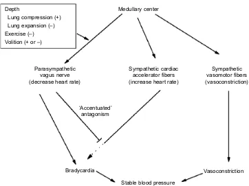
Here we focus on three primary factors that may contribute to heart rate regulation during dives: depth, exercise and volitional control. For this discussion, we have made the assumptions that (1) both parasympathetic and sympathetic cardiac fibers are active during the dive response, (2) the parasympathetic system is dominant, (3) the parasympathetic system is regulated by depth, exercise and volitional control and (4) adjustments in parasympathetic tone primarily account for the observed benign fluctuations in heart rate. Although depth and exercise have been suggested to contribute to heart rate regulation in diving marine mammals via parasympathetic and sympathetic responses, respectively (Davis and Williams, 2012; Williams et al., 2015a,b), we think that any such effects are mediated primarily by the parasympathetic system, given the discussion above. Pulmonary stretch receptor reflexes represent one possible mechanism by which depth may exert an effect on heart rate. These receptors are located in the walls of the tracheo-bronchial tree, and they signal the extent of lung inflation to the brain via the vagus nerve (Schelegle and Green, 2001; Widdicombe, 2006). Inflation of the lung inhibits further inspiration, a response termed the‘Hering–Breuer reflex’(Widdicombe, 2006). This response is associated with cardiovascular reflexes in which lung inflation promotes an increase in heart rate through a decrease in vagal nerve activity to the heart (Looga, 1997; Shepherd, 1981). These responses are intact in phocid seals and represent a vagal Depth
Lung compression (+) Lung expansion (–) Exercise (–) Volition (+ or –)
Medullary center
Sympathetic cardiac accelerator fibers (increase heart rate)
Sympathetic vasomotor fibers (vasoconstriction) Parasympathetic
vagus nerve (decrease heart rate)
‘Accentuated’ antagonism
Stable blood pressure
[image:4.612.126.488.55.326.2]Vasoconstriction Bradycardia
Fig. 1. Proposed roles of the parasympathetic and sympathetic nervous systems during the cardiovascular dive response include activation of the vagus nerve, the sympathetic cardiac accelerator fibers and the sympathetic vasomotor fibers.We propose that (1) the vagus nerve produces bradycardia despite activation of cardiac accelerator fibers as part of the dive response, (2) cardiac accelerator fiber activity is maintained and may even increase as part of the general sympathetic response to constrict arteries and maintain blood pressure during the bradycardia of dives, (3) the potential effects of cardiac accelerator fiber activity on the heart are blunted by accentuated antagonism from the vagus nerve as proposed by Jones and colleagues (see Neuroregulation of the dive response), (4) adjustments in vagal tone (both directly on the heart and via antagonism of the cardiac accelerator fibers) in the presence of elevated sympathetic cardiac tone primarily account for changes in heart rate and for heart rate fluctuations during dives and (5) effects of depth, exercise and volition on heart rate are primarily exerted through changes in vagal tone. Evidence is presented in the text for maintenance or initiation of cardiac accelerator fiber activity during the dive response, and for the potential effects of depth, exercise and volition on parasympathetic output from the medullary center. This hypothesis emphasizes the dominance of the parasympathetic system in the dive response, the lack of autonomic conflict in the dive response of marine mammals, and the common occurrence of benign irregular heartbeats in marine mammals as a consequence of vagal adjustments in the presence of elevated sympathetic tone. Activation is indicated by→; inhibition or inactivation is indicated by┴.
Journal
of
Experimental
mechanism whereby lung volume changes, which are associated with changes in dive depth (owing to Boyle’s law; see Glossary), may contribute to the heart rate response and heart rate fluctuations recorded during dives (Angell-James et al., 1981).
We also propose that any modulation of the dive response by exercise is primarily mediated by the parasympathetic system. Based on our review of heart rate regulation during exercise, we find that most investigators consider the increase in heart rate at the initiation of exercise and at workloads up to 60% of maximal oxygen consumption to be mediated via the parasympathetic system (Borresen and Lambert, 2008; Carter et al., 2003). Given that (1) exercise is often initiated after prolonged glides in marine mammals, (2) peak post-dive metabolic rates of Weddell seals
(Leptonychotes weddellii) are only about twice resting metabolic
rate, far less than 60% maximal oxygen consumption, and (3) field metabolic rates and stroke rates of diving sea lions are typical of those of sea lions swimming in a flume at≤50% maximal oxygen consumption, we postulate that any exercise modulation of heart rate during dives occurs primarily via the parasympathetic system (Costa et al., 1991; Feldkamp, 1987a,b; Ponganis et al., 1991; Tift et al., 2017; Williams et al., 2000, 2004). Furthermore, in a study of free-diving muskrats, the increase in heart rate owing to underwater exercise (swimming against a current) and the increase in heart rate during final ascent were both mediated by parasympathetic withdrawal (Signore and Jones, 1996). In addition, during deep dives of sea lions when heart rate is near 5 beats min−1(McDonald and Ponganis, 2014), an increase in heart rate induced by a further elevation in sympathetic tone would be counterproductive for peripheral oxygen delivery; increased sympathetic tone would cause the proximal arteries responsible for peripheral vasoconstriction in pinnipeds (White et al., 1973) to constrict even more, further limiting any peripheral blood flow (i.e. to the exercising muscle). This would result in hypertension secondary to both an increase in cardiac output (owing to an increased heart rate) and an increase in systemic vascular resistance (owing to increased vasoconstriction). Although we agree that exercise may modulate heart rate during dives (Bergman et al., 1972; Davis and Williams, 2012; Lindholm and Lundgren, 2009), we propose that exercise-induced increases in heart rate during a dive would be primarily due to vagal withdrawal, and not further sympathetic activation.
The higher cortex of the brain (responsible for central command and volitional control) may also influence the parasympathetic system and regulate the degree of bradycardia in sea lions and dolphins (Elmegaard et al., 2016; Ridgway et al., 1975). Such exquisite control of heart rate has also been observed in studies of phocid seals (Fedak et al., 1988; Grinnell et al., 1942; Jobsis et al., 2001; Kooyman and Campbell, 1972); thus, a contribution of volitional control to heart rate regulation during any phase of a dive of marine mammals in the wild can probably never be excluded, no matter how sophisticated the analysis.
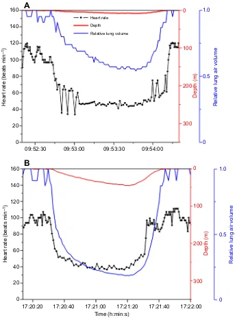
Heart rate, depth and relative lung volume profiles of diving sea lions
Heart rate, depth and relative lung volume profiles in four dives of California sea lions (McDonald and Ponganis, 2014) demonstrate both the complexity of heart rate responses and the potential to utilize the sea lion as a model to investigate heart rate regulation in a marine mammal. In the shallow dives shown in Fig. 2, the degree of bradycardia was similar despite a twofold greater reduction in relative lung volume in the deeper dive, suggesting that lung volume did not contribute significantly to the reduction in heart rate. Given the tracheo-bronchial location of the stretch receptors, and the lower
compliance of the airway relative to that of the alveoli (Cozzi et al., 2005; Fahlman et al., 2009; Kooyman et al., 1970), considerable lung volume reduction may be required before pulmonary stretch receptor activity is affected. Alternatively, different exercise patterns may also contribute to these heart rate profiles and mask the effects of depth on lung volume. We attribute the rapid oscillations and changes in heart rate in these shallow dives to adjustments in vagal tone, possibly secondary to exercise or to volitional control. During these dives, we suspect sympathetic tone is only mildly elevated because with a constant stroke volume (Elsner et al., 1964) and an approximately 50% reduction in heart rate in these dives, systemic vascular resistance (an index of sympathetic tone) should be increased approximately twofold in order to maintain blood pressure during the dives.
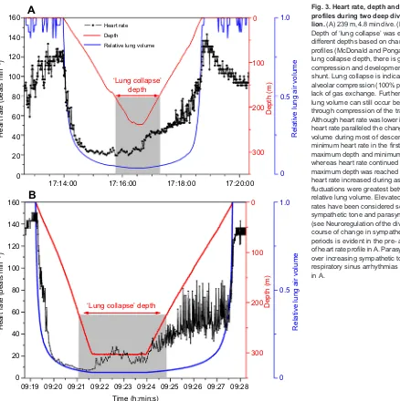
Deep dives of California sea lions (Fig. 3) were characterized by more intense bradycardia, and by an often variable increase in heart rate during the bottom and ascent phases of the dive. Again, we propose that these changes in heart rate are primarily due to changes in vagal tone. We suspect that sympathetic tone, including activity of the cardiac accelerator fibers, is high at the lowest heart rates in order to maintain systemic vascular resistance and blood pressure. Relative to a heart rate of 80 beats min−1, systemic vascular resistance at a heart rate of 10 beats min−1 would be eight times higher, fourfold greater than that in the shallow dives shown in Fig. 2. At these low heart rates during the late descent phase of the dive, heart rate control was tightest; beat-to-beat fluctuations were minimal (Fig. 3) and there was no evidence of autonomic conflict. We conclude that the vagus nerve dominates over the sympathetic cardiac response.
Interpretation of the effects of depth on pulmonary stretch receptors and heart rate is not straightforward in these dives, but is aided by knowledge of the depth of lung collapse (see Glossary) in the sea lion (Fig. 3). Although rapid changes in heart rate during descent and late ascent paralleled changes in depth and relative lung volume (Fig. 3), the decrease in heart rate in the 239-m dive stopped before minimum relative lung volume and maximum depth were reached, whereas heart rate in the 305-m dive continued to decrease almost to the bottom of the dive. Such differences may be due to differences in absolute lung volume (and depth of lung collapse) between the dives to different depths.
An increase in heart rate in these two deep dives (Fig. 3) occurred at the end of descent or early in the bottom phase of the dive before there was any increase in relative lung volume. Thus, pulmonary stretch receptors would not appear to have a role in this initial increase in heart rate during the bottom phases of these two dives. Exercise may exert an effect, especially because prolonged glides end at the start of the bottom phase of deep dives (Tift et al., 2017). Although difficult to evaluate, the increase in heart rate may also be secondary to volitional control at the start of the bottom phase and ascent phase of the dive.
During ascent from deep dives, absolute heart rate and fluctuations in heart rate both increased (Fig. 3). Large beat-to-beat variations in heart rate occurred primarily between 10 and 50% relative lung volume, consistent with lung re-expansion and vagal adjustments secondary to changes in pulmonary stretch receptor activity. Such re-expansion, especially in the tracheo-bronchial tree, would probably occur even if some air were exhaled in ascent, as in fur seals (Hooker et al., 2005). Exercise may also decrease vagal activity and increase heart rate and cardiac output during this phase of the dive (this is under current investigation). At the same time, sympathetic tone, although still elevated, would be expected to decrease if blood pressure were to remain constant and peripheral
Journal
of
Experimental
blood flow were to increase. Thus, in our view, heart rate variability during ascent in sea lions appears to be greatest under conditions of decreasing vagal tone and decreasing (but still elevated) sympathetic tone.
Conclusions
Autonomic conflict is considered arrhythmogenic in humans and rats (Shattock and Tipton, 2012). Irregular heartbeats are not uncommon in human divers and also occur in rats at the initiation of forced submersions, when sympathetic tone is thought to be elevated secondary to pre-dive restraint (Ferrigno and Lundgren, 2003; Olsen et al., 1962; Panneton et al., 2010; Shattock and Tipton, 2012). Life-threatening arrhythmias during cold water immersion of humans may be the ultimate example of such autonomic conflict, with simultaneous activation of the sympathetic cold shock response and the parasympathetic dive response (Shattock and Tipton, 2012).
However, in our view, the fluctuations or oscillations in heart rate of diving sea lions do not represent conflict or predispose the animal
to risk of morbid arrhythmias. Indeed, such fluctuations in heart rate are common, not only in swimming seals, dolphins and sea lions, but also in seals during sleep apnea (see Glossary), in stationary Baikal seals (Phoca sibirica) spontaneously breath-holding underwater, in long dives of grey seals (Halichoerus grypus) and in deep-diving emperor penguins (Aptenodytes forsteri) (Andrews et al., 1997; Ponganis et al., 1997; Thompson and Fedak, 1993; Wright et al., 2014). Benign heart rate fluctuations also regularly occur in other mammals, e.g. hibernators (Milsom et al., 1999). We know of only one report of a potentially life-threatening arrhythmia in a marine mammal–a transient episode of ventricular tachycardia in a struggling, forcibly submerged seal (Murdaugh et al., 1961). Based on our interpretations, autonomic control of heart rate in the sea lion is exquisitely adapted to the animal’s lifestyle. Heart rate fluctuations are minimal at the lowest heart rates when we think that sympathetic and parasympathetic activation are highest. This conclusion contrasts starkly with statements made in the lay press asserting that marine mammals demonstrate high-risk behaviors and 160
140
120
100
80
60
40
20
0
160
140
120
100
80
60
40
20
0
Heart rate (beats min
–1
)
Heart rate (beats min
–1
)
09:52:30 09:53:00 09:53:30 09:54:00
17:20:20 17:20:40 17:21:00 17:21:20 17:21:40 17:22:00 Time (h:min:s)
Heart rate
Depth
Relative lung volume
0
100
200
300
Depth (m)
0
100
200
300
Depth (m)
Relative lung air volume
1.0
0.5
0
Relative lung air volume
1.0
0.5
0
A
[image:6.612.49.395.57.521.2]B
Fig. 2. Heart rate, depth and relative lung volume profiles during two shallow dives of a California sea lion.(A) 7 m, 1.3 min dive. (B) 45 m, 1.3 min dive. Both dives are characterized by mild bradycardias to 40–50 beats min−1, which represent an
approximately twofold reduction in cardiac output and presumably a twofold increase in systemic vascular resistance (an index of sympathetic tone) to maintain blood pressure. Irregular beats and abrupt increases in heart rate are proposed to be secondary to adjustments in vagal tone. Heart rate does not appear to directly parallel changes in relative lung volume in these dives; minimum heart rates are similar despite a more than twofold greater reduction in relative lung volume in the second dive. Data in Figs 2 and 3 are from a prior publication (McDonald and Ponganis, 2014). Note that all axis scales on abscissa are identical in both figures; however, time on the ordinate varies in each graph. Relative lung volume was calculated as 1/(1+depth/10) with depth in m. Transient changes in relative lung volume at the surface in Figs 2 and 3 are secondary to short, very shallow submersions that are not evident on the 350-m depth scale.
Journal
of
Experimental
that they risk serious arrhythmias while hunting. Heart rate variability analysis (Dong, 2016; Pichot et al., 2016) and on-going analyses of the relationship of heart rate with relative lung volume, depth of lung collapse and stroke effort in the sea lion may provide further insight into the roles of these factors in heart rate regulation.
Lastly, in investigations of the etiology of the strandings and deaths of beaked whales after exposure to naval sonar (Fernández et al., 2005; Jepson et al., 2003), it has been found that some beaked whales respond to sonar exposure by performing longer-duration dives with higher fluke stroke rates (DeRuiter et al., 2013). Could fatal arrhythmias occur in these whales solely because of maximum parasympathetic–sympathetic cardiac stimulation (Shattock and Tipton, 2012) or conflict resulting from the combination of exercise and diving (Williams et al., 2015b)? Our interpretation of the literature on heart rate regulation in marine mammals would argue against this possibility.
Alternatively, our proposed adjustments of vagal gain in the presence of elevated sympathetic tone during a dive might be considered as an actual example of autonomic conflict and might be thought to predispose diving marine mammals to serious cardiac
arrhythmias (Shattock and Tipton, 2012). Conceivably, extreme exercise or stress during disturbances might also elevate sympathetic tone and promote such conflict. Based on our analysis of the literature, however, we suspect that the parasympathetic response will predominate in marine mammals during stress. In every case of which we are aware, when a diving animal was stressed, altered ascent and dove deeper, or was unable to access the surface to breathe, the animal maintained the apneic bradycardia or responded with a further decrease in heart rate (Andrews et al., 1997; Dormer et al., 1977; Fedak et al., 1988; Furilla and Jones, 1987; Jobsis et al., 2001; Kvadsheim et al., 2010; Lyamin et al., 2016; Meir et al., 2008; Murdaugh et al., 1961). Despite an increase in body acceleration and prolonged breath-holding on exposure to pinger-like sounds, a marked bradycardia also occurred in harbor porpoises (Phocoena phocoena) (Teilmann et al., 2006). In addition, a stressed beluga whale (Delphinapterus
leucas), diving with harpoon ECG leads and towing a 1200-pound
boat, had a bradycardia of 12 to 24 beats min−1(King et al., 1953). Clearly, further investigations of marine mammal dive responses would be beneficial to evaluate the hypotheses set out here. As illustrated in this Commentary, we propose that the California sea 160
140
120
100
80
60
40
20
0
160
140
120
100
80
60
40
20
0
Heart rate (beats min
–1
)
Heart rate (beats min
–1
)
17:14:00 17:16:00 17:18:00 17:20:00
09:19 09:20 09:21 09:22 09:23 09:24
Heart rate
Depth
Relative lung volume
0
100
200
300
Depth (m)
0
100
200
300
Depth (m)
Relative lung air volume
1.0
0.5
0
Relative lung air volume
1.0
0.5
0
A
B
Time (h:min:s)
09:25 09:26 09:27 09:28
‘Lung collapse’ depth
[image:7.612.54.492.54.494.2]‘Lung collapse’ depth
Fig. 3. Heart rate, depth and relative lung volume profiles during two deep dives of a California sea lion.(A) 239 m, 4.8 min dive. (B) 305 m, 8.5 min dive. Depth of‘lung collapse’was estimated for dives to different depths based on changes in arterial oxygen profiles (McDonald and Ponganis, 2012). Above the lung collapse depth, there is gradual alveolar compression and development of a pulmonary shunt. Lung collapse is indicative of complete alveolar compression (100% pulmonary shunt) and a lack of gas exchange. Further changes in relative lung volume can still occur below this threshold through compression of the tracheo-bronchial tree. Although heart rate was lower in the deeper dive, and heart rate paralleled the change in relative lung volume during most of descent and late ascent, minimum heart rate in the first dive occurred prior to maximum depth and minimum relative lung volume whereas heart rate continued to decrease until maximum depth was reached in the second dive. As heart rate increased during ascent, heart rate fluctuations were greatest between 10 and 50% relative lung volume. Elevated surface period heart rates have been considered secondary to increased sympathetic tone and parasympathetic withdrawal (see Neuroregulation of the dive response). The time course of change in sympathetic tone during these periods is evident in the pre- and post-dive portions of heart rate profile in A. Parasympathetic dominance over increasing sympathetic tone is evident in the respiratory sinus arrhythmias of the pre-dive period in A.
Journal
of
Experimental
lion is an ideal model for further analysis of the contribution of stroke rate, relative lung volume and depth of lung collapse to the degree of bradycardia and the occurrence and mechanisms of heart rate fluctuations during dives.
Acknowledgements
We commend T. M. Williams and co-workers for their research which stimulated this
analysis, and acknowledge the late Dave Jones’curiosity and insight into
cardiovascular regulation in diving animals. We also appreciate the comments of the anonymous reviewers; their efforts improved the manuscript greatly.
Competing interests
The authors declare no competing or financial interests.
Author contributions
P.J.P. conceived the commentary and performed analyses. P.J.P., B.I.M., M.S.T. and C.L.W. co-wrote and edited manuscript.
Funding
This work was supported by the Office of Naval Research (N00014-14-1-0404) and the National Science Foundation Division of Integrative Organismal Systems (IOS-1121428).
References
Andrews, R. D., Jones, D. R., Williams, J. D., Thorson, P. H., Oliver, G. W., Costa, D. P. and Le Boeuf, B. J.(1997). Heart rates of northern elephant seals
diving at sea and resting on the beach.J. Exp. Biol.200, 2083-2095.
Angell-James, J. E., de Burgh Daly, M. and Elsner, R. (1978). Arterial baroreceptor reflexes in the seal and their modification during experimental
dives.Am. J. Physiol.234, H730-H739.
Angell-James, J. E., Elsner, R. and De Burgh Daly, M.(1981). Lung inflation: effects on heart rate, respiration, and vagal afferent activity in seals.
Am. J. Physiol.240, H190-H198.
Antzelevitch, C. and Burashnikov, A.(2011). Overview of basic mechanisms of
cardiac arrhythmia.Card. Electrophysiol. Clin.3, 23-45.
Bergman, S. A., Campbell, J. K. and Wildenthal, K.(1972).“Diving reflex”in man:
its relation to isometric and dynamic exercise.J. Appl. Physiol.33, 27-31.
Blix, A. S. and Folkow, B. (1983). Cardiovascular adjustments to diving in
mammals and birds. InHandbook of Physiology. The Cardiovascular System.
Peripheral Circulation and Organ Blood Flow, Vol. 3 (J. T. Shepherd, F. M. Abboud and S. R. Geiger), pp. 917-945. Bethesda, MD: American Physiology Society. Blix, A. S., Elsner, R. W. and Kjekhus, J. K.(1983). Cardiac output and its
distribution through capillaries and A-V shunts in diving seals. Acta Physiol.
Scand.118, 109-116.
Blix, A. S., Walloe, L., Messelt, E. B. and Folkow, L. P.(2010). Selective brain
cooling and its vascular basis in diving seals.J. Exp. Biol.213, 2610-2616.
Borresen, J. and Lambert, M. I.(2008). Autonomic control of heart rate during and
after exercise.Sports Med.38, 633-646.
Bryden, M. M. and Molyneux, G. S.(1978). Arteriovenous anastomoses in the skin
of seals II. The California sea lion (Zalopohus californianus) and the northern fur
seal (Callorhinus ursinus) (Pinnipedia: Otariidae).Anat. Rec.191, 253-260.
Butler, P. J. and Jones, D. R.(1968). Onset of and recovery from diving bradycardia
in ducks.J. Physiol.196, 255-272.
Butler, P. J. and Jones, D. R.(1997). The physiology of diving of birds and
mammals.Physiol. Rev.77, 837-899.
Carter, J. B., Banister, E. W. and Blaber, A. P.(2003). Effect of endurance exercise
on autonomic control of heart rate.Sports Med.33, 33-46.
Cherepanova, V., Neshumova, T. V. and Elsner, R.(1993). Muscle blood flow in
diving mammals.Comp. Biochem. Physiol. A106, 1-6.
Christensen, N. J. and Galbo, H.(1983). Sympathetic nervous activity during
exercise.Annu. Rev. Physiol.45, 139-153.
Costa, D. P., Antonelis, G. A. DeLong, R. L.(1991). Effects of El Nino on the
foraging energetics of the California sea lion. In Pinnipeds and El Nino.
Responses to Environmental Stress (ed. F. Trillmich and K. A. Ono), pp. 156-165. Berlin: Springer.
Cozzi, B., Bagnoli, P., Acocella, F. and Costantino, M. L.(2005). Structure and
biomechanical properties of the trachea of the striped dolphin Stenella
coeruleoalba: evidence for evolutionary adaptations to diving.Anat. Rec.284, 500-510.
Davis, R. W. and Williams, T. M.(2012). The marine mammal dive response is
exercise modulated to maximize aerobic dive duration.J. Comp. Physiol. A198,
583-591.
de Burgh Daly, M., Elsner, R. and Angell-James, J. E.(1977). Cardiorespiratory control by carotid chemoreceptors during experimental dives in the seal.
Am. J. Physiol.232, H508-H516.
DeRuiter, S. L., Southall, B. L., Calambokidis, J., Zimmer, W. M. X., Sadykova, D., Falcone, E. A., Friedlaender, A. S., Joseph, J. E., Moretti, D., Schorr, G. S.
et al.(2013). First direct measurements of behavioural responses by Cuvier’s
beaked whales to mid-frequency active sonar.Biol. Lett.9, 20130223.
Dong, J.-G.(2016). The role of heart rate variability in sports physiology.Exp. Ther. Med.11, 1531-1536.
Dormer, K. J., Denn, M. J. and Stone, H. L.(1977). Cerebral blood flow in the sea
lion (Zalophus californianus) during voluntary dives.Comp. Biochem. Physiol. A
Physiol.58, 11-18.
Durham, D. and Worthley, L. I. G.(2002). Cardiac arrhythmias: diagnosis and
management. The tachycardias.Crit. Care Resusc.4, 35-53.
Elliott, N. M., Andrews, R. D. and Jones, D. R.(2002). Pharmacological blockade of the dive response: effects on heart rate and diving behaviour in the harbour seal (Phoca vitulina).J. Exp. Biol.205, 3757-3765.
Elmegaard, S. E., Johnson, M., Madsen, P. T. and McDonald, B. I.(2016).
Cognitive control of heart rate in diving harbor porpoises. Curr. Biol. 26,
R1167-R1176.
Elsner, R.(1965). Heart rate response in forced versus trained experimental dives of
pinnipeds.Hvalradets Skrifter48, 24-29.
Elsner, R. and de la Lande, I. S.(1998). Heterogeneous cholinergic reactions of
ringed seal coronary arteries.Comp. Biochem. Physiol. A119, 1019-1025.
Elsner, R. W., Franklin, D. L. and Van Citters, R. L.(1964). Cardiac output during
diving in an unrestrained sea lion.Nature202, 809-810.
Elsner, R., Kenney, D. W. and Burgess, K.(1966). Bradycardia in the trained
dolphin.Nature212, 407.
Elsner, R., Angell-James, J. E. and de Burgh Daly, M.(1977). Carotid body
chemoreceptor reflexes and their interactions in the seal.Am. J. Physiol.232,
H517-H525.
Elsner, R., Wartzok, D., Sonafrank, N. B. and Kelly, B. P.(1989). Behavioral and
physiological reactions of arctic seals during under-ice pilotage.Can. J. Zool.67,
2506-2513.
Fagius, J. and Sundölf, G.(1986). The diving response in man: effects on
sympathetic activity in muscle and skin nerve fascicles.J. Physiol.377, 429-443.
Fahlman, A., Hooker, S. K., Olszowka, A., Bostrom, B. L. and Jones, D. R. (2009). Estimating the effect of lung collapse and pulmonary shunt on gas
exchange during breath-hold diving: the Scholander and Kooyman legacy.Respir.
Physiol. Neurobiol.165, 28-39.
Fedak, M. A., Pullen, M. R. and Kanwisher, J.(1988). Circulatory responses of seals to periodic breathing: heart rate and breathing during exercise and diving in
the laboratory and open sea.Can. J. Zool.66, 53-60.
Feldkamp, S. D.(1987a). Forelimb propulsion in the California sea lionZalophus californianus.J. Zool. Soc. Lond.212, 4333-4357.
Feldkamp, S. D.(1987b). Swimming in the California sea lion: morphometrics, drag
and energetics.J. Exp. Biol.131, 117-135.
Fernández, A., Edwards, J., Martin, V., Rodrı́guez, V., Espinosa de los Monteros, A., Herráez, P., Castro, P. R., Jaber, M. R. and Arbelo, M.(2005).
“Gas and fat embolic syndrome”involving a mass stranding of beaked whales
exposed to anthropogenic sonar signals.J. Vet. Pathol.42, 446-457.
Ferrante, F. L. and Opdyke, D. F.(1969). Mammalian ventricular function during
submersion asphyxia.J. Appl. Physiol.26, 561-570.
Ferrigno, M. and Lundgren, C. E.(2003). Breath-hold diving. InPhysiology and Medicine of Diving(ed. A. O. Brubakk and T. S. Neuman), pp. 153-180. Edinburgh: Saunders.
Fisher, J. P., Seifert, T., Hartwich, D., Young, C. N., Secher, N. H. and Fadel, P. J. (2010). Autonomic control of heart rate by metabolically sensitive skeletal muscle
afferents in humans.J. Physiol.588, 1117-1127.
Furilla, R. A. and Jones, D. R.(1987). The relationship between dive and pre-dive
heart rates in restrained and free dives by diving ducks.J. Exp. Biol.127, 333-348.
Grinnell, S. W., Irving, L. and Scholander, P. F.(1942). Experiments on the
relation between blood flow and heart rate in the living seal.J. Cell. Comp. Physiol.
19, 341-350.
Hammel, H. T., Elsner, R. W., Heller, H. C., Maggert, J. A. and Bainton, C. R. (1977). Thermoregulatory responses to altering hypothalamic temperature in the
harbor seal.Am. J. Physiol.232, R18-R26.
Hance, A. J., Robin, E. D., Halter, J. B., Lewiston, N., Robin, D. A., Cornell, L., Caligiuri, M. and Theodore, M.(1982). Hormonal changes and enforced diving in
the harbor seal,Phoca vitulina. II. Plasma catecholamines.Am. J. Physiol.242,
R528-R532.
Hochachka, P. W., Liggins, G. C., Guyton, G. P., Schnieider, R. C., Stanek, K. S., Hurford, W. E., Creasy, R. K., Zapol, D. G. and Zapol, W. M.(1995). Hormonal
regulatory adjustments during voluntary diving in Weddell seals.Comp. Biochem.
Physiol.112B, 361-375.
Hooker, S. K., Miller, P. J. O., Johnson, M. P., Cox, O. P. and Boyd, I. L.(2005). Ascent exhalations of Antarctic fur seals: a behavioural adaptation for breath-hold
diving?Proc. R. Soc. Lond. B Biol. Sci.272, 355-363.
Houdi, A. A., Dowell, R. T. and Diana, J. N.(1995). Cardiovascular responses to
cigarette smoke exposure in restrained conscious rats.J. Pharmacol. Exp. Ther.
275, 646-653.
Irving, L., Scholander, P. F. and Grinnell, S. W.(1941). Significance of the heart
rate to the diving ability of seals.J. Cell. Comp. Physiol.18, 283-297.
Journal
of
Experimental
Jepson, P. D., Arbelo, M., Deaville, R., Patterson, I. A. P., Castro, P., Baker, J. R., Degollada, E., Ross, H. M., Herráez, P., Pocknell, A. M. et al.(2003).
Gas-bubble lesions in stranded cetaceans.Nature425, 575-576.
Jobsis, P. D., Ponganis, P. J. and Kooyman, G. L.(2001). Effects of training on
forced submersion responses in harbor seals.J. Exp. Biol.204, 3877-3885.
Jones, D. R.(1981). The control of cardiovascular adjustments to diving in birds and
mammals. InAdvances in Animal and Comparative Physiology(ed. G. Pethes
and V. L. Frenyo), pp. 307-314. Oxford: Pergamon Press.
Joyner, M. J.(2006). Baroreceptor function during exercise: resetting the record.
Exp. Physiol.91, 27-36.
Kadowaki, A., Matsukawa, K., Wakasugi, R., Nakamoto, T. and Liang, N.(2011). Central command does not decrease cardiac parasympathetic efferent nerve
activity during spontaneous fictive motor activity in decerebrate cats.
Am. J. Physiol.300, H1373-H1385.
Kaufman, M. P. and Forster, H. V. (1996). Reflexes controlling circulatory,
ventilatory and airway responses to exercise. InHandbook of Physiology, section
12, Chapter 10, Exercise: Regulation and Integration of Multiple Systems(ed. L. B. Rowell and J. T. Shepherd), pp. 381-227. Bethesda, MD: American Physiological Society.
King, R. L., Jenks, J. L. and White, P. D.(1953). The electrocardiogram of a beluga
whale.Circulation8, 387-393.
Kooyman, G. L. and Campbell, W. B.(1972). Heart rates in freely diving Weddell
seals,Leptonychotes weddelli.Comp. Biochem. Physiol. A Physiol.43, 31-36.
Kooyman, G. L., Hammond, D. D. and Schroeder, J. P.(1970). Bronchograms
and tracheograms of seals under pressure.Science169, 82-84.
Kvadsheim, P. H., Sevaldsen, E. M., Folkow, L. P. and Blix, A. S.(2010).
Behavioural and physiological responses of hooded seals (Cystophora cristata) to
1 to 7 kHz sonar signals.Aquat. Mamm.36, 239-247.
Levy, M. N.(1971). Brief Reviews: sympathetic-parasympathetic interactions in the
heart.Circ. Res.29, 437-445.
Levy, M. N. and Zieske, H.(1969). Autonomic control of cardiac pacemaker activity
and atrioventricular transmission.J. Appl. Physiol.27, 465-470.
Lindholm, P. and Lundgren, C. E.(2009). The physiology and pathophysiology of
human breath-hold diving.J. Appl. Physiol.106, 284-292.
Looga, R.(1997). Reflex cardiovascular responses to lung inflation: a review.
Respir. Physiol.109, 95-106.
Lyamin, O. I., Korneva, S. M., Rozhnov, V. V. and Mukhametov, L. M.(2016).
Cardiorespiratory responses to acoustic noise in belugas. InThe Effects of Noise
on Aquatic Life II(ed. A. N. Popper and A. Hawkins), pp. 665-672. New York, NY: Springer.
Maceel, B. C., Gallo, L., Marin Neto, J. A., Lima Filho, E. C. and Martins, L. E. B. (1986). Autonomic nervous control of the heart rate during dynamic exercise in
normal man.Clin. Sci.71, 457-460.
Matsukawa, K.(2012). Central command: control of cardiac sympathetic and vagal efferent nerve activity and the arterial baroreflex during spontaneous motor
behaviour in animals.Exp. Physiol.97, 20-28.
Matsukawa, K., Wall, P. T., Wilson, L. B. and Mitchell, J. H.(1992). Neurally mediated renal vasoconstriction during isometric muscle contraction in cats.
Am. J. Physiol.262, H833-H838.
McDonald, B. I. and Ponganis, P. J.(2012). Lung collapse in the diving sea lion:
hold the nitrogen and save the oxygen.Biol. Lett.8, 1047-1049.
McDonald, B. I. and Ponganis, P. J.(2014). Deep-diving sea lions exhibit extreme
bradycardia in long-duration dives.J. Exp. Biol.217, 1525-1534.
McPhail, L. T. and Jones, D. R.(1999). The autonomic nervous control of heart rate
in ducks during voluntary diving.Physiol. Biochem. Zool.72, 164-169.
Meir, J. U., Stockard, T. K., Williams, C. L., Ponganis, K. V. and Ponganis, P. J. (2008). Heart rate regulation and extreme bradycardia in diving emperor
penguins.J. Exp. Biol.211, 1169-1179.
Milsom, W. K., Zimmer, M. B. and Harris, M. B.(1999). Regulation of cardiac
rhythm in hibernating mammals.Comp. Biochem. Physiol. A Mol. Integr. Physiol.
124, 383-391.
Mitchell, J. H., Kaufman, M. P. and Iwamoto, G. A.(1983). The exercise pressor reflex: its cardiovascular effects, afferent mechanisms, and central pathways.
Annu. Rev. Physiol.45, 229-242.
Morrison, S. F.(2011). 2010 Carl Ludwig Distinguished Lectureship of the APS Neural Control and Autonomic Regulation Section: Central neural pathways for
thermoregulatory cold defense.J. Appl. Physiol.110, 1137-1149.
Murdaugh, H. V., Seabury, J. C. and Mitchell, W. L.(1961). Electrocardiogram of
the diving seal.Circ. Res.9, 358-361.
Nalivaiko, E., De Pasquale, C. G. and Blessing, W. W. (2003). Electrocardiographic changes associated with the nasopharyngeal reflex in
conscious rabbits: vago-sympathetic co-activation. Auton. Neurosci. 105,
101-104.
Noren, S. R., Kendall, T., Cuccurullo, V. and Williams, T. M.(2012). The dive response redefined: underwater behavior influences cardiac variability in freely
diving dolphins.J. Exp. Biol.215, 2735-2741.
O’Leary, D. S.(1993). Autonomic mechanisms of muscle metaboreflex control of
heart rate.J. Appl. Physiol.74, 1748-1754.
Olsen, C. R., Fanestil, D. D. and Scholander, P. F.(1962). Some effects of breath
holding and apneic underwater diving on cardiac rhythm in man.J. Appl. Physiol.
17, 461-466.
Panneton, W. M.(2013). The mammallian diving response: an enigmatic reflex to
preserve life?Physiology28, 284-297.
Panneton, W. M., Gan, Q. and Dahms, T. E.(2010). Cardiorespiratory and neural
consequences of rats brought past their aerobic dive limit.J. Appl. Physiol.109,
1256-1269.
Paton, J. F. R., Boscan, P., Pickering, A. E. and Nalivaiko, E.(2005). The yin and
yang of cardiac autonomic control: Vago-sympathetic interactions revisited.Brain
Res. Rev.49, 555-565.
Paton, J. F. R., Nalivaiko, E., Boscan, P. and Pickering, A. E.(2006). Reflexly evoked coactivation of cardiac vagal and sympathetic motor outflows:
observations and functional implications.Clin. Exp. Pharmacol. Physiol. 33,
1245-1250.
Petro, J. K., Hollander, A. P. and Bouman, L. N.(1970). Instantaneous cardiac
acceleration in man induced by a voluntary muscle contraction.J. Appl. Physiol.
29, 794-798.
Pichot, V., Roche, F., Celle, S., Barthélémy, J.-C. and Chouchou, F.(2016).
HRVanalysis: a free software for analyzing cardiac autonomic activity.Front.
Physiol.7, 557.
Ponganis, P. J.(2015).Diving Physiology of Marine Mammals and Seabirds. Cornwall: Cambridge University Press.
Ponganis, P. J., Kooyman, G. L. and Zornow, M. H.(1991). Cardiac output in
swimming California sea lions, Zalophus californianus. Physiol. Zool. 64,
1296-1306.
Ponganis, P. J., Kooyman, G. L., Baronov, E. A., Thorson, P. H. and Stewart, B. S.(1997). The aerobic submersion limit of Baikal seals, Phoca sibirica.
Can. J. Zool.75, 1323-1327.
Ridgway, S. H., Carder, D. A. and Clark, W.(1975). Conditioned bradycardia in the
sea lionZalophus californianus.Nature256, 37-38.
Rowell, L. B. and O’Leary, D. S.(1990). Reflex control of the circulation during
exercise: chemoreflexes and mechanoreflexes.J. Appl. Physiol.69, 407-418.
Schelegle, E. S. and Green, J. F.(2001). An overview of the anatomy and
physiology of slowly adapting pulmonary stretch receptors.Respir. Physiol.125,
17-31.
Scholander, P. F.(1940). Experimental investigations on the respiratory function in
diving mammals and birds.Hvalradets Skrifter22, 1-131.
Scholander, P. F., Irving, L. and Grinnell, S. W.(1942). Aerobic and anaerobic
changes in seal muscle during diving.J. Biol. Chem.142, 431-440.
Shattock, M. J. and Tipton, M. J.(2012).“Autonomic conflict”: a different way to die
during cold water immersion?J. Physiol.590, 3219-3230.
Shepherd, J. T.(1981). The lungs as receptor sites for cardiovascular regulation.
Circulation63, 1-10.
Signore, P. E. and Jones, D. R.(1995). Effect of pharmacological blockade on
cardiovascular responses to voluntary and forced diving in muskrats.J. Exp. Biol.
198, 2307-2315.
Signore, P. E. and Jones, D. R.(1996). Autonomic nervous control of heart rate in
muskrats during exercise in air and under water.J. Exp. Biol.199, 1563-1568.
Smith, E. E., Guyton, A. C., Manning, R. D. and White, R. J.(1976). Integrated mechanisms of cardiovascular response and control during exercise in the normal
human.Prog. Cardiovasc. Dis.18, 421-443.
Smith, S. A., Mitchell, J. H. and Garry, M. G.(2006). The mammalian exercise
pressor reflex in health and disease.Exp. Physiol.91, 89-102.
Stramba-Badiale, M., Vanoli, E., De Ferrari, G. M., Cerati, D., Foreman, R. D. and Schwartz, P. J. (1991). Sympathetic-parasympathetic interaction and
accentuated antagonism in conscious dogs.Am. J. Physiol.260, H335-H340.
Teilmann, J., Tougaard, J., Miller, L. A., Kirketerp, T., Hansen, K. and Brando, S. (2006). Reactions of captive harbor porpoises (Phocoena phocoena) to
pinger-like sounds.Mar. Mamm. Sci.22, 240-260.
Thompson, D. and Fedak, M. A.(1993). Cardiac responses of grey seals during
diving at sea.J. Exp. Biol.174, 139-164.
Tift, M. S., Hückstädt, L. A., McDonald, B. I., Thorson, P. H. and Ponganis, P. J. (2017). Flipper stroke rate and venous oxygen levels in free-ranging California sea
lions.J. Exp. Biol.220, 1533-1540.
Tse, G.(2016). Mechanisms of cardiac arrhythmias.J. Arrhythmia32, 75-81. Tsuchimochi, H., Hayes, S. G., McCord, J. L. and Kaufman, M. P.(2009). Both
central command and exercise pressor reflex activate cardiac sympathetic nerve
activity in decerebrate cats.Am. J. Physiol.296, H1157-H1163.
Tulppo, M. P., Makikallio, T. H., Takala, T. E., Seppanen, T. and Huikuri, H. V. (1996). Quantitative beat-to-beat analysis of heart rate dynamics during exercise.
Am. J. Physiol.271, H244-H252.
Tulppo, M. P., Mäkikallio, T. H., Seppänen, T., Laukkanen, R. T. and Huikuri, H. V.(1998). Vagal modulation of heart rate during exercise: effects of age and
physical fitness.Am. J. Physiol.274, H424-H429.
Tulppo, M. P. P., Kiviniemi, A. M. M., Hautala, A. J. P., Kallio, M. M. D., Seppanen, T. P., Makikallio, T. H. M. D. and Huikuri, H. V. M. D.(2005). Physiological
background of the loss of fractal heart rate dynamics.Circulation112, 314-319.
Journal
of
Experimental
Van Citters, R. L., Franklin, D. L., Smith, O. A., Jr, Watson, N. W. and Elsner, R. W.(1965). Cardiovascular adaptations to diving in the northern elephant seal
Mirounga angustirostris.Comp. Biochem. Physiol.16, 267-276.
Vatner, S. F. and Pagani, M.(1976). Cardiovascular adjustments to exercise:
hemodynamics and mechanisms.Prog. Cardiovasc. Dis.19, 91-108.
Victor, R. G., Seals, D. R. and Mark, A. L.(1987). Differential control of heart rate and sympathetic nerve activity during dynamic exercise. Insight from intraneural
recordings in humans.J. Clin. Investig.79, 508-516.
Waldrop, T. G. and Iwamoto, G. A. (2006). Point:Counterpoint: supraspinal locomotor centers do/do not contribute significantly to the hyperpnea of dynamic
exercise.J. Appl. Physiol.100, 1077-1083.
Waldrop, T. G., Eldridge, F. L., Iwamoto, G. A. and Mitchell, J. H.(1996). Central
neural control of locomotion, respiration, and circulation during exercise. InHandbook
of Physiology. Section 12, Exercise: Regulation and Integration of Multiple Systems, Vol. 12, pp. 333-380. Bethesda, MD: American Physiological Society.
White, F. N., Ikeda, M. and Elsner, R. W.(1973). Andrenergic innervation of large
arteries in the seal.Comp. Gen. Physiol.4, 271-276.
Widdicombe, J.(2006). Reflexes from the lungs and airways: historical perspective.
J. Appl. Physiol.101, 628-634.
Williams, T. M., Davis, R. W., Fuiman, L. A., Francis, J., Boeuf, B. L. and Horning, M.(2000). Sink or swim: strategies for cost-efficient diving by marine
mammals.Science288, 133-136.
Williams, T. M., Fuiman, L. A., Horning, M. and Davis, R. W.(2004). The cost of
foraging by a marine predator, the Weddell sealLeptonychotes weddellii: pricing
by the stroke.J. Exp. Biol.207, 973-982.
Williams, T. M., Bengston, P., Steller, D. L., Croll, D. A. and Davis, R. W.(2015a).
The healthy heart: lessons from nature’s elite athletes.Physiology30, 349-357.
Williams, T. M., Fuiman, L. A., Kendall, T., Berry, P., Richter, B., Noren, S. R., Thometz, N., Shattock, M. J., Farrell, E., Stamper, A. M. et al.(2015b). Exercise at depth alters bradycardia and incidence of cardiac anomalies in deep-diving
marine mammals.Nat. Commun.6, 6055.
Wright, A. K., Ponganis, K. V., McDonald, B. I. and Ponganis, P. J.(2014). Heart rates of emperor penguins diving at sea: implications for oxygen store
management.Mar. Ecol. Prog. Ser.496, 85-98.
Zapol, W. M., Liggins, G. C., Schneider, R. C., Qvist, J., Snider, M. T., Creasy, R. K. and Hochachka, P. W.(1979). Regional blood flow during simulated diving
in the conscious Weddell seal.J. Appl. Physiol.47, 968-973.