DETECTION OF TEMPORAL BONE PATHOLOGIES BY HIGH RESOLUTION COMPUTED
Consultant Radiology, J and K Health Services
ARTICLEINFO ABSTRACT
Background:
tissue, is the best method for evaluating the mastoid air cell system. It helps in accurate asses pathology prior to surgical exploration regarding location, extent and complication of the disease. HRCT is now established as the most useful and versatile procedure for showing bony detail in the petrous pyramid, soft tissue abnormalities in the
cranial cavity.
HRCT in diagnosis and detection of pathologies of the temporal bone. conducted i
Imaging in co
thick slices were obtained using an ultra high algorithm obtained perpendicular to the axial
Intravenous contrast were administered wherever necessary to study for intracranial or extracranial extension of middle ear disease.
to 64 years.
study population was 2:1.
mastoiditis was seen in 9 cases. Combination of both cholesteatoma and mastoiditis was seen in 7 cases. Total 27 cases of cholesteatoma (20+7) and 16 cases of mastoiditis (9+7) were seen. diagnosed opacified mastoid air cells were seen in 16 cases
cholesteatoma were operated and 8 cases of mastoiditis alone were treated conservatively.
Conclusion:
outdated
ideal for evaluation of Temporal Bone lesions.
Copyright©2019Dr. Arti Khurana. This is an open access distribution, and reproduction in any medium, provided
INTRODUCTION
Chronic otitis media (COM) is a long standing inflammation of the middle ear cleft without reference to the etiology or pathogenesis.It is important for otolaryngologists differentiate between the two types of Chronic Otitis Media (COM): The chronic mucosal disease and the COM with cholesteatoma; this is because of higher risk of complications associated with the cholesteatoma group, which
threatening conditions. It may, however, be useful for showing evidence of bony erosion in acute and chronic mastoiditis, extent of pneumatization of the temporal bone and relationship of the pathology to adjacent critical neurovascular s
such as the dura, internal carotid artery, lateral sinus and facial nerve. It is now being claimed that a cholesteatoma as small as 3mm in size can be diagnosed much earlier by the use of CT. With its excellent imaging quality and the ability to
bone and soft tissue, HRCT is the best method for evaluating the mastoid air cell system.
ISSN: 0975-833X
Article History:
Received 09th February, 2019
Received in revised form
22nd March, 2019
Accepted 11th April, 2019
Published online 30th May, 2019
Citation: Dr. Arti Khurana. 2019. “Detection of temporal bone pathologies by high resolution computed tomography
Research, 11, (05), 4110-4115.
Availableonlineathttp://www.journal
Key Words:
High Resolution Computed Tomography, Chronic Otitis Media,
Mastoiditis.
*Corresponding author: Dr. Arti Khurana
REVIEW ARTICLE
DETECTION OF TEMPORAL BONE PATHOLOGIES BY HIGH RESOLUTION COMPUTED
TOMOGRAPHY
*
Dr. Arti Khurana
Consultant Radiology, J and K Health Services
ABSTRACT
Background: HRCT with its excellent imaging quality and the ability to eliminate bone and soft tissue, is the best method for evaluating the mastoid air cell system. It helps in accurate asses pathology prior to surgical exploration regarding location, extent and complication of the disease. HRCT is now established as the most useful and versatile procedure for showing bony detail in the petrous pyramid, soft tissue abnormalities in the middle ear and the extension of the disease into the cranial cavity. Objective: The objective of the study is primarily to understand the capability of HRCT in diagnosis and detection of pathologies of the temporal bone.
conducted in 60 patients with symptoms of mastoiditis, in the department of Radio Imaging in co-ordination with the department of Otolaryngology GM
thick slices were obtained using an ultra high algorithm in the axial axis.
obtained perpendicular to the axial plane from the cochlea to the posterior semicircular canal. Intravenous contrast were administered wherever necessary to study for intracranial or extracranial extension of middle ear disease. Results: In our study population,
to 64 years. There were 40 males ( 66.5%). There were 20 (33.5%) females. Male to female ratio of study population was 2:1. Patients with infections (n=36), cholesteatoma was seen in 20 cases mastoiditis was seen in 9 cases. Combination of both cholesteatoma and mastoiditis was seen in 7 cases. Total 27 cases of cholesteatoma (20+7) and 16 cases of mastoiditis (9+7) were seen. diagnosed opacified mastoid air cells were seen in 16 cases however only 8 cases with associated cholesteatoma were operated and 8 cases of mastoiditis alone were treated conservatively.
Conclusion: HRCT produced high quality images of soft tissue lesions in air spaces. HRCT has outdated the conventional modalities of investigations by providing
ideal for evaluation of Temporal Bone lesions.
access article distributed under the Creative Commons Attribution the original work is properly cited.
Chronic otitis media (COM) is a long standing inflammation of the middle ear cleft without reference to the etiology or otolaryngologists to differentiate between the two types of Chronic Otitis Media (COM): The chronic mucosal disease and the COM with cholesteatoma; this is because of higher risk of complications associated with the cholesteatoma group, which can lead to life threatening conditions. It may, however, be useful for showing evidence of bony erosion in acute and chronic mastoiditis, extent of pneumatization of the temporal bone and relationship of the pathology to adjacent critical neurovascular structures such as the dura, internal carotid artery, lateral sinus and facial is now being claimed that a cholesteatoma as small as 3mm in size can be diagnosed much earlier by the use of CT. With its excellent imaging quality and the ability to eliminate bone and soft tissue, HRCT is the best method for evaluating
Newer high resolution multidetector spiral imaging system can generate nearly isotropic voxels for multiplanar reconstruction, making need for multiple series with direct imaging in several planes unnecessary. HRCT has the advantage of excellent topographic visualization, devoid of artifacts from superimposition of structures. It helps in accurate assessment of pathology prior to surgical exploration rega
extent and complication of the disease. HRCT is now established as the most useful and versatile procedure for showing bony detail in the petrous pyramid, soft tissue abnormalities in the middle ear and the extension of the disease into the cranial cavity. CT scanning is of vital importance in the detection of intracranial complications
MATERIALS AND METHODS
The study was conducted in 60 patients with symptoms of mastoiditis, in the department of Radio
International Journal of Current Research Vol. 11, Issue, 05, pp. 4110-4115, May, 2019
DOI: https://doi.org/10.24941/ijcr.35631.05.2019
Detection of temporal bone pathologies by high resolution computed tomography
Availableonlineathttp://www.journalcra.com
z
DETECTION OF TEMPORAL BONE PATHOLOGIES BY HIGH RESOLUTION COMPUTED
HRCT with its excellent imaging quality and the ability to eliminate bone and soft tissue, is the best method for evaluating the mastoid air cell system. It helps in accurate assessment of pathology prior to surgical exploration regarding location, extent and complication of the disease. HRCT is now established as the most useful and versatile procedure for showing bony detail in the middle ear and the extension of the disease into the The objective of the study is primarily to understand the capability of HRCT in diagnosis and detection of pathologies of the temporal bone. Methods: The study was in the department of Radio-Diagnosis and GMC Jammu. Contiguous 0.5-1mm in the axial axis. Coronal images were plane from the cochlea to the posterior semicircular canal. Intravenous contrast were administered wherever necessary to study for intracranial or extracranial , age of patients ranged from 7 years 40 males ( 66.5%). There were 20 (33.5%) females. Male to female ratio of Patients with infections (n=36), cholesteatoma was seen in 20 cases and mastoiditis was seen in 9 cases. Combination of both cholesteatoma and mastoiditis was seen in 7 cases. Total 27 cases of cholesteatoma (20+7) and 16 cases of mastoiditis (9+7) were seen. HRCT however only 8 cases with associated cholesteatoma were operated and 8 cases of mastoiditis alone were treated conservatively. images of soft tissue lesions in air spaces. HRCT has providing better soft tissue contrast and is
License, which permits unrestricted use,
Newer high resolution multidetector spiral imaging system can generate nearly isotropic voxels for multiplanar reconstruction, series with direct imaging in several planes unnecessary. HRCT has the advantage of excellent topographic visualization, devoid of artifacts from superimposition of structures. It helps in accurate assessment of pathology prior to surgical exploration regarding location, extent and complication of the disease. HRCT is now established as the most useful and versatile procedure for showing bony detail in the petrous pyramid, soft tissue abnormalities in the middle ear and the extension of the disease cranial cavity. CT scanning is of vital importance in the detection of intracranial complications (Phelps PD 1997).
AND METHODS
conducted in 60 patients with symptoms of mastoiditis, in the department of Radio-Diagnosis and Imaging
INTERNATIONAL JOURNAL OFCURRENTRESEARCH
Figure 1. Normal HRCT anatomy of Temporal Bone
promontory, basal turn of the cochlea, cochlear duct. The descending facial nerve canal, sigmoid sinus, and
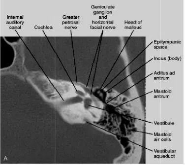
Figure 2. Axial CT image shows the geniculate ganglion and horizontal portions of the facial nerve canal in continuity with t vestibule, vestibular aqueduct and endolymphatic sac. The
4111 Dr. Arti Khurana et al. Detection
Normal HRCT anatomy of Temporal Bone Axial CT section shows external auditory canal, long process of the malleus, cochlear promontory, basal turn of the cochlea, cochlear duct. The descending facial nerve canal, sigmoid sinus, and mastoid air cells are also shown
Figure 2. Axial CT image shows the geniculate ganglion and horizontal portions of the facial nerve canal in continuity with the internal auditory canal, cochlea, vestibule, vestibular aqueduct and endolymphatic sac. The epitympanic space holds the head of the malleus and the body of the incus.
Detection of temporal bone pathologies by high resolution computed tomography
Axial CT section shows external auditory canal, long process of the malleus, cochlear mastoid air cells are also shown
he internal auditory canal, cochlea, epitympanic space holds the head of the malleus and the body of the incus.
[image:2.595.103.480.424.759.2]Figure 3. Axial CT scan of the left temporal bone centered at the neck of the malleus in the epitympanic space shows long process of th and the stapes posteriorly. The three turns of the cochlea, descending facial nerve canal is also seen. Other labeled st
Figure 4. Coronal CT section made at the level of the oval window shows stapes, incudostapedial joint, horizontal portion of the facial nerve, short process of the incus. The internal auditory canal (IAC), middle ear, external auditory canal (EAC), stapes, and
4112 International Journal
Axial CT scan of the left temporal bone centered at the neck of the malleus in the epitympanic space shows long process of th and the stapes posteriorly. The three turns of the cochlea, descending facial nerve canal is also seen. Other labeled st
apex, carotid canal, and sigmoid sinus
Coronal CT section made at the level of the oval window shows stapes, incudostapedial joint, horizontal portion of the facial nerve, short process of the incus. The internal auditory canal (IAC), middle ear, external auditory canal (EAC), stapes, and
of the cochlea are also shown
Journal of Current Research, Vol. 11, Issue, 05, pp.4110-4115, May, 201
Axial CT scan of the left temporal bone centered at the neck of the malleus in the epitympanic space shows long process of the malleus and the stapes posteriorly. The three turns of the cochlea, descending facial nerve canal is also seen. Other labeled structures include the petrous
Coronal CT section made at the level of the oval window shows stapes, incudostapedial joint, horizontal portion of the facial nerve, short process of the incus. The internal auditory canal (IAC), middle ear, external auditory canal (EAC), stapes, and basal turn
[image:3.595.121.505.442.750.2]in co-ordination with the department of Otolaryngology GMC Jammu. Patients with electric devices at the skull base, such as cochlear implants or any other metallic implants, were excluded from the study. Contiguous 0.5-1mm thick slices were obtained using an ultra high algorithm in the axial axis, Scanning commenced from the lower margin of the external auditory meatus and extendedupward to the arcuate eminence of the superior semicircular canal as seen on lateral topogram. Coronal images were obtained perpendicular to the axialplane from the cochlea to the posterior semicircular canal. Intravenous contrast were administered wherever necessary to study for intracranial or extracranial extension of middle ear disease.
Aims and Objectives
To study the extent of middle ear infections and their complications.
To correlate imaging findings with surgical findings, wherever possible.
RESULTS
[image:4.595.314.551.85.340.2]In our study population, age of patients ranged from 7 years to 64 years. There were 40 males (66.5%). There were 20 (33.5%) females. Male to female ratio of study population was 2:1.
Table 1. Distribution of the patients according to age (n=60).
Sex No. of Patients Percentage
Male 40 66.5
Female 20 33.5
Table 2. Etiology of the disease (n=60)
Disease No. of Patients Percentage
Infection 36 60
Trauma 10 16.6
Tumor 7 11.7
Congenital 3 5
[image:4.595.326.541.368.468.2]Others 4 6.7
Table 3. Summarizes the symptoms of the patients
S.No. Clinical complaints* No. of Patients Percentage
1. Hearing Loss 27 45
2. Ear pain 21 35
3. Headache 18 30
4. Fever 21 35
5. Ear discharge 10 16.7
6. Tinnitus 10 16.7
7. Diplopia 5 8.3
8. Facial nerve weakness 4 6.7
9. Cerebellar signs 3 5
*One patient may have more than one complaint
Table 4. Distribution of infection according to age (n=36)
Age in years No. of patients Percentage
0-10 3 5
11-20 9 15
21-30 12 20
31-40 7 11.7
41-50 3 5
51-60 0 0
61-70 2 3.3
Total 36
Table 5. Distribution according to ear affected, type of infection Extent of the cholesteatoma and Complications of cholesteatoma
(a) Infection according to ear affected (n=36)
1. Right 15 41.7
2. Left 13 36.1
3. Bilateral 8 22.2
(b) Type of Infection(n=36)
1. Cholesteatoma 20 55.5
2. Mastoiditis 9 25
3. Both cholesteatoma and mastoiditis 7 19.4
(c) Extent of the cholesteatoma (n=27)
1. Prussack’s space 20 74
2 Epitympanum 16 59.2
3 Antrum 15 55.5
4 Protympanum 12 44.4
5 Mesotympanum 12 44.4
6 Hypotympanum 11 40.7
7 Facial recess 11 40.7
8 Sinus tympani 10 37
9 Mastoid Air cells 7 25.9
(d) Complications of cholesteatoma (n=27)CT Findings
Ossicular chain destruction 18 66.7
Facial Canal Dehiscence 4 14.8
Tegmen tympani erosion 4 14.8
Mastoid Cortex dehiscence 6 22.2
Carotid canal dehiscence 2 7.4
[image:4.595.52.271.475.535.2]Dural plate dehiscence 0 0
Table 6. HRCT findings and operative findings in infections (n=36)
CT findings in Patients
No. of
patients
Patients with same operative findings
Cholesteatoma 27 27
Mastoid air cells
Opacification 16
8 (8 treated conservatively).
Ossicular erosion 18 16
Tegmen erosion 4 3
Facial canal erosion 4 4
Scutum erosion 12 12
[image:4.595.48.279.570.669.2]Carotid canal erosion 2 2
Table 7. HRCT findings compared with operative results for involvement of ear ossicles (n=18)
CT Findings Cases diagnosed
by HRCT
Patients with same operative findings (%)
Only Incus
involvement 12 10 (83.3%)
Malleus, Incus
and Stapes
involved
6 6 (100%)
Table 8. Part of temporal bone fractured (n=10)
Part of temporal bone
fractured* No. of patient Percentage
Squamous 10 100
Petrous 8 80
Mastoid 6 60
Tympanic 4 40
Styloid 0 0
*In majority of patients of trauma more than one bone was fractured.
Table 9. CT findings associated with Temporal bone trauma (n=10)
Associated findings No. of patients Percentage
Sinus opacification 7 70
External/middle ear
opacification 8 80
Other fractures 7 70
Local pneumocephalus 5 50
Cerebral contusion/EDH 4 40
[image:4.595.322.546.617.689.2] [image:4.595.71.253.714.803.2]Patients with infections (n=36), cholesteatoma was seen in 20 cases and mastoiditis was seen in 9 cases. Combination of both cholesteatoma and mastoiditis was seen in 7 cases. Total 27 cases of cholesteatoma (20+7) and 16 cases of mastoiditis (9+7) were seen. HRCT diagnosed opacified mastoid air cells were seen in 16 cases however only 8 cases with associated cholesteatoma were operated and 8 cases of mastoiditis alone were treated conservatively. Results of our study are elaborated in table 1 to 10.
DISCUSSION
Radiographic assessment of temporal bone is difficult owing to complicated anatomy of middle and inner ear. Computed Tomography is acquiring an increasingly important role in the radiographic assessment of temporal bone and is helpful in diagnosis and treatment of temporal bone diseases (Clement P et al 1982 and Watts S et al 2000). The most important advantage of spiral CT in temporal bone imaging is its perfect visualization of the contrast between bony structures and the air in the middle ear. In addition to detailed evaluation of the bony structures it also permits assessment of soft tissue components as well (Maffe MF et al 1983). CSOM is prevalent in all age groups, particularly affecting the patients in pediatric and younger age groups (WHO, 2004). One of the important pathologies affecting the temporal bone during CSOM was cholesteatoma. Acquired cholesteatomas are commonly seen in patients less than 30 years (Gomaa MA et al, 2013). In present study we too found that the majority of our patients were less than 30 years. Datta et al. (2014) in their study also found the maximum number of patients with unsafe cholesteatoma to be less than 30 years (76%) and mean age to be 26.74 years. Studying the anatomical variation of vestibular aqueduct of temporal bone Poursadegh M et al (2000) also reported the mean age of patients to be 24.2 years. Mean age of patients in the series of Gerami et al. (2009) was 27.9±16.3 years. Thus, most of the studies targeted to study temporal bone pathologies or normal variations had reported the mean age of patients between 25 to 30 years. However, Paparella MM et al (1977) claim an average of about 35.1 years, this variation was because of CSOM is more common amongst children in our country. Maffee et al. (1988) studied cholesteatoma in 48 patients with Computed Tomography preoperatively. Operative reports of these patients were correlated with CT findings in all the patients.
Comparing the imaging changes in the attic with findings at operation they found agreement between the CT and surgical findings in 90% of the cases. In present study majority of cases were males (n=40; 66.5%), females constituted 20 (33.5%) cases. Male to female ratio in infective cases was 2:1. With respect to CSOM being the major temporal bone pathology no gender wise difference in prevalence of CSOM has been reported in community studies, however, in hospital based studies. Poursadegh M et al (2000) reported a male to female ratio of 1.39:1 which is quite similar to present study. Contrarily, Datta G et al (2014) and Gomaa MA et al (2013) found a higher proportion of females as compared to males. With respect to gender wise differences these could be purely incidental and could generally be attributed to gender biased healthcare seeking practices in our settings. Out of 60 patients, in the infective group 36 belonged to low socio economic groups due to poor nutrition and poor hygiene coupled with illiteracy. The common presenting symptoms in present study were hearing loss (n=27; 45%) otorrhoea (n=10; 16.7%) and otalgia (n=21; 35%). The discharge was scanty, foul smelling and purulent. In the series of Gomaa et al. (2013) too chronic ear discharge with hearing loss and otalgia were the main clinical presentation (60.7%). Increased ear discharge, persistent earache, fever, post auricular swelling and facial weakness heralded complications of cholesteatoma. The presence of vomiting, headache, drowsiness and altered sensorium indicated more sinister threat of a lurking intracranial complications. Bilateral cholesteatoma were rare. According to our study prussack’s space (74%) and epitympanum (59.2%) were the most common sites in middle ear to be involved in cholesteatoma which were similar to findings by Sirigiri et al. (2011). In the antrum, HRCT detection rate was 55.5 %, which is less than the observations by Sirigiri et al. (2011). In case of mesotympanum, HRCT detection rate was only 44.4%, which is less than the findings by Walshe et al. (2002) however involvement of protympanum (44.4%) and hypotympanum (40.7%) is in accordance with study of Sirigiri et al. (2011). Extension of cholesteatoma into the mastoid air system was seen in 7 cases (25.9%) which is similar to observations by Gerami H et al (2009). Ossicular chain involvement was seen in 18 cases out of which 12 cases (66.6%) having involvement of incus only while 6 (33.4%) had involvement of all the three bones of ossicular chain. However, in the series of Gomma MA et al (2013) all the cases had involvement of one or more bones in the ossicular chain.
Table 10. Distribution of tumour according to age, gender and its findings on CT
(a) Incidence of tumors according to age (n=7)
Age (in years) No of patients Percentage
0-20 1 14.2
20-40 0 0
40-60 3 42.8
60-80 3 42.8 (b) Distribution of tumors according to gender (n=7)
Sex No. of patients Percentage
Male 4 57.14
Female 3 42.85
Distribution of Tumor Patients (%) CT features Operation
/ biopsy findings
Acoustic neuroma 3(40%)
Hypodense to slightly hyperdense cerebellopontine
angle mass showing contrast enhancement with widened internal auditory canal
+
Metastasis 2 (20%) Destruction of apex of petrous bone on left side
Meningioma 1(20%) Densely calcified, heterogeneously enhancing lesion in left CP angle +
Rhabdomyosarcoma 1 (20%) Mildly enhancing soft tissue density lesion in left ear causing
destruction of surrounding bones.
+
The higher level of involvement of ossicular bones and temporal bone erosion and presence of cranial pathologies in these case series might be attributed to choleastoma being the key finding in these series of patients (Gomaa MA et al, 2013). In present study, HRCT could detect involvement of incus in 12 cases only whereas intraoperatively, it was confirmed in 10 cases, thus HRCT overdiagnosed the involvement of incus in two case. The agreement between HRCT and surgical findings was 83.3% for incus involvement. Similar observations were also made by Chee NW et al (2001). Tegmen erosion was reported in 4 (14.8%) cases whereas carotid canal erosion was seen in 2 (7.4%) case. Scutum erosion was seen in 12 (44.4%) cases of cholesteatoma which is less than that seen by Gaurano JL et al (2004) who found it in 86%. However, HRCT detected scutum erosion was accurate in all cases. Hence, HRCT is 100% sensitive and specific to detect scutum erosion as per this study. This is in accordance with the study carried by Rocher P et al (1995). In present study mastoiditis was seen in 16 cases. Mastoiditis is a common finding in cases with suspected temporal bone pathology and CSOM (Chole RA et al, 2005). Mastoiditis is usually caused by a middle ear infection (acute otitis media). The infection may spread from the ear to the mastoid bone of the skull. The prevalence of mastoiditis in present study was 44.4% whereas Keskin S et al (2011) who in their series reported its prevalence to be 73.2%.
Conclusion
HRCT produced high quality images of soft tissue lesions in air spaces. HRCT has outdated the conventional modalities of investigations by providing better soft tissue contrast and is ideal for evaluation of Temporal Bone lesions. For the assessment of middle ear infections, a close clinical correlation is essential to evaluate the nature of middle ear soft tissue masses, as cholesteatoma is mimicked by many other pathologies. In these cases, HRCT is advantageous in assessing the complications of infection. It also helps in postoperative cases, to comment regarding the extent of surgery, and the general overall condition of the postoperative temporal bone including the internal auditory canal. The residual/recurrent disease and status of the inner ear and scan be assessed.
REFERENCES
Chee NW., Tan TY. 2001. The value of pre-operative high resolution CT scans in cholesteatoma surgery. Singapore Med J., 42:155-9.
Chole RA., Sudhoff H. 2005. Chronic otitis media, mastoiditis and petrositis. In: Cummings CW, Fredriskon MJ, Harker L. Otolaryngology head and neck surgery. 4th ed. Philadelphia (PA): Mobsy; pp.2988-3008
Clement P., DeSmedt E. 1982. “High resolution tomographic scans of the normal and abnormal ear”. Am J Otolaryngol.,
3: 286-94
Datta G., Mohan C., Mahajan M., Mendiratta V. 2014. Correlation of preoperative HRCT findings with surgical findings in Unsafe CSOM. J Dent Med Sci., 13:120-5. Gaurano JL., Joharjy IA. 2004. Middle ear cholesteatoma:
Characteristic CT findings in 64 patients. Ann Saudi Med., 24:442-7.
Gerami H., Naghavi E., Wahabi-Moghadam M., Forghanparast K., Akbar MH. 2009. Comparison of pre-operative computerized tomography scan imaging of temporal bone with the intra-operative findings in patients undergoing mastoidectomy. Saudi Med J., 30:104-8.
Gomaa MA., Abdel Karim AR., Abdel Ghany HS., Elhiny AA., Sadek AA. 2013. Evaluation of temporal bone cholesteatoma and the correlation between high resolution computed tomography and surgical finding. Clin Med Insights Ear Nose Throat.6:21-8.
Keskin S., Çetin H., Töre HG. 2011. The Correlation of temporal bone CT with surgery findings in evaluation of chronic inflammatory diseases of the middle ear. Eur J Gen Med., 8:24-30.
Mafee MF., Levin BC., Applebaum EL., Campos M., James CF.1988. Cholesteatoma of the middle ear and mastoid. A comparison of CT scan and operative findings. Otolaryngol Clin North Am., 21:265-93.
Mafee, M., Kumar, A., Yanniss, D. 1983. Computed tomography of the middle ear in the evaluation of cholesteatoma and other soft tissue mass; comparison with pleuri-direction tomography. Radiology. 148: 465–72.
Paparella MM., Kim CS. 1977. Mastoidectomy update. Laryngoscope 87:1977-88.
Phelps PD. 1997. Radiology of ear. In: Otology, Scott Browns Otolaryngology. 6th ed., Vol. 3, Ch. 2. Boca Raton, Florida: CRC Press; p. 1-9.
Poursadegh M., Hashemi G., Jalali M. 2000. Evaluation of Anatomical Variations of vestibular aqueduct dimensions in temporal bone CT scan. MJIRI.14(3):199-202.
Rocher P., Carlier R., Attal P., Doyon D., Bobin S. 1995. Contribution and role of the scanner in the preoperative evaluation of chronic otitis. Radiosurgical correlation apropos of 85 cases. Ann Otolaryngol Chir Cervicofac.,
112:317-23
Sirigiri RR., Dwaraknath K. 2011. Correlative study of HRCT in attico-antral disease. Indian J Otolaryngol Head Neck Surg., 63:155-8
Walshe P., McConn Walsh R., Brennan P., Walsh M. 2002. The role of computerized tomography in the preoperative assessment of chronic suppurative otitis media. Clin Otolaryngol Allied Sci., 27:95-7.
Watts S., Flood LM., Clifford K. 2000. A systematic approach to interpretation of computed tomography scans prior to surgery of middle ear cholesteatoma. J Laryngol Otol.,
114:248-53.
4115 Dr. Arti Khurana et al. Detection of temporal bone pathologies by high resolution computed tomography