NAILS: A WINDOW TO SYSTEMIC DISEASES
1
Dr. Pooja,
1, *Dr. Vijay Kumar Vijayran,
1
Dental Officer, Kiriburu General Hospital, Jharkhand, Steel Authority of India
2
Oral Medicine and Radiology Department, PDM Dental College and Research Institute, Bahadurgarh, India
ARTICLE INFO ABSTRACT
Abnormal changes in nails can be due to
changes can be a presenting feature before other signs of a systemic disease become clinically evident. The purpose of this review is not to address localized trauma or nail infections but offers ex
nail abnormalities that may occur with systemic disease.
nail changes like koilonychia, longitudinal and transverse grooves, nail pitting, clubbing, nicotine stained nails, cyanosis, and jaundice,
dyschromo
Copyright © 2018, Pooja et al. This is an open access distribution, and reproduction in any medium, provided
INTRODUCTION
Many of the nail abnormalities have been found to be associated with the systemic diseases. Nail examination makes a very important part of the general examination with which an oral diagnostician should also be familiar with.
fingernails can help the clinician to detect a number of general and specific factors, including overall vitality, inner emotional state, cerebral dominance, occupation and hobbies, medical history, nutritional status and other systemic illness.Some nail changes can occur concomitantly with the changes in oral cavity or even before that. Thus Oral Diagnosticians must acquaint themselves with these nail changes as they can be a helpful clue in diagnosing systemic diseases
Mayayise, 2010; Sheokand, 2014).
Nails Examination: Human fingernails, located on the dorsal aspect of the terminal 40% of the distal phalanx of each finger, are complex structures involving 3 different layers: the nail plate (the nail-keratinized structure, which grows throughout life), the nail bed (the vascular bed that is responsible for nail growth and support); and the eponychium (the epidermal layer between the proximal nail fold and the dorsal aspect of the nail plate). Examination of the nails is part of a full dermatological examination and nail changes may alert one to the diagnosis of
*Corresponding author: Dr. Vijay Kumar Vijayran,
Dental Officer, Kiriburu General Hospital, Jharkhand, Steel Authority of India
DOI: https://doi.org/10.24941/ijcr.30862.06.2018
ISSN: 0975-833X
Article History:
Received 20th March, 2018
Received in revised form 18th April, 2018
Accepted 27th May, 2018
Published online 30th June, 2018
Citation: Dr. Pooja, Dr. Vijay Kumar Vijayran, Dr. Mamta Malik, Dr. Sanjeev Laller and Dr. Navneet
International Journal of Current Research, 10, (6), 70591
Key words:
Nails, Systemic diseases, Koilonychia, Onycholysis.
RESEARCH ARTICLE
NAILS: A WINDOW TO SYSTEMIC DISEASES
Vijay Kumar Vijayran,
2Dr. Mamta Malik,
2Dr. Sanjeev Laller and
General Hospital, Jharkhand, Steel Authority of India
Oral Medicine and Radiology Department, PDM Dental College and Research Institute, Bahadurgarh, India
3Resident at AFMC, Pune
ABSTRACT
Abnormal changes in nails can be due to diseases primarily affecting nails and some of these nail changes can be a presenting feature before other signs of a systemic disease become clinically evident. The purpose of this review is not to address localized trauma or nail infections but offers ex
nail abnormalities that may occur with systemic disease. This paper
nail changes like koilonychia, longitudinal and transverse grooves, nail pitting, clubbing, nicotine stained nails, cyanosis, and jaundice, information on the -nychias (leuko
dyschromo-), onycholysis and onychoschizia, yellow nail syndrome, Terry’s nails etc.
access article distributed under the Creative Commons Attribution License, the original work is properly cited.
Many of the nail abnormalities have been found to be diseases. Nail examination makes a very important part of the general examination with which an oral diagnostician should also be familiar with. Examination of fingernails can help the clinician to detect a number of general overall vitality, inner emotional state, cerebral dominance, occupation and hobbies, medical history, nutritional status and other systemic illness.Some nail changes can occur concomitantly with the changes in oral agnosticians must acquaint themselves with these nail changes as they can be a
helpful clue in diagnosing systemic diseases(Motswaledi and
Human fingernails, located on the dorsal aspect of the terminal 40% of the distal phalanx of each finger, are complex structures involving 3 different layers: the nail keratinized structure, which grows throughout ascular bed that is responsible for nail growth and support); and the eponychium (the epidermal layer between the proximal nail fold and the dorsal aspect of the nail plate). Examination of the nails is part of a full dermatological anges may alert one to the diagnosis of
Dental Officer, Kiriburu General Hospital, Jharkhand, Steel Authority of India.
a systemic disease. Individual's manicure can reveal state of health, nutritional status, past events, personality, occupation, and one's inner state. Thus the examination of nails should be done critically in adequate light. The nail should be rotated gently in the light so that all the aspects of nail gets highlighted by reflection. It is always useful to follow the following sequence when examining the nails
Dausber et al., 1998):
Check the nail shape
Examine the nail color
Survey processes around
Compare hands
Note skin conditions
Before going to alteration in nails, one should always check to see whether the nails are normal by looking at softness and flexibility of free edge; shape and color; quality of paronychial tissue; and growth rate.
Figure-Significance of nail growth
Nail growth rate varies among different individuals and among the different digits of the same individual. It depends on the turnover rate of the nail matrix cells and is influenced by several physiologic and pathologic conditions. Nail growth rate is slow at birth, increases slightly during childhood, and usually reaches its maximum between the second and the third
International Journal of Current Research
Vol. 10, Issue, 06, pp.70591-70596, June, 2018
Dr. Pooja, Dr. Vijay Kumar Vijayran, Dr. Mamta Malik, Dr. Sanjeev Laller and Dr. Navneet, 2018. “NAILS: A window to systemic diseases 70591-70596.
Available online at http://www.journalcra.com
Dr. Sanjeev Laller and
3Dr. Navneet
General Hospital, Jharkhand, Steel Authority of India Limited, India
Oral Medicine and Radiology Department, PDM Dental College and Research Institute, Bahadurgarh, India
diseases primarily affecting nails and some of these nail changes can be a presenting feature before other signs of a systemic disease become clinically evident. The purpose of this review is not to address localized trauma or nail infections but offers examples of This paper includes information on common nail changes like koilonychia, longitudinal and transverse grooves, nail pitting, clubbing,
nicotine-nychias (leuko-, pachy-, melano- and ), onycholysis and onychoschizia, yellow nail syndrome, Terry’s nails etc.
License, which permits unrestricted use,
Individual's manicure can reveal state of health, nutritional status, past events, personality, occupation, and one's inner state. Thus the examination of nails should be done critically in adequate light. The nail should be rotated that all the aspects of nail gets highlighted It is always useful to follow the following sequence when examining the nails (Pratt and Kaplan, 2005;
processes around the nails
Before going to alteration in nails, one should always check to see whether the nails are normal by looking at softness and flexibility of free edge; shape and color; quality of paronychial
-1 shows the normal nail.
Nail growth rate varies among different individuals and among the different digits of the same individual. It depends on the turnover rate of the nail matrix cells and is influenced by ral physiologic and pathologic conditions. Nail growth rate is slow at birth, increases slightly during childhood, and usually reaches its maximum between the second and the third
INTERNATIONAL JOURNAL OF CURRENT RESEARCH
decades of life. It sharply decreases after the age of 50 years. Conditions that have been associated with a slow growth rate include systemic illness, malnutrition, peripheral vascular or neurologic diseases, and treatment with antimitotic drugs. An arrest of nail growth is a typical feature of yellow nail syndrome. Conditions that have been associated with accelerated nail growth include pregnancy, finger trauma. psoriasis, and treatment with oral retinoids or itraconazole. Due to their slow growth rate, the nails may provide information on pathologic conditions that have occurred up to
several months before the time of observation (Tosti et al.,
2003).
Abnormalities of nails and systemic diseases
Abnormalities of Nail Shape: The most common abnormalities of nail shape are koilonychia and clubbing followed by pincer nail, macronychia and micronychia.
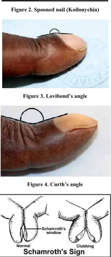
Koilonychia: (Greek: Koilos = hollow, Onyx = nail)
It is the spoon-shaped concavity of nails or presence of reverse curvature in the transverse and longitudinal axis which results in concave dorsal aspect of the nail (Figure-2).To determine whether a nail is spooned, perform the water drop test. Place a drop of water on the nail. If the drop does not slide off, then the nail shows sign of spooning. Koilonychia is commonly associated with iron deficiency anemia and haemochromatosis. The presence of koilonychia is an alarming sign for clinician to look for other signs of anemia and to do a full blood count and
serum iron studies (Dausber et al., 1998; Braunwald et al.,
2005).
Clubbing: It isbullous enlargement of the distal segments of the fingers due to proliferation of connective tissue, particularlyon the dorsal surface. The pathophysiology of clubbing says that when platelet precursors fail to become fragmented into platelets within the pulmonary circulation, they are easily trapped in the peripheral vasculature, releasing platelet derived growth factor and vascular endothelial growth factor, promoters of vascularity and this ultimately results in clubbing.To determine whether nails are clubbed, have the patient place both forefinger nails together and look between them. If you can see a small diamond space between them (Schamroth's window) then the nails are not clubbed
(Schamroth's sign) (Sheokand, 2014; Spicknall et al., 2005). 3
forms of geomatric analysis can be checked for clubbing-
1) Lovibond’s angle (found at the junction between the
nail plate and proximal nail fold). Normally less than 160º. In clubbing, this is increased to over 180º (Figure-3).
2) Curth’s angle (found at the distal interphalangeal joint).
It is normally approximately 180º. This is less than 1600
in clubbing. (Figure-4).
3) Schamroth’s window. It is a small diamond space seen
between when the patient place both the fingernails together. It is obliterated in clubbing and seen in normal individuals(Schamroth’s sign) (Figure-5).
Causes of clubbing include: Lung cancer, sarcoidosis, Beryllium poisoning, pulmonary arteriovenous fistula, subacute bacterial endocarditis, gastrointestinal causes: inflammatory bowel disease, sprue, neoplasms (esophagus, liver, bowel), Hyperthyroidism(Motswaledi and Mayayise, 2010; Sheokand, 2014; Spicknall et al., 2005): Pincer nail is a curved in growth of nail with bilateral penetration to the nail
folds and recently reported to be seen in end stage renal
disease secondary to diabetes (Motswaledi and Mayayise,
2010; Kirkland and Sheth, 2009). Macronychia (too large nails) is seen in gigantism and Micronychia (too small nails) is seen in plexiform neuromas (Motswaledi and Mayayise, 2010).
Abnormalities of Nail Colour
Leukonychia Striae: These are white splotches or whitish discoloration of nails. In total leukonychia (Figure-6) all the nails are porcelain white; this is seen in chronic liver disease. In subtotal leukonychia the proximal two-thirds of the nail is white and this is caused by delay in keratin maturation. Transverse white lines are seen in systemic problems such as chemotherapy or poisoning (Motswaledi and Mayayise, 2010;
Dausber et al., 1998).
Cyanosis: It is bluish discoloration of the nails which can be central and peripheral types. Peripheral cyanosis is seen in cases of cold exposure, shock, congestive cardiac failure and peripheral vascular disease. Central cyanosis is seen in congenital heart diseases such as Tetralogy of Fallot. Both the varieties are seen in cardiogenic shock with pulmonary edema (Motswaledi and Mayayise, 2010; Braunwald, 2005; Tally and O’Connors, 2008).
Jaundice: Jaundice or icterus is a yellowish discoloration of nails which results due to deposition of bilirubin. Nails are
only affected in severe cases (Motswaledi and Mayayise,
2010).
Nicotine-stained nails: Here yellow staining of the nails due to nicotine is seen in heavy cigarette smokers (Figure-7). These stains are usually but not always seen in patients with cigarette-smoking associated cardiovascular disease and other
diseases such as lung cancer(Motswaledi and Mayayise, 2010;
Sheokand, 2014).
Melanonychia: Here longitudinal or transverse brownish to black pigmentation of the nails are seen (Figure 8). It can occur usually in association with Lichen planus, drug therapy with minocycline and zidovudine (Motswaledi and Mayayise, 2010).
Splinter haemorrhages: Splinter haemorrhages are caused by haemorrhages of the distal capillary loop (Figure 9). These haemorrhages in nails and petechiae on the skin may occur in patients with Infective endocarditis. Other rare causes of splinter haemorrhages include vasculitis, as in rheumatoid arthritis, polyarteritis nodosa and anti-phospholipid syndrome. They can be due to nail trauma also, especially if seen distally (Motswaledi and Mayayise, 2010; Karchmer, 2005).
Terry’s half and half nails: Here the nails are white proximally (edema and anemia) and normal distally, usually seen in liver cirrhosis, congestive cardiac failure and
adult-onset diabetes mellitus (Motswaledi and Mayayise, 2010)
(Figure 10).
HIV-associated dyschromonychia: Dyschromonychia is a change in nail and nail bed color and is a term generally applied to patients with increased nail pigmentation. Many patients with dyschromonychia in association with advanced human immunodeficiency virus (HIV) infection are seen
(Motswaledi and Mayayise, 2010; Leppard, 1990; Levay et al.,
Figure-1: The Normal Nail
Figure 2. Spooned nail (Koilonychia)
Figure 3. Lovibond’s angle
Figure 4. Curth’s angle
[image:3.595.354.514.54.146.2]Figure 5. Schamroth’s window and sign
[image:3.595.354.515.179.282.2]Figure 6. Leukonychia (white discoloration of nails)
[image:3.595.74.252.376.787.2]Figure 7. Nicotine-stained nails
[image:3.595.357.512.440.604.2]Figure 8. Melanonyhia
Figure 9. Splinter haemorrhages
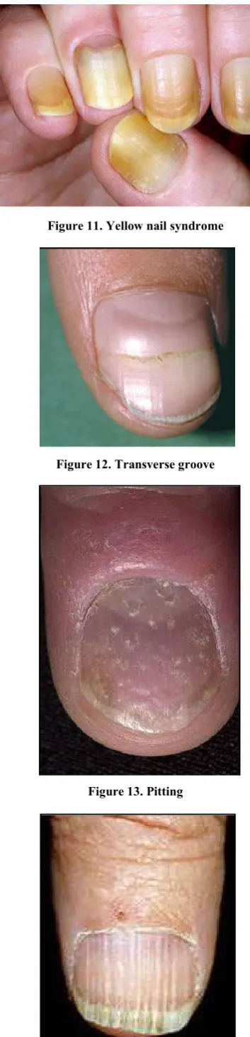
[image:3.595.359.509.629.787.2]Yellow Nail Syndrome: In yellow nail syndrome the nails are yellow due to thickening, sometimes with a tinge of green, possibly due to secondary infection (Figure 11). It may be associated with lymphoedema at one or more sites and
respiratory or nasal sinus disease(Motswaledi and Mayayise,
2010; Dausber et al., 1998).
Green nails: Bacteria are not capable of attacking a healthy nail plate. The Gram-negative bacterium Pseudomonas aeruginosa may colonize the dorsal or ventral nail plate under propitious conditions, such as chronic paronychia or onycholysis. The presence of Pseudomons is revealed by a characteristic green-black nail pigmentation due to pyocyanin
staining(Tosti et al., 2003).
Abnormalities of Nail Attachment
Onycholysis: It is defined as the distal or lateral separation of the nail plate from the nail bed with appearance of white or yellow areas of seperation,due to air beneath the nail and sequestered debris. Onycholysis is reported to be commonly seen in certain skin diseases and other conditions like hypo and
hyperthyroidism(Motswaledi and Mayayise, 2010; Sheokand,
2014).
Ptergium: It is seen in graft versus host disease and systemic sclerosis where a central fibrotic band divides a nail into two which obstructs the normal growth of nail (Motswaledi and Mayayise, 2010).
Abnormalities of Nail Surface
Longitudinal grooves: These are full or partial thicknees grooves which run along the longitudinal axis of all or part of the length of the nail. Longitudinal grooves may be seen in patients with the use of oral retinoids or may be familial also
(Motswaledi and Mayayise, 2010; Dausber et al., 1998).
Transverse grooves: These are full or partial thicknees grooves which run along the transverse axis of all or part of the length of the nail and reflects systemic disease such as
coronary thrombosis, mumps and pneumonia(Motswaledi and
Mayayise, 2010; Dausber et al., 1998) (Figure 12)
Nail pitting: Nail pitting are punctuated erosions on the nail surface which can be shallow or deep having continous or irregular outline (Figure-13). Nail pitting is common in skin diseases such as Psoriasis and Lichen planus (Motswaledi and Mayayise, 2010; Eisen, 1999).
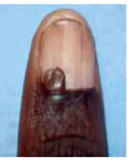
Nail Beading: With nail beading, the beads seem to drip down the nail like wax. It is associated with endocrine conditions, including Diabetes mellitus, Thyroid disorders, Addison's
disease and Vitamin B deficiency (Figure-14) (Sheokand,
2014).
Onychoschizia: It is defined as the transverse splitting of the nail plate into layers at or near the free edge in fingers and toes (Figure 15). And due to sequestration of debris between the layers, discoloration of nails is also seen. Onychoschizia has been reported in association with polycythemia (Motswaledi
and Mayayise, 2010; Dausber et al., 1998).
[image:4.595.337.514.51.783.2]Rough Nail Surface: Sandpapered and dull appearance of nails is seen in patients of Psoriasis, Chemical exposure and Lichen planus (Sheokand, 2014).
Figure 11. Yellow nail syndrome
[image:4.595.322.547.52.194.2]Figure 12. Transverse groove
Figure 13. Pitting
[image:4.595.364.507.381.787.2]Figure 15. Onychoshizia
[image:5.595.42.286.107.438.2]Figure 16. Pachyonychia congenita
Figure 17. Koenen tumour
Brittle nails: History of frequent immersion of hands in water is often associated with brittle nails. Other common causes are iron deficiency anaemia and impaired peripheral circulation (Motswaledi and Mayayise, 2010).
Genodermatoses With Nail Abnormalities
Localised areas of hyperkeratoses on the palm and soles with grossly thickened wedge shape nails (Figure-16) is seen in
autosomal dominant rare genodermatosis known as
Pachyonychia congenita.Oral lesions like leukokeratosis of the
tongue, and oral mucosa, hoarseness due to involvement of
larynx are seen in Type-I whereas Type-II shows neonatal teeth and cutaneous cysts and hair abnormalities but without invovement of oral cavity (Tosti, 2003).
Koenen tumors (Figure-17) are seen in other rare autosomal dominant disease-tuberous sclerosis complex, as periungual fibromas that occur on the nails of the fingers and toes. Other cutaneous manifestations include adenoma sebaceum, which is pathognomonic of Tuberos sclerosis complex, ash leaf macules and shagreen patches (Motswaledi and Mayayise, 2010;
Griffiths et al., 1998; McKee et al., 2005).
Conclusion
Examination of nails provide a helpful clinical road map of nail changes that may be associated with systemic disease and will be a helping hand to the clinician to identify and verify these clinical signs to act as an adjunct in proper and prompt diagnosis. Nail changes provide a window of opportunity as a profound knowledge of the associations may help in a complete and targeted work up of the patient.
“You can tell a lot from a person’s nail…..when a life starts to unravel, they are among the first to go..”
REFERENCES
Braunwald E. 2005. Hypoxia and cyanosis. In: Kasper DL, Fauci AS, Longo DL, et al, eds. Harrison’s principles of internal medicine. 16th edition. New York: McGraw-Hill; 209–12.
Dausber RPR, Baran R, De Berker D. 1998. Disorders of nails. In: Champion RH, Burton JL, Burns DA, Breathnach SM, eds. Roock/Wilkinson/Ebling textbook of dermatology. 6th
edition. Oxford: Blackwell Science, 2815–68.
Eisen, D.1999The evaluation of cutaneous, genital, scalp, nail, esophageal, and ocular involvement in patients with oral
lichen planus. Oral Surg Oral Med Oral Pathol Oral
Radiol Endod., 88:431-6.
Griffiths WAD, Judge MR, Leigh IM. 1998. Disorders of keratinization. In: Champion RH, Burton JL, Burns DA, Breathnach SM, eds. Rook/Wilkinson/Ebling textbook of dermatology. 6th edition. Oxford: Blackwell Science, 1483–588.
Karchmer AW. 2005. Infective endocartitis. In: Kasper DL, Fauci AS, Longo DL, et al, eds. Harrison’s principles of internal medicine. 16th edition. New York: McGraw-Hill; 731–40.
Kirkland CR, Sheth P. 2009. Acquired pincer nail deformity associated with end stage renal disease secondary to
diabetes. Dermatol Online J., 15(4):1–3.
Leppard B. 1990. Blue nails are a sign of HIV infection. Int J STD AIDS, 10:479–82.
Levay P.F., Botes M.E. and Malela E. 2005.
Dyschromonychia: clinical significance in a South African population. SA Fam Pract, 47(9):54–9.
McKee P.H., Calonje E. and Granter S.R. 2005. Diseases of collagen and elastic tissue. In: Pathology of the skin with clinical correlations. 3rd edition. Elsevier: Mosby, 1023– 59.
Motswaledi M.H. and Mayayise M.C. 2010. Nail changes in
systemic diseases. SA Fam Pract., 52(5):409-413.
[image:5.595.99.229.471.638.2]internal medicine. 16th edition. New York: McGraw-Hill, 238–43.
Sheokand P. 2014. Nail changes in systemic diseases. Journal
of Innovative Dentistry, Vol 4, Issue 2 & 3.
Spicknall K.E., Zirwas M.J. and English J.C. 2005. Clubbing:
an update on diagnosis, differential diagnosis,
pathophysiology and clinical relevance. J Am Acad
Dermatol., 56(6):1020–7.
Tally N.J. 2008. ‘O’Connors S. The cardiovascular system. In: Clinical examination: a systemic guide to physical diagnosis. 5th edition. Sydney: Churchill Livingstone, 26– 91.
Tosti A et al. 2003. Hair and nail abnormalities. In :Wolf R, Goldsmith L et al, Eds. Fitzpatrick’s Dermatology in
General Medicine.7th edition vol 1 and 2. New York:
McGraw Hill, 778-794.