ISSN Online: 2160-8717 ISSN Print: 2160-8709
DOI: 10.4236/ojst.2019.99021 Sep. 6, 2019 199 Open Journal of Stomatology
Implantosuported Rehabilitation after
Marginal Recession of Mandibular Calcifying
Epithelial Odontogenic Tumor: Case Report
Bruna Barcelos Ferreira, Victor Tieghi Neto, Andréa Guedes Barreto Gonçales,
Denise Tostes Oliveira, Fábio Sanches Magalhães Tunes, Eduardo Sanches Gonçales
*Department of Surgery, Stomatology, Pathology and Radiology of the Sao Paulo University, Bauru School of Dentistry, Bauru, Brazil
Abstract
The Calcifying Epithelial Odontogenic Tumor (CEOT) also called the Pind-borg’s Tumor represents a rare benign epithelial odontogenic neoplasm with locally aggressive behavior. Surgical excision with a safety margin is one of the treatment options, requiring bone and soft tissue reconstruction for im-plant-supported rehabilitation, providing the individual with a return to adequate oral functions. The objective of the present study is to report on the case of the treatment stages and the factors involved in the rehabilitation of a 23-year-old man with a history of mandibular lesion diagnosed as CEOT. A marginal resection of the mandible lesion was performed, with posterior re-construction with iliac crest bone graft and soft tissue graft to allow the im-plant-supported rehabilitation. Currently, in the postoperative control, the individual shows no signs of tumor recurrence and is rehabilitated without functional and/or aesthetic complaints. Surgical excision should be framed as a form of treatment, aiming for better resolution in the more complex cases of these invasive tumors.
Keywords
Oral Pathology, Bone Reconstruction, Dental Implants, Oral Rehabilitation
1. Introduction
The Calcifying Epithelial Odontogenic Tumor (CEOT) was first described by Pindborg in 1955. It is an uncommon Odontogenic lesion and occurs less than
1% of all odontogenic tumors [1][2]. It is locally aggressive and the surgical
ex-How to cite this paper: Ferreira, B.B., Neto, V.T., Gonçales, A.G.B., Oliveira, D.T., Tunes, F.S.M. and Gonçales, E.S. (2019) Implantosuported Rehabilitation after Marginal Recession of Mandibular Calcifying Epithelial Odontogenic Tumor: Case Report. Open Journal of Stomatology, 9, 199-207.
https://doi.org/10.4236/ojst.2019.99021
Received: July 26, 2019 Accepted: September 3, 2019 Published: September 6, 2019
Copyright © 2019 by author(s) and Scientific Research Publishing Inc. This work is licensed under the Creative Commons Attribution International License (CC BY 4.0).
DOI: 10.4236/ojst.2019.99021 200 Open Journal of Stomatology When the option of surgical excision is made, the main purpose of bone re-construction is restoring the anatomical features, masticatory function and aes-thetic at the end of the treatment. In addition, implant-supported rehabilitation should be planned, providing the patient suitable masticatory function and aes-thetics [6][7][8][9].
In order to perform osseous reconstructions after mandibular resection, dif-ferent approaches are described, either in relation to the kind of reconstruction or the best time to do so and also different treatment protocols for prosthetic
rehabil-itation, which in many cases involve reconstructive surgeries of soft tissue [7].
The objective is to report a case diagnosed as CEOT, demonstrating the stages of treatment from diagnosis to implant-supported rehabilitation and the factors involved in rehabilitation after marginal resection of the tumor.
2. Case Report
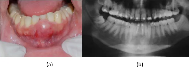
Male, 23 years old, had a history of mandibular lesion showing swelling with reddish spots in some areas and whitish in others, resilient consistency and smooth texture, asymptomatic, involving vestibular and lingual ridge and
ante-rior mandibular teeth, approximately 4.0 cm in diameter (Figure 1(a)). Teeth 31
[image:2.595.210.539.550.667.2]and 41 presented mobility and teeth 32, 33 and 42 without pulp vitality when evaluated. Radiographic examination revealed a radiolucent image with irregular borders extending from the dental elements 32 to 43, teeth 31 and 41 with chan-nel treated (Figure 1(b)).
Figure 1. (a) Initial clinical aspect of the lesion. Swelling with reddish spots in some areas
and whitish in others, resilient consistency and smooth texture with approximately 3.5 cm in diameter in the anterior region of mandible; (b) Initial panoramic radiograph. Multilocular radiolucent image of the irregular border region of mandibular symphysis.
DOI: 10.4236/ojst.2019.99021 201 Open Journal of Stomatology
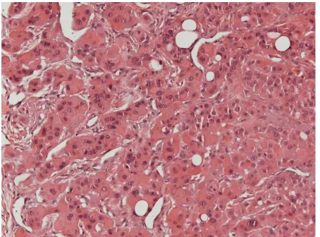
Figure 2. Histological section demonstrating the presence of
cords of polyhedral epithelial cells with eosinophilic cytop-lasm, intercellular bridges, few mitotic figures and discrete pleomorphism. The presence of homogeneous hyaline materi-al was observed with severmateri-al foci of dystrophic cmateri-alcification.
The biopsy of the lesion was performed and the microscopic sections showed polyhedral epithelial cell cords with eosinophilic cytoplasm, intercellular bridges, few mitotic figures and discrete pleomorphism. In the periphery of the lesion, there was presence of vacuolated cells with clear cytoplasm. From within the ep-ithelial cell strands, there was a presence of hyaline material, homogeneous and
with several foci of dystrophic calcification (Figure 2). In addition, neoplastic
epithelial cells were negative. Through the association of clinical, imaging and histopathological aspects, the final diagnosis was Calcifying Epithelial Odonto-genic Tumor (Tumor of Pindborg).
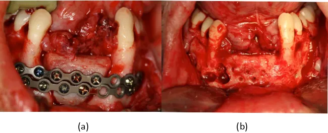
The clinical and radiographic characteristics of the lesion determined the marginal resection of the lesion, under general anesthesia, with local infiltration of 2% xylocaine and vasoconstrictor epinephrine at 1:200,000. An intra-oral in-cision was performed for exposure and resection of the lesion and preservation of the mandibular base allowing the maintenance of its shape. At the same sur-gical time, the extra-oral incision in the sub-mental region was chosen for adap-tation of the reconstruction plate. Through osteotomies, 1 cm away from the tumor margins, the surgical specimen was resected and the 2.4 reconstruction
plate was adapted (Figure 3(a) and Figure 3(b)).
After 1 year, local reconstruction with iliac bone graft was performed and
fixed with 2.0 plates and screws (Figure 4(a)). About 1 month after the
proce-dure, infection was observed in the grafted site, with exit of purulent secretion, requiring drainage and antibiotic therapy. After 6 months of reconstruction, bone graft fixation plates were removed and dental implant installation was
per-formed (Figure 4(b)).
DOI: 10.4236/ojst.2019.99021 202 Open Journal of Stomatology
Figure 3. (a) Extra-oral incision and extensive jaw exposure; (b) Resected part with safety
margin and margin of preserved mandible and reconstruction plate 2.4 adapted.
Figure 4. (a) Reconstruction of the previously resected area with iliac crest graft and
fixa-tion with plates and screws system 2.0; (b) Removal of fixafixa-tion plates after 6 months of reconstruction before installation of dental implants.
objective of increasing the amount of keratinized mucosa and improve the pe-ri-implant seal and consequently increase the success of the dental implants.
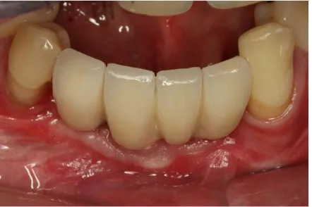
The bone graft and dental implants were good and without signs of tumor re-currence and a re-established masticatory function with definitive prostheses in
the 8-year follow-up from the tumor removal (Figure 5).
3. Discussion
CEOT occurs most commonly in the posterior mandible region and is associated
with retained teeth [3], however it can affect anterior region with or without
as-sociation with retained teeth [2] as observed in the case reported.
Although CEOT is benign and has slow growth [1] it is a tumor with high
growth potential and local tissue invasion which implies in increase of the
re-currence rates [7][10] especially related to more conservative treatments, such
as curettage [11][12].
The literature is controversial about to the best surgical management. Some authors reported that in lesions of great proportions and indefinite margins,
re-section is the best treatment [1][8][9][13]. In cases of small proportions and
defined margins, enucleation by curettage can be employed [14] [15]. In the
present case, we choose to perform marginal resection of the mandible with a safety margin, because it was an extensive lesion with indefinite limits that was
[image:4.595.212.537.244.376.2]DOI: 10.4236/ojst.2019.99021 203 Open Journal of Stomatology
Figure 5. Final restoration with definitive prostheses.
involving soft tissue, bone and also teeth. Therefore, knowing that the tumor has high potential for growth and local tissue invasion, proceeding enucleation by curettage of the lesion alone would not be a resolutive treatment and probably it will be favourable to tumor recurrence.
The predominant factors in this case, in order to opt for marginal resection, were the multilocular aspect of the lesion, the absence of clearly defined borders and the involvement of teeth within the tumor mass. Obviously when we re-sected a bone or even part of it, especially when teeth are involved in the resec-tion, it creates a new problem for the individual, which is the need to replace missing teeth. When there is intention to replace the lost teeth by means of sup-ported implants, it is also necessary to reconstruct the hard and soft tissues aim-ing for the installation of dental implants and their longevity.
The main objective of all reconstruction is the restoration of form and func-tion. The first involves preserving the patient’s labial competence and maintain-ing favourable speech articulation, and the second is to return the best possible masticatory function to the patient, allowing him to have a relatively normal diet [16].
Many options are available for mandibular reconstruction, such as recon-struction plates isolated or associated with bone grafts, vascularized or not, or osteogenic distraction devices incorporated into the fixation system using
re-maining healthy bone [17].
Bone grafts allow the treatment of a series of alveolar bone defects, either in
thickness or height defects [18]. An important determinant for bone graft
pre-dictability is the presence of autogenous bone as a component of the graft, since it has the property of bone formation from cells transplanted from the spongy bone, in addition, it contributes to the growth of the bone with various growth factors that are released during the incorporation of the graft, forming bone by
means of induction [19].
DOI: 10.4236/ojst.2019.99021 204 Open Journal of Stomatology the bone tissue [19].
In general, dental implants are good option for the rehabilitative treatment of
total and partial edentulous patients [16][17] and the success rate in this kind of
case is about 96% [21] although the 10-year longitudinal study of Keller [22]
evaluated 400 implants installed in the anterior region of the mandible after au-togenous iliac crest bone grafting in 91.7% of cases.
Regardless of the area being grafted or not, in order to achieve success and greater predictability of treatment, factors such as quality and quantity of bone tissue, location of the implant, adequate initial stabilization of the dental im-plants, absence of habits, good general hygiene condition and the presence of soft tissue around the implant are fundamental to the aforementioned objectives
(masticatory, phonetic and aesthetic function) were achieved [16][17].
The relationship between the condition of the periodontal tissue and the bone
tissue is directly related to the longevity of the dental implants [18] and the
ab-sence of keratinized mucosa on the reduced or thin border increases the chances
of failure of the implants [18]. During planning it is important to determine if
soft tissue augmentation therapy will be performed before, during or after
im-plant placement [23]. In general, the optimal time for using peri-implant plastic
techniques is preferably prior to or at the time of implant installation [23].
The sub-epithelial connective tissue graft is indicated to be performed when the absence of keratinized mucosa, loss of soft tissue thickness, exposure of the
prosthetic component and papilla alteration exist [23]. This graft, when well
performed and indicated, has the capacity to increase the metabolic activity at the receptor site, increasing and preserving the amount of keratinized mucosa, since it induces the keratinization of the epithelial cells that will proliferate on
the graft, obtaining, in this way, a better peri-implant marginal seal [17].
Among the many difficulties encountered in reconstruction cases, post-resection and post-graft soft tissue characteristics are prominent. In the case in question, soft tissue was lost during tumor resection, with the result that, after bone re-construction with an iliac crest graft, soft tissue was deficient. In addition, the correct adaptation and prognosis of soft tissues is quite unpredictable, and in many cases there is a need for the association of local grafts for the future
pros-thesis present better conditions [16][19], which was performed in this case. We
obtain-DOI: 10.4236/ojst.2019.99021 205 Open Journal of Stomatology ing better peri-implant marginal sealing, consequently increasing the chances of success of the implants installed.
Factors such as unsatisfactory soft tissue closure, contamination of grafts by endogenous bacteria, or failure of aseptic surgical technique may lead to the loss
of any graft performed [18]. When there is exposure of the fixation material, an
invasion of bacteria occurs to the grafted region, generating local inflammation
and decreased bone formation [24]. Therefore, the primary closure of the soft
tissue is determinant for the success of the graft, ensuring healing by first
inten-tion in addiinten-tion to reduce the patient’s postoperative discomfort [19].
In case of contamination, the treatment of this type of complication depends mainly on the presence of local exudate or not. When the infection is small, with only local erythema, one should proceed with physical debridement, through irri-gation with 0.12% chlorhexidine or physiological solution. In cases with purulent secretion, in addition to the local debridement, the use of antimicrobials should be
associated [18]. In the reported case, the iliac crest bone graft was successful
de-spite a postoperative infection, which required local drainage, irrigation with saline solution and rigorous patient follow-up plus antimicrobial prescription.
Following all the steps for prosthetic rehabilitation (bone reconstruction, soft tissue reconstruction, installation of dental implants) it is essential to periodical-ly follow these patients. Despite the possibility of relapse being reduced in cases where mandibular resection was performed, it is not ruled out. In the case ported, the patient presented a postoperative period of 8 years after the tumor re-moval and good prognosis. In addition, because it is an implant-supported reha-bilitation, there is a need for periodontal, occlusal and stability of the implants, al-ways seeking the motivation of the patient to perform adequate oral hygiene.
4. Conclusion
Knowing the aggressive behavior of CEOT and its potential for relapse, surgical excision should be considered. However, when opted for this treatment, plan-ning for bone reconstruction and restoration of the normal anatomy, function and aesthetics it is important to keep in mind the necessities for rehab the pa-tient and be able to treat possible complications that may arise during all proce-dures.
Consent
The patient had given his consent for this case report to be published. This Statement is in the cover of the chart at the Bauru School of Dentistry.
Conflicts of Interest
The authors declare no conflicts of interest regarding the publication of this paper.
References
DOI: 10.4236/ojst.2019.99021 206 Open Journal of Stomatology
32-35.
[4] Abrams, A.M. and Howell, F.V. (1967) Calcifying Epithelial Odontogenic Tumors: Report of Four Cases. The Journal of the American Dental Association, 74, 1231-1240.https://doi.org/10.14219/jada.archive.1967.0426
[5] Hicks, M.J., Flaitz, C.M., Wong, M.E., et al. (1994) Clear Cell Variant of Calcifying Epithelial Odontogenic Tumor: Case Report and Review of the Literature. Head & Neck, 16, 272-277.https://doi.org/10.1002/hed.2880160311
[6] Abrahão, A.C., Camisasca, D.R., Bonelli, B.R.M.V., Cabral, M.G., Lourenço, S.Q.C., Torres, S.R. and Pinto, J.R. (2009) Recurrent Bilateral Gingival Peripheral Calcifying Epithelial Odontogenic Tumor (Pindborg Tumor): A Case Report. Oral Surgery,
Oral Medicine, Oral Pathology, Oral Radiology, and Endodontology, 108, 66-71. https://doi.org/10.1016/j.tripleo.2009.04.037
[7] Rydin, K., Sjöström, M. and Warfvinge, G. (2016) Clear Cell Variant of Intraosseous Calcifying Epithelial Odontogenic Tumor: A Case Report and Review of the Litera-ture. Oral and Maxillofacial Pathology, 122, e125-e130.
https://doi.org/10.1016/j.oooo.2016.01.001
[8] Serrilho, R.P., Tolentino, L.S. and Pinto, G.N.S. (2015) Soft Tissue Grafting as an Option to Address Peri-Implant Defects. Clinical Case Report. Revista da Asso-ciação Paulista de Cirurgiões Dentistas, 69, 350-354.
[9] Afrogheh, A., Schneider, J., Mohamed, N. and Hille, J. (2014) Calcifying Epithelial Odontogenic Tumor with Clear Langerhans Cells: A Novel Variant, Report of a Case and Review of the Literature. Head and Neck Pathology, 8, 214-219.
https://doi.org/10.1007/s12105-013-0490-8
[10] Ai-Ru, L., Zhen, L. and Jian, S. (1982) Calcifying Epithelial Odontogenic Tumors: A Clinicopathologic Study of Nine Cases. Journal of Oral Pathology & Medicine, 11, 399-406.https://doi.org/10.1111/j.1600-0714.1982.tb00181.x
[11] Cheng, Y.S.L., Wright, J.M., Walstad, W.R. and Finn, M.D. (2002) Calcifying Epi-thelial Odontogenic Tumor Showing Microscopic Features of Potentially Malignant Behavior. Oral Surgery, Oral Medicine, Oral Pathology, Oral Radiology, and
Endo-dontology, 93, 287-295.https://doi.org/10.1067/moe.2002.121991
[12] Oliveira, M.G., Chaves, A.C.M., Visioli, F., Rojas, E.U., Moure, S.P., Romanini, J., Mariath, J.E.A., Rados, P.V. and Filho, M.S. (2009) Peripheral Clear Cell Variant of Calcifying Epithelial Odontogenic Tumor Affecting 2 Sites: Report of a Case. Oral Surgery, Oral Medicine, Oral Pathology, Oral Radiology, and Endodontology, 107, 407-411.https://doi.org/10.1016/j.tripleo.2008.11.004
DOI: 10.4236/ojst.2019.99021 207 Open Journal of Stomatology
E. (2013) Tratamiento conservador de tumor epitelial odontogénico calcificante asociado al canino inferior retenido: Relato de caso clínico. Revista Española de Ci-rugía Oral y Maxilofacial, 35, 123-127.
https://doi.org/10.1016/j.maxilo.2011.09.007
[15] Munteanu, C., Pirici, D., Stepan, A.E., Camen, A. and Margaritescu, C. (2016) Max-illary Calcifying Epithelial Odontogenic Tumor with Sinus and Buccal Vestibule Extension: A Case Report and Immunohistochemical Study. Diagnostic Pathology, 11, Article No. 134.https://doi.org/10.1186/s13000-016-0582-3
[16] Baker, A., Macmabon, J. and Parmar, S. (2001) Immediate Reconstruction of Con-tinuity Defects of the Mandible after Tumor Surgery. Journal of Oral and Maxillofa-cial Surgery, 59, 1333-1339.https://doi.org/10.1053/joms.2001.27825
[17] Goh, B.T., Lee, S., Tideman, H. and Stoelinga, P.J. (2008) Mandibular Reconstruc-tion in Adults: A Review. International Journal of Oral and Maxillofacial Surgery, 37, 597-605.https://doi.org/10.1016/j.ijom.2008.03.002
[18] Mazonetto, R. (2009) Reconstruções em implantodontia: Protocolos clínicos para o sucesso e previsibilidade. Napoleão, Nova Odessa, 268.
[19] Misch, C.E. (2005) Dental Implant Prosthetics. Mosby, St Louis.
[20] Osborna, T.M., Helalb, D. and Mehraa, P. (2018) Iliac Crest Bone Grafting for Mandibular Reconstruction: 10-Year Experience Outcomes. Journal of Oral Biology and Craniofacial Research, 8, 25-29.https://doi.org/10.1016/j.jobcr.2017.12.001
[21] Tolman, T.E. (1995) Reconstructive Procedures with Endosseous Implants in Grafted Bone: A Review of the Literature. The International Journal of Oral & Maxillofacial Implants, 10, 275.
[22] Keller, E.E. (1995) Reconstruction of the Severely Atrophic Edentulous Mandible with Endosseous Implants: A 10-Year Longitudinal Study. Journal of Oral and
Maxillofacial Surgery, 53, 305-320.https://doi.org/10.1016/0278-2391(95)90231-7
[23] Pinto, F.R., Menezes, K.M., Fernandes, A.N., Moranza, T.A., Ciotti, D.L. and Gurgel, B.C.V. (2014) Connective Tissue Graft in Patient with Dental Implants in the Anterior Region Case Report. Revista da Associação Paulista de Cirurgiões Den-tistas, 68, 106-111.