Survey: Lung Nodule Detection
Kaustubh Padamwar1, Gokul Sabu2, Krishna Piple3, Abhitabh Arjariya4
1, 2, 3, 4
Department of computer Engineering , sinhgad academy of engineering Pune , Maharashtra , India
Abstract: Early identification of lung cancer includes detection of uncertain nodules and classifying them into different condition of disease. The identification stage includes pattern matching and confirmation to increase accuracy, performed by fuzzy logic, support vector machine, statistical classifiers. The categorization stage involves matching characters (texture, shape and density) of the detected nodules to characters of normal cells (texture, shape and density) of nodules with known condition of disease (confirmed by sample extraction techniques). The nodule detection is mainly considered as it plays an important role in cancer detection nodules extracted are classified using neural network classifiers to differentiate between normal and abnormal lung cancer.
Keywords: Lung Nodule, Image Acquisition, preprocessing, segmentation, feature extraction
I. INTRODUCTION
The term cancer is a technical term refers to uncontrolled cell growth in tissues leading to malfunctioning of body organs at extreme state of influence cause major suffering and even death.
Cancer leads to excessive multiplication of abnormal cells without control and are able to affect other tissues. Cancer cells infect neighbouring part of human body through connective tissue. Most of the cancers are named for the organ where cancer starts – rapid growth of abnormal cancer cells in the colon is called colon cancer; that of skin is called basal carcinoma.
Cancer types are grouped into following categories:
1) Carcinoma – is a cancer in which cells of Skin is affected.
2) Sarcoma - is a cancer in which cells of cartilage, fat, muscle, bone, blood vessels, supportive or connective tissue is affected. 3) Leukemia is a cancer in which cells of blood generation tissue like bone marrow is affected.
4) Lymphoma and myeloma - is a cancer in which cells of the immunity of human is affected. Central nervous system cancers - is
a cancer in which tissues of the brain and spinal cord is affected.
A. What is Lung Nodule?
A nodule is a "spot on the lung," seen on an X-ray or computed tomography (CT) scan. In fact, a nodule shows up on about one in every 500 chest X-rays. Normal lung tissue surrounds this small round or oval solid overgrowth of tissue. It may be a single or solitary pulmonary nodule. Or, you may have multiple nodules.
Your lung nodule is more likely to be benign if:
1) If you are younger than age 40.
2) There is calcium in the nodule.
3) The nodule is small.
Figure: Lung CT Scan Image.
Figure 1: Anatomy of lung.
There are numerous systems to analyse the lung malignancy, for example, Chest Radiograph (x-beam), Computed Tomography (CT), Magnetic Resonance Imaging (MRI sweep) and Sputum Cytology. Be that as it may, the greater part of these procedures is costly and tedious. In this way, there is an extraordinary requirement for another innovation to analyse the lung malignancy in its initial stages. Picture handling procedures give a decent quality device to enhancing the manual investigation. The utilization of picture handling strategies can help radiologists and specialists in diagnosing illnesses and to offer a fast access to restorative data picked up significance in a brief timeframe. In this paper, MATLAB has been utilized through each techniques made.
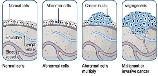
Figure2: The beginning of cancer.
II. BACKGROUND METHODOLOGY
A general description of lung cancer nodules segmentation and feature extraction system that contains five basic steps. The first step starts with taking a collection of CT images (normal and abnormal) from the available database. The second step applies image preprocessing, to get best level of quality and clearness. The third step is image
Segmentation which plays an effective role in image processing steps, and the fourth step contains feature extraction. The final step gives image diagnosis result of the indicators of normality or abnormality of lung images.
A. Image Acquisition
Figure: Block diagram of Lung Cancer Nodule Segmentation and Feature Extraction System.
B. Image Preprocessing
Picture preprocessing is one of the classes of picture taking care of, try to make definite more plainly obvious. Picture preprocessing is a way to deal with upgrade the way of picture, so that the vital picture is better than the first. The channel and center channel are displayed for preprocessing of selecting the CT pictures. The center channel is a non-straight instrument, while the ordinary channel is an immediate one. Mean isolating is a clear, instinctual and easy to realize of smoothing pictures, i.e. reducing the measure of force assortment between one pixel and the accompanying. The center channel is ordinarily used to diminish salt-and-pepper upheaval in a photo. It as often as possible makes a better appearing with respects than the mean channel of defending accommodating unpretentious component in the photo.
Figure: Outputs of image preprocessing.
C. Image Segmentation
Image segmentation is a fundamental step in Computerized image analysis and it deals with separating classes in an image into continuous and separate regions. Scan may contain chest wall, heart segments in addition to lung tissues. The goal of segmentation is to isolate the lung tissue in image processing, image segmentation is extremely rich area in the image analysis and computer vision. Yet, broadly speaking, image segmentation methods may be classified into three distinct approaches, a) statistical b) geometric c) variation. Statistical methods model the image information and cast the region process as mapping from raw images. Geometric methods exploit object shape descriptions in order to separate the image contents into classes. Variation methods (e.g., level sets) create an implicit description of class boundaries in terms of a curve/surface that evolves and cuts the particular object at its boundary (zero-level set). Under each category, there exists an enormous body of theoretical and algorithmic foundation.
D. Feature Extraction
After the segmentation is performed on lung region, the segmented nodules are used for feature extraction. Feature extraction is one of the most important steps in this system. A feature is a significant piece of information extracted from an image which provides more detailed understanding of the image. A feature is defined as a function of one or more measurements, the values of some quantifiable property of an object, computed so that it quantifies some significant characteristics of the object.
The features like geometric and intensity-based statistical features are extracted. This measurement information is very helpful in detecting lung nodule as cancer or not. Shape measurements are physical dimensional measures that characterize the appearance of an object. Only these features were considered to be extracted; area, perimeter and eccentricity.
III. LITERATURE REVIEW
“D. J. Brenner” et al proposed Computed tomography-an increasing source of radiation exposure "This paper is focus on an automatic segmentation approach of sub-pleural lung nodules from Computed Tomography (CT) scans based on morphological operations. Because the extraction of sub-pleural nodules is challenging and a computer-aided diagnosis system is, hence, indispensable. The proposed system is divided into three steps: pre-processing, initial detection of sub-pleural lung nodule and post-processing.
“J. G. Fletcher”, et al proposed Perspective on radiation risk in CT imaging" The aim of proposed system is to develop an efficient Computer Aided Diagnosis (CAD) for detection of lung nodules from parenchyma region of lung and classify the nodule into either cancerous (Malignant) or non-cancerous (Benign). The proposed system consists of following steps: i) the image taken is enhanced initially and then the region of interest is cropped, where the user can select the area to be cropped. ii) Morphological operation is performed to suppress the blood vessels and enhance the nodules. iii) Nodules are identified by labeling. iv) Features Extraction. v) Neural networks are implemented for features classification. The proposed work was able to detect the lung nodule that falls in close proximity to the lung wall. The system is able to achieve an overall accuracy of 92.2%.
“A. J. Einstein”, et al Estimating risk of cancer associated with radiate exposure from 64-slice computed tomography coronary angiography “The proposed system implements a computer-aided detection (CAD) system that detects small size nodules (larger 3 mm) in High Resolution CT (HRCT) images. It used a cylindrical filter for filtering nodule cases from other objects in images. They use a lung LIDC image database.
“D. L. Donoho” Compressed sensing "This paper proposes a method to detect lung nodule of lung CT images using 3D convolutional neural networks. It combines the traditional morphological preprocessing methods and 3D convolutional neural networks are applied to lung CT images.
“Yihuan Lu” et al investigation of Sub Centimeter Lung Nodule Quantification for Low-Dose PET " In this paper, we thoroughly investigated the combined impact of respiratory motion, size of nodule, reconstruction voxel size, nodule contrast, and noise level on lung nodule quantification. In addition, most of the studies cited above evaluated nodules larger than 10 mm in diameter, whereas we are focusing on sub-centimeter lung nodules. A Mini-Derenzo phantom study was also performed to investigate the impact of voxel size. In addition, interpolation methods were also compared when up sampling of the reconstruction to a finer voxel size was performed. Our goal was to evaluate the feasibility of imaging sub-centimeter nodules under different low-dose protocols, to determine the upper-bound performance for quantification of such nodules, and to provide a guideline for future patient studies. “Jue Jiang”, et al proposed Multiple Resolution Residually Connected Feature Streams For Automatic Lung Tumor Segmentation From CT Images " Our work extends the FRRN by residually combining features computed at multiple image resolutions, whereby, a dense feature representation is computed by simultaneously combining feature maps at multiple image resolutions and feature levels. Such a dense feature representation increases the network capacity and ultimately enables the network to recover the input image spatial resolution better than the existing methods.
IV. CONCLUSION
REFERENCES
[1] D. J. Brenner and E. J. Hall, “Computed tomography-an increasing source of radiation exposure”. N. Engl. J. Med. vol. 357, pp. 2277-2284, 2007.
[2] J. G. Fletcher, J. M. Koer, J. A. Coburn, D. H. Bruining, and C. H. Mccollough, “Perspective on radiation risk in CT imaging”. Abdominal Imaging, vol. 38, pp. 22-31,2013.
[3] A. J. Einstein, M. J. Henzlova, and S. Rajagopalan, “Estimating risk of cancer associated with radiation exposure from 64-slice computed tomography coronary angiography”. JAMA, vol. 298, pp. 317-323, 2007.
[4] D. L. Donoho, Compressed sensing. IEEE Transactions on Information Theory, vol. 52, pp. 1289-1306, 2006. [5] M. Dahiya “A Tool of Conversation: Chatbot” International Journal of Computer Sciences and Engineering (IJCSE) 2017.
[5] Yihuan Lu , Kathryn Fontaine, Mary Germino, Tim Mulnix, Michael E. Casey, Richard E. Carson, and Chi Liu “Investigation of Sub-Centimeter Lung Nodule Quantification for Low-Dose PET" IEEE 2018.
[6] Lung Cancer Database Consortium, [online available],www.eddie.via.cornell.edu.com
[7] A.Amutha and R.S.D Wahidabanu, “A Novel Method for Lung Tumor Diagnosis and Segmentation using Level Set- Active Contour Modelling”, European Journal of Scientific Research, Vol.90, No.2, November 2012, pp.175-187.
[8] Mokhled S. Al-Tarawneh, “Lung Cancer Detection Using Image Processing Techniques”, Leonardo Electronic Journal of Practices and Technologies, Issue 20, January-June 2012, p.147-158. ISSN 1583-1078
[9] N.A. Memon et. al, “Segmentation of Lungs from CT Scan Images for Early Diagnosis of Lung Cancer,” World Academy of Science, Engineering and Technology. 2006.
[10] Nunes É.D.O., Pérez M.G., "Medical Image Segmentation by Multilevel Thresholding Based on Histogram Difference", presented at 17th International Conference on Systems, Signals and Image Processing, 2010. [12] Huang Q., Gao W., Cai W., "Thresholding technique with adaptive window selection for uneven lighting image", Pattern Recognition Letters, Elsevier, p. 801-808.
[11] Sudha.V, Jayashree.P.," Lung Nodule Detection in CT Images Using Thresholding and Morphological Operations", International Journal of Emerging Science and Engineering (IJESE) ISSN: 2319–6378, Volume-1, Issue-2, December 2012.
[12] Lei Fan, Zhaoqiang Xia, Xiaobiao Zhang, Xiaoyi Feng. “Lung Nodule Detection Based on 3D Convolutional Neural Networks” IEEE 2017.