Original Article
Effect of simulated microgravity on human
chondrocyte-like cells
Chenrong Ke, Yimin Weng, Jianzhong Kong, Xiaolong Shui, Jianjun Hong, Hua Chen
Department of Orthopedics, The Second Affiliated Hospital and Yuying Children’s Hospital of Wenzhou Medical University, Wenzhou, Zhejiang, China
Received January 9, 2017; Accepted September 10, 2018; Epub July 15, 2019; Published July 30, 2019
Abstract: Background: Cartilage tissue engineering shows a promising prospect for human cartilage reconstruc-tion. However, the effect of microgravity rotating culture system on the proliferation of tissue-engineered human chondrocyte-like cells remains poorly understood. Materials and methods: Chondrocyte-like cells differentiated from human multipotent adult progenitor cells were seeded in the three-dimensional composite scaffold. They were cultured for three weeks under simulated microgravity environment by rotating cell culture system or under the static environment. Structure of complexes was determined by scanning electron microscopy, while HE-staining, immunohistological staining for type II collagen and MTT assay of both group samples were conducted after three-week culture. Results: Micrographs under scanning electron microscopy represented the structure of scaffold, in which chondrocyte-like cells appeared. HE-staining analysis exhibited more cells within the scaffold in the simulated microgravity group than that in the control group. The immunohistological staining result showed that the contents of collagen type II were higher in chondrocyte-like cells exposed to simulated microgravity. The chondrocyte-like cells proliferated faster in the simulated microgravity group than that in normal group as demonstrated by MTT assay. Conclusions: Simulated microgravity promotes the proliferation and matrix production of tissue-engineered human chondrocyte-like cells, suggesting the promising prospect of simulated microgravity by rotating cell culture system combined with tissue-engineered cartilage for human cartilage reconstruction.
Keywords: Rotating cell culture system, simulated microgravity, cartilage tissue engineering, articular cartilage defect, three-dimensional composite scaffold
Introduction
With the acceleration of social changes and aged tendency of population, articular cartilage injury frequently occurs resulting from exces-sive activity, trauma or degeneration of chon-drocytes. However, articular cartilage is com-posed of hyaline cartilage without blood ves- sel, nerves and lymphoidtissue, thus leading to limitation of self-repair potential. Conse- quently, repair or replacement is generally nec-essary for the treatment of damaged cartilage. Currently, treatments for damaged articular cartilage include drugs and physical therapy [1, 2], arthroscopic lavage and debridement [3, 4], cartilage graft [5]. Nevertheless, several disad-vantages are found in these treatments, such as deficient and inconsistent in long-term repair and especially incompletely resolve the prob-lems such as immune rejection after allogeneic transplantation.
Cartilage tissue engineering using a cell-scaf-fold approach has emerged as a new multidisci-plinary field, showing potential for the effective regeneration and repair of damaged articular cartilage. Cartilage tissue engineering aims at recovering the function of tissue by recombin-ing the chondrocyte and extracellularmatrix. Cartilage tissue engineering develops so rapid-ly and has been brought into clinical trial or applications in repairing all kinds of cartilage defects of large area, e.g. articular cartilage and thyroidcartilage [6]. Cellular selection, scaf-fold design and biological stimulation are the key points of tissue engineering, and also bring the challenges [7].
[8, 9]. It has been reported that MAPCs could be induced to be differentiated into various types of cells such as smooth muscle cells [10] and neurons [11]. Therefore, it may be a prom-ising candidate of seed cell for cartilage tissue engineering.
Three-dimensional osteochondral composite scaffold was generated by a TheriFormTM
three-dimensional printing process [12]. Cartilage region of the scaffold was 90% porous and composed of D, L-PLGA/L-PLA, with macro-scopic staggered channels to facilitate the seeding of chondrocytes into the center of the cartilage portion and allow the transport of nutrients to the cells as well as removal of cel-lular and polymer degradation by-products. The bone portion was made up of L-PLGA/TCP com-posite with the aim to maximize bone in growth and maintain critical mechanical properties. The transition region between these two sec-tions was designed to prevent delamination. This scaffold serves as a fully resorbable bipha-sic synthetic scaffold with good biocompatibili-ty, the structure of which resembles the normal articular cartilage. The rotating cell culture reactor (RCCS) can simulate the microgravity environment for the chondrocytes in vitro and be more effective than routine three-dimen-sional static culture. RCCS, developed by NASA, is often referred to a rotating wall vessel biore-actor. The vessel for cell culture rotates around the horizontal axis, permitting gas exchange through a permeable hydrophobic membrane. Besides, culture medium is mixed gradually through the rotation, leading to a uniform inter-nal environment, ensuring sufficient material transfer [13-15]. Many studies have also dem-onstrated that the microgravity environment is effective for the construction of tissue-engi-neered cartilage in vitro [16-19].
In this study, chondrocyte-like cells differenti-ated from human MAPCs were seeded in the three-dimensional composite scaffold and cul-tured in simulated microgravity environment by rotating cell culture system. The biological char-acteristics of chondrocyte-like cells were evalu-ated and the expression of a specific cartilage extracellular matrix protein (collagen type II) in the different culture groups was determined to investigate the effect of the microgravity rotat-ing culture system on the proliferation of tis-sue-engineered human chondrocyte-like cells.
Materials and methods
Differentiation and cultivation of
chondrocyte-like cells
Chondrocyte-like cells were differentiated from human multipotent adult progenitor cells (MAPCs) in vitro which were obtained from the bone marrow of clinical healthy volunteers [20]. This study was performed with the approval of the Second Hospital of Wenzhou Medical University Research Ethics Committee (L2014- 05). Informed consents of volunteers before experiments were collected, and all the rele-vant data in the experiment are anonymous with no violations of individuals’ health, safety and privacy. Cells were cultivated in a specific medium, and then subjected to magnetic acti-vated cell sorting, as our previous work de- scribed [21]. The third passage of cells in good condition was sorted out, and cultured in DMEM/F12 basal medium (Gibco) containing 10% fetal bovine serum (Gibco), 1 ng/ml of TGF-β1 and 5 ng/ml of FGF-2 (Peprotech) at 37°C with 5% CO2.
Seeding of three-dimensional scaffold
Three-dimensional D, L-PLGA/L-PLA composite scaffold (3 mm × 3 mm × 4 mm) were obtained from Laser Rapid Prototyping Center in the Tsinghua University. Before use, scaffolds were pre-wetted in 75% ethanol for 30 min for th- ree times, followed by repeated washings with PBS. Afterwards, scaffolds were packed by poly lysine solution to promote cellular attach-ment, disinfected by ultravioletlight and pre-treated in DMEM/F12 medium supplemented with fetal bovine serum (FBS), TGF-β1 and FGF-2 overnight.
Each scaffold was seeded for 6 h with above liquid mixture in incubator at 37°C to facilitate cellular adhesion to the scaffold.
Culture of chondrocyte-like cells
spe-cific medium for three weeks at 37°C, 5% CO2, saturated humidity.
The solutions were changed every other day for the first three days, then changed every day thereafter.
Observation under scanning electron micros-copy
Micrographs of chondrocyte-like cells which attached to the scaffold under scanning elec-tron microscopy (SEM) were taken following three weeks of culture. Samples were fixed in 2.5% glutaraldehyde and 2% paraformalde- hyde in PBS, dehydrated in a graded ethanol series and transferred to isopropyl acetate for substitution. Then, the samples were dried with a critical point dryer (Samdri-790; Tousimis Research Corp, Rockville, MD) and sputter-coated with gold using a Sputter Coater (Desk II, Denton Vacuum, Cherry Hill, NJ, USA). A JEOL 100S (Tokyo, Japan) electron microscope was utilized to image samples.
Histology
The samples were fixed in 4% neutral buffered formalin, dehydrated, and embedded in paraf-fin. The sections were cut at a thickness of 5 μm, stained with Hematoxylin-eosin solution, and observed under the light microscope. The expression of type II collagen was analyzed by means of immunohistological staining. The sections were dewaxed in xylene and hydrated in a graded series of alcohol. 0.3% hydrogen peroxide in PBS was used to block endogenous peroxidase activity. The sections were incubat-ed with a mouse polyclonal antibody (Gibco) overnight at 4°C after blocking with goat serum
(1:100 dilution). After being rinsed three times in PBS, the sections were incubated with sec-ondary anti-mouse IgG for 1 h at 37°C. The staining was developed in DAB solution, and the sections were counter-stained with hema-toxylin and viewed under the light microscope. MTT assay
An MTT-based assay was employed to estimate cellular activity and spatial distribution. In brief, samples were incubated in MTT solution (0.5 mg/ml in 2% fetal bovine serum culture medi-um) (Sigma) at 37°C with 5% CO2 for 2 h and washed in PBS. The insoluble precipitant was extracted in isopropanol for 24 h at room tem-perature, and the optical density (OD) was determined at a wavelength of 540 nm. Growth curve was drawn with time as abscissa and average optical density as ordinate. Chon- drocyte-like cells seeded in three-dimensional D, L-PLGA/L-PLA composite scaffold were cul-tured statically as a control.
Statistical analysis
Data were analyzed using SPSS software 19.0 (SPSS Inc., Chicago, IL, USA) and presented as means ± standard deviations (SD). Student
t-test was performed for comparison between the static and RCCS cultured samples. P < 0.05 was considered statistically significant.
Results
Chondrocyte-like cells under scanning electron microscopy
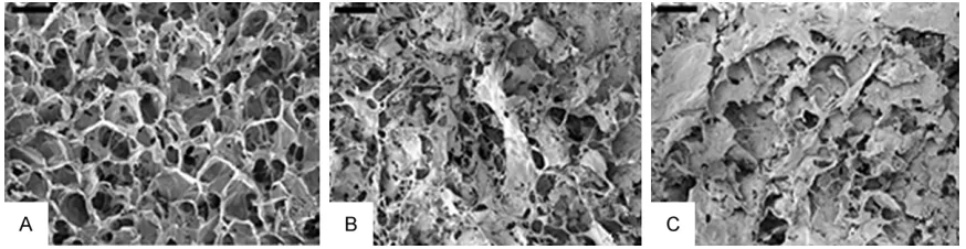
[image:3.612.88.525.71.184.2]macroscopic staggered channels. SEM analy-sis clearly showed the attachment of seeded chondrocyte-like cells to the scaffold after culture of three weeks (Figure 1B, 1C). Figure 1B represented part of porous channel of scaf-fold where the rest channel was taken up by cells. Besides, it seemed difficult to identify the structure of scaffold in Figure 1C becau- se of the attachment of seeded chondrocyte-like cells on the scaffold and the massive
depo-simulated microgravity promotes matrix secre-tion and deposisecre-tion of surrounding cells. Evaluation of cell vitality by MTT assay
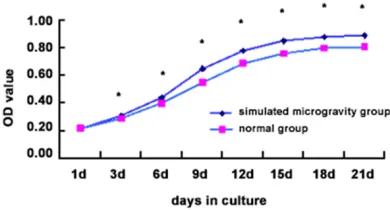
MTT method was used to estimate the vitality of the chondrocyte-like cells within microgravity after incubation of 1, 3, 6, 9, 12, 15, 18, 21 days. The result indicated that cellular prolifera-tion was remarkably enhanced under simulat-Figure 2. HE staining of chondrocyte-like cells/scaffold complexes after
[image:4.612.91.379.72.193.2]three-week culture. A: In simulated microgravity environment (× 200); B: In normal static environment (× 200).
Figure 3. Type II collagen immunohistochemical staining of chondrocyte-like cells/scaffold after three-week culture. A: In simulated microgravity environ-ment (× 200). B: In normal static environenviron-ment (× 200). Compared with Stim-ulated microgravity, **P < 0.01.
sition of extracellular matrix. Our data indicated that the proliferation of chondrocyte-like cells in the simulated microgravity culture was re- markably increased. In addi-tion, equal distribution of cells with more matrix deposition was observed, compared with that in the control group. HE staining and
immuno-histochemical staining of
collagen type II
[image:4.612.90.374.249.531.2]ed microgravity (Figure 4). There was signifi-cant difference (P < 0.05) between simulated microgravity groups and control groups except the first day.
Discussion
Based on previous findings that chondrocytes in articular cartilage are inclined to be dedif-ferentiated into fibroblastic cells with limited chondrogenic potential in two-dimensional cul-tivation and yet chondrocytes maintain their chondrogenic potential in a three-dimensional environment [22, 23], we used three-dimen-sional D, L-PLGA/L-PLA composite scaffold according to a previous study [12]. Our data demonstrated that chondrocyte-like cells pref-erentially seeded into composite scaffold and proliferated in good condition during three-week culture, reflecting favorable biocompati-bility between chondrocyte-like cells and com-posite scaffold. D, L-PLGA degraded into harmless DL-lactic and glycolic acids, while PLA gradually degraded and finally turned into CO2 and H2O. Nevertheless, the degradation of composite scaffold was accompanied with the accumulation of these degradation products, which may lead to the gradual decrease in the pH and further impede cellular growth. We did not observe any evident scaffold degradation characteristics during this study.
Rotating cell culture system (RCCS) is often used for simulation of the microgravity environ-ment. In addition, maintenance of a fluid orbit provides low-shear and low-turbulence environ-ment which reduces mechanical damage to the cells and allows more effective transport of material, thus facilitating cellular proliferation
[image:5.612.90.285.73.178.2]and function [15]. In contrast, cells in static three dimension system in vitro are unevenly distributed and the growth is relatively slow. Cells outside of the scaffold receive adequate nutrition with more matrix secretion and depo-sition, thus impeding air exchange and limiting the penetration of nutrients to the center of scaffold to some extent. Furthermore, cellular metabolic waste in the center cannot be promptly discharged, resulting in certain cyto-toxicity which hampers the cellular proliferation [24, 25].
It has been demonstrated that microgravity rotating culture system significantly enhanced the chondrogenic effect of TGF-b1 on rabbit bone marrow mesenchymal stem cells in vitro
as indicated by increased expression of mRNAs and proteins of collagen II and aggrecan [18]. Previous evidence showed that microgravity rotating bioreactor promoted the re-differentia-tion of rat chondrocytes which were seeded within PLGA sponge, resulting in formation of hyaline-like cartilage in vitro [26]. Yu et al. [27] also reported that the dedifferentiated human articular chondrocytes could regain the differ-entiated phenotype if cultured in the micrograv-ity environment using the rotational cell culture system (RCCS). Based on above findings, we believed that microgravity rotating culture sys-tem in our study may prevent the de-differenti-ation of chondrocyte-like cells and favor the maintenance of their phenotype. Our study elu-cidated that higher rate of chondrocyte-like cells proliferation was performed under micro-gravity circumstance with growing amounts of type II collagen in contrast to static environ-ment, which is in agreement with the previous study showing that simulation of microgravity with a rotating bioreactor enhanced prolifera-tion and metabolic activity of chondrocytes stem from bovine and human [28].
Regarding the culture conditions, transforming growth factor-β (TGF-β), as a multifunctional cytokine, plays an essential role in regulating cellular growth and differentiation. It was previ-ously reported that TGF-β1 could promote chondrocyte proliferation, differentiation as well as the synthesis and deposition of the extracellular matrix [29, 30]. In addition, chon-drocyte growth was stimulated in the presence of fibroblast growth factor 2 (FGF-2) [31]. However, the exact mechanism by how TGF-β1 Figure 4. The growth curve of human
and FGF-2 are involved in promoting the grow- th of human chondrocyte-like cells in vitro cul-ture remains unclear and requires further investigation.
The combination of simulated microgravity by RCCS and scaffold in our study created a three dimensional dynamic condition for human ch- ondrocyte-like cells in vitro which promoted proliferation and growth of cells, suggesting their promising prospect for human cartilage repair and reconstruction. Further long-term investigations are required to identify the pro- liferation of implanted tissue engineered chon-drocytes and degradation of corresponding scaffold in vivo after simulated microgravity culture.
Acknowledgements
This work was supported by Wenzhou Scien- ce and Technology Bureau Foundation (No. Y20140714).
Disclosure of conflict of interest
None.
Address correspondence to: Dr. Yimin Weng, De-
partment of Orthopedics, The Second Affiliated
Hospital and Yuying Children’s Hospital of Wenzhou Medical University, 109 W Xueyuan Road, Wenzhou 325000, Zhejiang, China. Tel: +86-577-88002823; Fax: +86-577-88002823; E-mail: yiminweng5@sina. com
References
[1] Diehl P, Gerdesmeyer L, Schauwecker J, Kreuz PC, Gollwitzer H and Tischer T. Conservative therapy of osteoarthritis. Orthopade 2013; 42: 125-139.
[2] McAlindon TE, Bannuru RR, Sullivan MC, Arden NK, Berenbaum F, Bierma-Zeinstra SM, Hawk-er GA, Henrotin Y, HuntHawk-er DJ, Kawaguchi H, Kwoh K, Lohmander S, Rannou F, Roos EM and Underwood M. OARSI guidelines for the non-surgical management of knee osteoarthri-tis. Osteoarthritis Cartilage 2014; 22: 363-388.
[3] Jackson RW and Dieterichs C. The results of arthroscopic lavage and debridement of osteo-arthritic knees based on the severity of degen-eration: a 4- to 6-year symptomatic follow-up. Arthroscopy 2003; 19: 13-20.
[4] Shannon FJ, Devitt AT, Poynton AR, Fitzpatrick
P and Walsh MG. Short-term benefit of ar
-throscopic washout in degenerative arthritis of the knee. Int Orthop 2001; 25: 242-245. [5] Hoffman JK, Geraghty S and Protzman NM.
Ar-ticular cartilage repair using marrow stimula-tion augmented with a viable chondral al-lograft: 9-month postoperative histological evaluation. Case Rep Orthop 2015; 2015: 617365.
[6] Fulco I, Largo RD, Miot S, Wixmerten A, Martin I, Schaefer DJ and Haug MD. Toward clinical application of tissue-engineered cartilage. Fa-cial Plast Surg 2013; 29: 99-105.
[7] Kuo CK, Li WJ, Mauck RL and Tuan RS. Carti-lage tissue engineering: its potential and uses. Curr Opin Rheumatol 2006; 18: 64-73. [8] Jacobs SA, Roobrouck VD, Verfaillie CM and
Van Gool SW. Immunological characteristics of human mesenchymal stem cells and multipo-tent adult progenitor cells. Immunol Cell Biol 2013; 91: 32-39.
[9] Schwartz RE, Reyes M, Koodie L, Jiang YH, Blackstad M, Lund T, Lenvik T, Johnson S, Hu WS and Verfaillie CM. Multipotent adult pro-genitor cells from bone marrow differentiate into functional hepatocyte-like cells. J Clin In-vest 2002; 109: 1291-1302.
[10] Ross JJ, Hong ZG, Willenbring B, Zeng LP, Isen-berg B, Lee EH, Reyes M, Keirstead SA, Weir EK, Tranquillo RT and Verfaillie CM. Cytokine-induced differentiation of multipotent adult progenitor cells into functional smooth muscle cells. J Clin Invest 2006; 116: 3139-3149. [11] Zhang N, Zhou H, Wang H, Yu L, Li Z, Xu H and
Wang M. Cerebral function of bone marrow multipotent adult progenitor cells after trans-plantation in parkinson’s disease rat models. Transplant Proc 2013; 45: 719-725.
[12] Sherwood JK, Riley SL, Palazzolo R, Brown SC,
Monkhouse DC, Coates M, Griffith LG, Lan -deen LK and Ratcliffe A. A three-dimensional osteochondral composite scaffold for articular cartilage repair. Biomaterials 2002; 23: 4739-4751.
[13] Liu TQ, Li XQ, Sun XY, Ma XH and Cui ZF. Analy-sis on forces and movement of cultivated par-ticles in a rotating wall vessel bioreactor. Bio-chemical Engineering Journal 2004; 18: 97-104.
[14] Hansmann J, Groeber F, Kahlig A, Kleinhans C and Walles H. Bioreactors in tissue engineer-ing-principles, applications and commercial constraints. Biotechnol J 2013; 8: 298-307. [15] Zwezdaryk KJ, Warner JA, Machado HL, Morris
CA and Bentrup KH. Rotating cell culture sys-tems for human cell culture: human tropho-blast cells as a model. J Vis Exp 2012. [16] Ishaug-Riley SL, Crane-Kruger GM, Yaszemski
biode-gradable polymers. Biomaterials 1998; 19: 1405-1412.
[17] Yu B, Yu DG, Cao L, Zhao X, Long T, Liu GW, Tang TT and Zhu ZN. Simulated microgravity using a rotary cell culture system promotes chondrogenesis of human adipose-derived mesenchymal stem cells via the p38 MAPK pathway. Biochem Biophys Res Commun 2011; 414: 412-418.
[18] Wu X, Li SH, Lou LM and Chen ZR. The effect of the microgravity rotating culture system on the chondrogenic differentiation of bone marrow mesenchymal stem cells. Mol Biotechnol 2013; 54: 331-336.
[19] Grimm D, Wehland M, Pietsch J, Aleshcheva G, Wise P, van Loon J, Ulbrich C, Magnusson NE, Infanger M and Bauer J. Growing tissues in real and simulated microgravity: new methods for tissue engineering. Tissue Eng Part B Rev 2014; 20: 555-566.
[20] Jiang YH, Vaessena B, Lenvik T, Blackstad M, Reyes M and Verfaillie CM. Multipotent progen-itor cells can be isolated from postnatal mu-rine bone marrow, muscle, and brain. Exp He-matol 2002; 30: 896-904.
[21] Yu LL, Weng YM, Shui XL, Fang WL, Zhang EG and Pan J. Multipotent adult progenitor cells from bone marrow differentiate into chondro-cyte-like cells. J Arthroplasty 2015; 30: 1273-1276.
[22] Martin I, Vunjak-Novakovic G, Yang J, Langer R and Freed LE. Mammalian chondrocytes
ex-panded in the presence of fibroblast growth
factor 2 maintain the ability to differentiate and regenerate three-dimensional cartilagi-nous tissue. Exp Cell Res 1999; 253: 681-688.
[23] Darling EM, Pritchett PE, Evans BA, Superfine
R, Zauscher S and Guilak F. Mechanical prop-erties and gene expression of chondrocytes on micropatterned substrates following dediffer-entiation in monolayer. Cell Mol Bioeng 2009; 2: 395-404.
[24] Granet C, Laroche N, Vico L, Alexandre C and Lafage-Proust MH. Rotating-wall vessels, promising bioreactors for osteoblastic cell cul-ture: comparison with other 3D conditions. Med Biol Eng Comput 1998; 36: 513-519. [25] Freed LE, Vunjak-Novakovic G and Langer R.
Cultivation of cell-polymer cartilage implants in bioreactors. J Cell Biochem 1993; 51: 257-264.
[26] Emin N, Koc A, Durkut S, Elcin AE and Elcin YM. Engineering of rat articular cartilage on porous sponges: effects of tgf-beta 1 and microgravity bioreactor culture. Artif Cells Blood Substit Im-mobil Biotechnol 2008; 36: 123-137.
[27] Yu F, Guo Q and Huang L. Redifferentiation of the dedifferentiated human articular chondro-cytes by the bioreactor culturing. Zhongguo Xiu Fu Chong Jian Wai Ke Za Zhi 2006; 20: 840-844.
[28] Akmal M, Anand A, Anand B, Wiseman M, Goodship AE and Bentley G. The culture of ar-ticular chondrocytes in hydrogel constructs within a bioreactor enhances cell proliferation and matrix synthesis. J Bone Joint Surg Br 2006; 88B: 544-553.
[29] Rosier RN, O’Keefe RJ, Crabb ID and Puzas JE. Transforming growth factor beta: an autocrine regulator of chondrocytes. Connect Tissue Res 1989; 20: 295-301.
[30] Redini F, Galera P, Mauviel A, Loyau G and Pu-jol JP. Transforming growth factor beta stimu-lates collagen and glycosaminoglycan biosyn-thesis in cultured rabbit articular chondrocytes. FEBS Lett 1988; 234: 172-176.
[31] Yan D, Chen D, Cool SM, van Wijnen AJ, Mikecz K, Murphy G and Im HJ. Fibroblast growth