Original Article
Geographical distribution affects survival of
patients with intracranial ependymoma
in the USA: a SEER based study
Xinxin Ji
1,3*, Xuedi Yu
1,3*, Ping Li
1,3, Li Ma
1, Xun Jin
2, Wenliang Li
11Department of Neurosurgery, 2Department of Biochemistry and Molecular Biology, Tianjin Medical University Cancer Hospital and Institute, Key Laboratory of Cancer Prevention and Therapy of Tianjin, National Clinical Research Center for Cancer, Tianjin 300060, China; 3Tianjin Medical University, Tianjin 300060, China.*Equal contributors.
Received January 1, 2019; Accepted April 19, 2019; Epub June 15, 2019; Published June 30, 2019
Abstract: Precision medicine, based on the relationship between personalized clinical characteristics and prog-nosis, leads to better patient therapeutic outcomes. Social factors directly affect patient prognosis in several can-cers. However, the relationship between social factors and prognosis of patients with intracranial ependymoma (IE) remains unclear. The aim of this retrospective study was to identify the association between social factors and
IE patient prognosis. Information was collected from patients histologically confirmed to have IE between 1973
and 2015, analyzing the relationship between patients and social factors using the Surveillance, Epidemiology, and End Results (SEER) database. Kaplan-Meier and Cox’s proportional hazards regression analyses were used to
evaluate patient survival. Present results revealed that geographical distribution was statistically significantly as -sociated with prognosis of IE patients, based on the SEER database. Patients from the Midwest region of the USA had a lower 5-year survival rate (64.8%) than those from the Northeast (74.8%), South (79.6%), and West (72.0%). Reducing bias, propensity score matching was further applied in analyzing relevant factors. Similarly, prognosis in the Midwest region (hazard ratio [HR] = 1) was poorer than that in the Northeast (HR = 0.62), South (HR = 0.55), and West (HR = 0.71). Therefore, geographical distribution may play an important role in the malignant progression
of IE and may be beneficial for the precise treatment of patients with IE.
Keywords: Intracranial ependymoma, geographical distribution, prognosis, SEER, 5-year survival rate
Introduction
Intracranial ependymomas (IEs) are rare
glio-mas found in the central nervous system [1].
IEs originate from cerebral ventricular walls
found inside the brain, the external ventricular
system, or in ependymal cells blocked in the
brain parenchyma during embryonic
develop-ment [2-4]. IEs account for 1.2%-7.8% of
intra-cranial tumors. They have a poorer overall
sur-vival, compared with spinal ependymomas [5,
6]. Incidence of IEs is greater in males than in
females [7]. IEs commonly occur in children,
accounting for 6%-10% of intracranial tumors
[6, 8]. The
2016
World Health Organization
(WHO) classification of ependymal tumors cat
-egorized sub-ependymomas and myxopapillary
ependymomas as Grade I. Ependymomas, in-
cluding papillary, clear cell, and tanycytic
vari-ants, are classified as Grade II. RELA
fusion-positive ependymomas are classified as Grade
II or III. Anaplastic ependymomas, the most
malignant histological type, are classified as
Grade III [9]. Epidemiological data shows that
5-year and 10-year overall survival rates are
83.4% and 79.1%, respectively [10].
Precision medicine is defined as treatment
according to distinguishing characteristics of
different patients with the same disease.
Characteristics may be unique but not limited
to one person. These include age, gender, race,
histological grade, tumor size, genomics,
micro-biome, and social factors, such as geography,
socioeconomic status, lifestyles, and
environ-mental exposure [11-13]. Prognostic factors
for IE include age, gender, histological grade, tu-
improve the accuracy of
sur-vival analysis. All patients with
IE were confirmed histological
-ly and tumor behavior was
identified as malignant.
Pri-mary tumors were confirmed
[image:2.612.90.371.73.213.2]according to international rules
and originated from the brain.
Cause of death was due to
brain and CNS incidents. Pa-
tients that were offered active
follow-ups and explicit survival
results were included. Patients
with benign or borderline tu-
mors, as well as those with
Figure 1. Inclusion and exclusion chart.extent of resection, and adjuvant radiotherapy
[14-17].
[
The present study reviewed clinical data from
the Surveillance, Epidemiology, and End Re-
sults (SEER) program of the National Cancer
Institute, comprising a large population of pa-
tients from the United States. The current study
investigated the effect of social factors on
over-all survival rates of patients with IE, identifying
geographical distribution as a potential
prog-nostic indicator of IE.
Materials and methods
Database and patient population
Retrospective analysis was conducted using
SEER Program (www.seer.cancer.gov) research
data: Incidence-SEER 18 Registries Resear-
ch Data, November 2017 Submission
(1973-2015), National Cancer Institute, Division of
Cancer Control and Population Sciences (DC-
CPS), Surveillance Research Program, Sur-
veillance Systems Branch, released in April
2018.
The SEER database follows-up patient survival
every year. It provides data, such as basic
infor-mation of patients, extent of the tumors, WHO
grades, metastasis at the time of diagnosis,
surgery conditions, and marital status. How-
ever, the database does not contain
informa-tion concerning patient physical condiinforma-tions,
clinical symptoms, postoperative recovery, or
other adjuvant treatments.
In the present study, patients diagnosed be-
tween 1973 and 2015 were selected, aiming to
more than three malignant tumors, were ex-
cluded.
Variable collection
Patients with IE were included from the SEER
database based on International
Classifica-tion of Disease in Oncology 3
rdedition (ICD-O-3)
with codes 9391, 9392, 9393, and 9394.
Prognostic factors were divided into three
cat-egories: (1) Personal basic factors: Age (>18
years or ≤ 18 years), gender, and race; (2)
Clinical factors: Primary laterality (one site or
paired site), histological grade, tumor size (>4
cm or ≤ 4 cm), tumor location (supratentorial,
infratentorial, ventricle, across the velarium,
and others), summary stage
(unknown/un-staged was excluded from analysis), surgical
resection (yes or no); and (3) Social factors:
Marital status (divorced, separated, and single
(never married or widowed) were combined to
form the single group), insurance status
(insured/no specifics were included in the
insured group), rural-urban continuum
condi-tions, and geographical distribution (Northeast
included Connecticut and New Jersey, Midwest
included Michigan and Iowa, South included
Georgia, Kentucky, and Louisiana, and West
included California, Utah, New Mexico, Wa-
shington, Hawaii, and Alaska).
Statistical analysis
regres-sion model was applied to univariate
and multivariate analyses. Hazard ratios
(HR) and 95% confidence intervals (CI)
were calculated. Propensity score ma-
tching (PSM) analysis was performed,
further adjusting potential baseline
con-founding factors. Two-sided
P
-values
<0.05 are considered statistically signifi
-cant. Statistical analysis was carried out
using SPSS version 22.0 (IBM Corp,
Armonk, NY) and GraphPad Prism
ver-sion 6 (GraphPad Software Inc.).
Results
Study characteristics
A total of 2,691 IE patients, diagnosed
between 1973 and 2015, were initially
eligible. Of these, 115 patients were
excluded due to unclear histology or
because they were inconsistent with
international rules. Another 25 patients
were lost to follow-up and 45 patients
had benign or borderline tumors. Two
patients had more than three malignant
tumors and 386 patients died of causes
not related to the brain or CNS. The
remaining 2,118 patients were included
in the current study (Figure 1). Patients
with incomplete IE information and
indi-vidual subgroup information were ex-
cluded. Therefore, the number of pa-
tients in each subgroup displayed
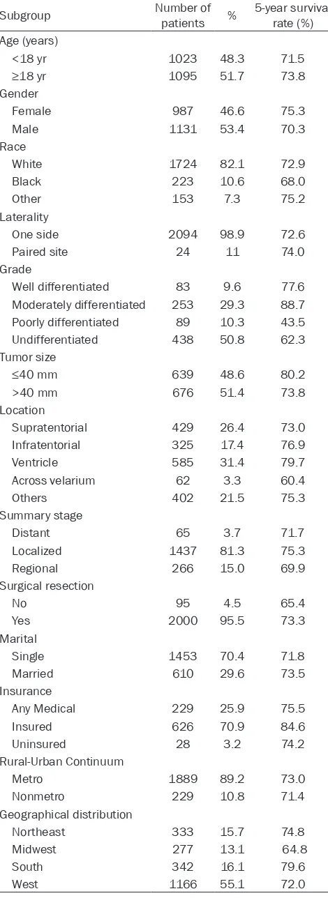
differ-ent statistics. Characteristics of patidiffer-ents
with IE are summarized in Table 1. Most
patients were white (82.1%), in which
the 5-year survival rate was 72.9%. Over
90% of patients received surgical
treat-ment. Five-year survival rates in the
Northeast, Midwest, South, and West
were 74.8%, 64.8%, 79.6%, and 72.0%,
respectively (Table 1).
Survival differences
A prognostic examination of personal,
clinical, and social factors was
conduct-ed, aiming to identify prognostic factors
in patients with IE. Consistent with
previ-ous reports, several personal and
clini-cal factors, including gender, race, tumor
size, tumor location, histological grade,
and surgical resection, were significantly
[image:3.612.93.328.86.730.2]correlated with IE patient survival [4, 14,
Table 1. Characteristics of patients with IE
Subgroup Number of patients % 5-year survival rate (%) Age (years)
<18 yr 1023 48.3 71.5
≥18 yr 1095 51.7 73.8
Gender
Female 987 46.6 75.3 Male 1131 53.4 70.3 Race
White 1724 82.1 72.9 Black 223 10.6 68.0 Other 153 7.3 75.2 Laterality
One side 2094 98.9 72.6 Paired site 24 11 74.0 Grade
Well differentiated 83 9.6 77.6 Moderately differentiated 253 29.3 88.7 Poorly differentiated 89 10.3 43.5 Undifferentiated 438 50.8 62.3 Tumor size
≤40 mm 639 48.6 80.2
>40 mm 676 51.4 73.8 Location
Supratentorial 429 26.4 73.0 Infratentorial 325 17.4 76.9 Ventricle 585 31.4 79.7 Across velarium 62 3.3 60.4 Others 402 21.5 75.3 Summary stage
Distant 65 3.7 71.7 Localized 1437 81.3 75.3 Regional 266 15.0 69.9 Surgical resection
No 95 4.5 65.4
Yes 2000 95.5 73.3 Marital
Single 1453 70.4 71.8 Married 610 29.6 73.5 Insurance
Any Medical 229 25.9 75.5 Insured 626 70.9 84.6 Uninsured 28 3.2 74.2 Rural-Urban Continuum
Metro 1889 89.2 73.0 Nonmetro 229 10.8 71.4 Geographical distribution
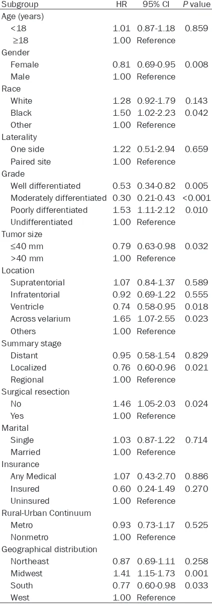
Table 2. Univariate Cox proportional hazard
regression analysis of risk factors among patients
with IE
Subgroup HR 95% CI P value Age (years)
<18 1.01 0.87-1.18 0.859
≥18 1.00 Reference
Gender
Female 0.81 0.69-0.95 0.008 Male 1.00 Reference Race
White 1.28 0.92-1.79 0.143 Black 1.50 1.02-2.23 0.042 Other 1.00 Reference Laterality
One side 1.22 0.51-2.94 0.659 Paired site 1.00 Reference Grade
Well differentiated 0.53 0.34-0.82 0.005 Moderately differentiated 0.30 0.21-0.43 <0.001 Poorly differentiated 1.53 1.11-2.12 0.010 Undifferentiated 1.00 Reference Tumor size
≤40 mm 0.79 0.63-0.98 0.032
>40 mm 1.00 Reference Location
Supratentorial 1.07 0.84-1.37 0.589 Infratentorial 0.92 0.69-1.22 0.555 Ventricle 0.74 0.58-0.95 0.018 Across velarium 1.65 1.07-2.55 0.023 Others 1.00 Reference Summary stage
Distant 0.95 0.58-1.54 0.829 Localized 0.76 0.60-0.96 0.021 Regional 1.00 Reference Surgical resection
No 1.46 1.05-2.03 0.024 Yes 1.00 Reference Marital
Single 1.03 0.87-1.22 0.714 Married 1.00 Reference Insurance
Any Medical 1.07 0.43-2.70 0.886 Insured 0.60 0.24-1.49 0.270 Uninsured 1.00 Reference Rural-Urban Continuum
Metro 0.93 0.73-1.17 0.525 Nonmetro 1.00 Reference Geographical distribution
Northeast 0.87 0.69-1.11 0.258 Midwest 1.41 1.15-1.73 0.001 South 0.77 0.60-0.98 0.033 West 1.00 Reference
15, 17, 18], according to univariate analysis.
Furthermore, two social factors were found to
play potential prognostic roles. Regional stage
patients showed poorer progress, compared
with patients with other stages. Patients from
the Midwest region showed a poorer prognosis,
compared with those from other regions (Table
2). The effects of geographical distribution on
IE patient survival were confirmed using
Kaplan-Meier survival analysis and log-rank
tests. As shown in Figure 2A, patients from the
Midwest region showed a significant risk for
poor survival, compared with those from the
Northeast (HR = 0.62, 95% CI = 0.47-0.82),
South (HR = 0.55, 95% CI = 0.41-0.72), and
West (HR = 0.71, 95% CI = 0.58-0.87).
Identifying independent risk factors for survival
of patients with IE, multivariate Cox regression
was conducted with significant factors (P<
0.05) from univariate analysis. Race,
histologi-cal grade, and geographihistologi-cal distribution were
found to be significant risk factors affecting
survival of patients (Table 3). Aiming to reduce
selection bias, PSM was applied for gender,
race, and surgical resection. According to
sur-vival analysis, patients from the Midwest region
had a shorter survival, compared with those
from the Northeast (HR = 0.62, P = 0.001),
South (HR = 0.55, P<0.0001), and West (HR =
0.71, P = 0.001). Similarly, after conducting
PSM for gender, race, and surgery status,
patients from the Midwest had worse survival
rates, compared with those from the Northeast
(HR = 0.50, P<0.0001), South (HR = 0.54, P<
0.0001), and West (HR = 0.52, P = 0.0001)
(Figure 2B-D). Although race and histological
grade have been reported to be independent
prognostic factors for IE [18, 19], the current
study identified geographical distribution as a
novel social independent prognostic factor for
IE.
Discussion
Most current cancer research has focused on
personal and clinical factors. The association
between social factors and prognosis of cancer
patients is often overlooked. The present study
analyzed personal, clinical, and social
charac-teristics of patients with IE selected from the
SEER database. The current study also
con-firmed current IE prognostic factors, including
suggest geographical distribution as a new
independent prognostic factor.
Gastric cancer is a typical regional high-risk
cancer in East Asia, including South Korea,
China, and Japan [20, 21]. However, incidence
of gastric cancer has sharply decreased in
Japanese immigrants in Hawaii, indicating that
different lifestyles and modifiable factors affect
the incidence frequency of gastric cancer [22].
Substantial evidence from cohort studies has
strongly suggested that salt intake and
Heli-
cobacter pylori
infections play synergistic roles
in the occurrence and development of gastric
cancer [21]. Environmental exposure has also
been associated with geological
characteris-tics of high mortality clusters, including
envi-ronmental exposure to naturally occurring
heavy metals [23, 24]. Moreover, lifestyle
[image:5.612.95.524.73.433.2]fac-tors, such as alcohol consumption, smoking,
and low fruit intake, have been reported as
other potential risk factors for gastric cancer
[22, 25].
Similarly, there is an obvious geographical
aggregation in the morbidity and mortality of
esophageal cancer [26]. Incidence and
mortal-ity rates of esophageal cancer are especially
high in Henan and Hebei Provinces of North
China [27]. The types and quality of the drinking
water, as well as dietary habits, are related to
the high morbidity and mortality rates of
esoph-ageal cancer in these areas. Use of tobacco
and the consumption of alcohol, hot food, hard
and rough food, and fast food have been
asso-ciated with an increased likelihood of
develop-ing esophageal cancer [28, 29]. Moreover,
N
-nitroso compounds, found in pickled
vegeta-Figure 2. Kaplan Meier curves of patients stratified by all different geographical regions (A) and survival probabilityTable 3. Multivariate Cox proportional hazard
regression analysis of risk factors among patients
with IE
Subgroup HR 95% CI P value Gender
Female 0.74 0.53-1.03 0.076 Male 1.00 Reference Race
White 1.89 0.98-3.66 0.058 Black 2.56 1.20-5.46 0.015 Other 1.00 Reference Grade
Well differentiated 0.51 0.24-1.05 0.067 Moderately differentiated 0.25 0.15-0.42 <0.001 Poorly differentiated 1.43 0.88-2.32 0.152 Undifferentiated 1.00 Reference Tumor size
≤40 mm 0.99 0.70-1.40 0.944
>40 mm 1.00 Reference Location
Supratentorial 1.32 0.59-2.99 0.502 Infratentorial 1.51 0.66-3.44 0.327 Ventricle 1.76 0.77-4.02 0.182 Across velarium 1.66 0.66-4.17 0.282 Others 1.00 Reference Summary stage
Distant 1.15 0.48-2.78 0.758 Localized 0.67 0.28-1.59 0.362 Regional 1.00 Reference Surgical resection
No 1.77 0.74-4.26 0.203 Yes 1.00 Reference Geographical distribution
Northeast 0.41 0.19-0.88 0.023 Midwest 1.88 0.51-1.54 0.657 South 0.82 0.53-1.27 0.373 West 1.00 Reference
bles and barbecued food, have also been
shown to play an important role in the induction
of esophageal cancer [30].
Nasopharyngeal carcinoma is a rare cancer.
However, it occurs frequently in South China. It
exhibits a unique morbidity and mortality in
Guangdong and Guangxi Provinces [31]. It is
believed to be related to geological
environ-ment properties and traditional lifestyles [32].
In these areas, patients with the disease are
frequently infected with the Epstein-Barr virus.
Moreover, the nickel-rich soil directly influences
morbidity and mortality rates of
nasopharyn-geal carcinoma [33, 34]. Commonly eaten
foods, such as salted fish, often contain a high
quantity of carcinogens (e.g., nitrosamines) th-
at increase incidence rates of nasopharyngeal
carcinoma in South China [35].
Differences in geographical distribution could
also be reflected in climate conditions. Many
studies have demonstrated the effects of
cli-mate on human health [36]. Specific clicli-mate
conditions, such as heat waves, sunshine, and
cold exposure, could increase mortality [36,
37]. The weakened physiology of cancer pa-
tients makes them more susceptible to
weath-er conditions, which may lead to death in pa-
tients with advanced cancers [38].
Precision medicine represents an important
focus of disease management. Individualized
treatment protocols have been developed,
based on specific factors, and the roles of
social factors may have been underestimated
[13]. However, the database used in the
pres-ent study lacked social information, including
lifestyles, dietary habits, family history,
microbi-ome, and environmental exposure. Therefore,
further data collection and research are re-
quired.
The current study had many limitations. First,
PSM analysis was not applied to all influential
factors. This was due to a low number of
patients with ependymomas. Therefore,
selec-tion bias could not be minimized. Second,
char-acteristic information was not available for
some IE patients in the SEER database. In
addi-tion, information may have been lacking for
other factors that cause diversity in survival,
such as family history, genetic profiles, and dis
-ease presentation. Third, there was no
informa-tion concerning radiotherapy. This may be a
significant prognostic factor in patients with IE.
Fourth, factors such as climate, lifestyle, and
environmental exposure may have confounded
present results.
In summary, present results suggest that race,
histological grade, and geographical
distribu-tion were significant prognostic factors for
pa-tients with IE. Papa-tients in Midwest USA show-
ed poorer survival, compared with those in the
Northeast, South, and West. This is the first
report showing the effects of geographical
Acknowledgements
We would like to acknowledge the Surveillance,
Epidemiology, and End Results (SEER) Program
for providing the dataset for analysis. This work
was supported by the General Program of the
National Natural Science Foundation of China
(No. 81572891), Youth Program of the National
Science Foundation of China (No. 81702481),
and Natural Science Foundation of Tianjin (No.
15JCQNJC44800).
Disclosure of conflict of interest
None.
Address correspondence to: Wenliang Li, Depart- ment of Neurosurgery, Tianjin Medical University Cancer Hospital and Institute, Key Laboratory of Cancer Prevention and Therapy of Tianjin, National Clinical Research Center for Cancer, Tianjin 300060, China. E-mail: liwenliang2338@163.com; Xun Jin, Department of Biochemistry and Molecular Biology, Tianjin Medical University Cancer Hospital and Institute, Key Laboratory of Cancer Prevention and Therapy of Tianjin, National Clinical Research Cen- ter for Cancer, Tianjin 300060, China. E-mail: jinx2345@163.com
References
[1] Chen L, Zou X, Wang Y, Mao Y and Zhou L. Cen-tral nervous system tumors: a single center pathology review of 34,140 cases over 60 years. BMC Clin Pathol 2013; 13: 14.
[2] Metellus P, Guyotat J, Chinot O, Durand A, Bar-rie M, Giorgi R, Jouvet A and Figarella-Branger D. Adult intracranial WHO grade II ependymo-mas: long-term outcome and prognostic factor analysis in a series of 114 patients. Neuro On-col 2010; 12: 976-984.
[3] Nowak A and Marchel A. Surgical treatment of intraventricular ependymomas and subepen-dymomas. Neurol Neurochir Pol 2012; 46: 333-343.
[4] Sayegh ET, Aranda D, Kim JM, Oh T, Parsa AT and Oh MC. Prognosis by tumor location in adults with intracranial ependymomas. J Clin Neurosci 2014; 21: 2096-2101.
[5] Dutzmann S, Schatlo B, Lobrinus A, Murek M, Wostrack M, Weiss C, Schaller K, Raabe A, Meyer B, Goldbrunner R, Franz K, Seifert V and Senft C. A multi-center retrospective analysis of treatment effects and quality of life in adult patients with cranial ependymomas. J Neu-rooncol 2013; 114: 319-327.
[6] Cage TA, Clark AJ, Aranda D, Gupta N, Sun PP, Parsa AT and Auguste KI. A systematic review of treatment outcomes in pediatric patients
with intracranial ependymomas. J Neurosurg Pediatr 2013; 11: 673-681.
[7] Ruda R, Reifenberger G, Frappaz D, Pfister SM, Laprie A, Santarius T, Roth P, Tonn JC, Soffietti
R, Weller M and Moyal EC. EANO guidelines for the diagnosis and treatment of ependymal tu-mors. Neuro Oncol 2018; 20: 445-456. [8] Lillard JC, Venable GT, Khan NR, Tatevossian
RG, Dalton J, Vaughn BN, Klimo P Jr. Pediatric supratentorial ependymoma: surgical, clinical, and molecular analysis. Neurosurgery 2018. [9] Louis DN, Perry A, Reifenberger G, von
Deim-ling A, Figarella-Branger D, Cavenee WK, Oh-gaki H, Wiestler OD, Kleihues P and Ellison DW. The 2016 world health organization
clas-sification of tumors of the central nervous sys -tem: a summary. Acta Neuropathol 2016; 131: 803-820.
[10] Ostrom QT, Gittleman H, Xu J, Kromer C, Wolin-sky Y, Kruchko C and Barnholtz-Sloan JS. CB-TRUS statistical report: primary brain and oth-er central noth-ervous system tumors diagnosed in the united states in 2009-2013. Neuro On-col 2016; 18: v1-v75.
[11] Metellus P, Barrie M, Figarella-Branger D, Chi-not O, Giorgi R, Gouvernet J, Jouvet A and Guy-otat J. Multicentric French study on adult in- tracranial ependymomas: prognostic factors analysis and therapeutic considerations from a cohort of 152 patients. Brain 2007; 130: 1338-1349.
[12] Reni M, Brandes AA, Vavassori V, Cavallo G, Casagrande F, Vastola F, Magli A, Franzin A, Basso U and Villa E. A multicenter study of the prognosis and treatment of adult brain epen-dymal tumors. Cancer 2004; 100: 1221-1229. [13] Sengupta R and Honey K. AACR cancer prog-ress report 2018: harnessing research
discov-eries for patient benefit. Clin Cancer Res 2018;
24: 4351.
[14] McGuire CS, Sainani KL and Fisher PG. Both location and age predict survival in ependy-moma: a SEER study. Pediatr Blood Cancer 2009; 52: 65-69.
[15] Massimino M, Buttarelli FR, Antonelli M, Gan-dola L, Modena P and Giangaspero F. Intracra-nial ependymoma: factors affecting outcome. Future Oncol 2009; 5: 207-216.
Deimling A, Lichter P, Taylor MD, Gilbertson R, Ellison DW, Aldape K, Korshunov A, Kool M and
Pfister SM. Molecular classification of ependy -mal tumors across all CNS compartments, his-topathological grades, and age groups. Cancer Cell 2015; 27: 728-743.
[17] Wani K, Armstrong TS, Vera-Bolanos E, Raghu-nathan A, Ellison D, Gilbertson R, Vaillant B, Goldman S, Packer RJ, Fouladi M, Pollack I,
Mikkelsen T, Prados M, Omuro A, Soffietti R,
Ledoux A, Wilson C, Long L, Gilbert MR, Aldape K; Collaborative Ependymoma Research Net-work. A prognostic gene expression signature in infratentorial ependymoma. Acta Neuro-pathol 2012; 123: 727-38.
[18] Bates JE, Choi G and Milano MT. Myxopapillary ependymoma: a SEER analysis of epidemiolo-gy and outcomes. J Neurooncol 2016; 129: 251-258.
[19] Ye J, Zhu J, Yan J, Chen P, Wan Z, Chen F, Zhang L, Qian J and Luo C. Analysis on therapeutic outcomes and prognostic factors of intracrani-al ependymoma: a report of 49 clinicintracrani-al cases in a single center. Neurol Sci 2015; 36: 2253-2261.
[20] Sugano K. Screening of gastric cancer in asia. Best Pract Res Clin Gastroenterol 2015; 29: 895-905.
[21] Shin JY, Kim J, Choi KS, Suh M, Park B and Jun JK. Relationship between salt preference and gastric cancer screening: an analysis of a na-tionwide survey in Korea. Cancer Res Treat 2016; 48: 1037-1044.
[22] Bray F, Ferlay J, Soerjomataram I, Siegel RL, Torre LA and Jemal A. Global cancer statistics 2018: GLOBOCAN estimates of incidence and mortality worldwide for 36 cancers in 185 countries. CA Cancer J Clin 2018; 68: 394-424.
[23] Montero-Oleas N, Nunez-Gonzalez S and Si-mancas-Racines D. The remarkable geograph-ical pattern of gastric cancer mortality in Ecua-dor. Cancer Epidemiol 2017; 51: 92-97. [24] Torres J, Correa P, Ferreccio C,
Hernandez-Su-arez G, Herrero R, Cavazza-Porro M, Domin-guez R and Morgan D. Gastric cancer inci-dence and mortality is associated with altitude
in the mountainous regions of pacific latin
America. Cancer Causes Control 2013; 24: 249-256.
[25] Choi YJ, Lee DH, Han KD, Kim HS, Yoon H, Shin CM, Park YS and Kim N. The relationship be-tween drinking alcohol and esophageal, gas-tric or colorectal cancer: a nationwide popula-tion-based cohort study of South Korea. PLoS One 2017; 12: e0185778.
[26] He YT, Hou J, Chen ZF, Qiao CY, Song GH, Meng FS, Jin HX and Chen C. Trends in incidence of esophageal and gastric cardia cancer in high-risk areas in China. Eur J Cancer Prev 2008; 17: 71-76.
[27] Li YY, Du LB, Hu XQ, Jaiswal S, Gu SY, Gu YX and Dong HJ. A suggested framework for con-ducting esophageal cancer screening in China. J Dig Dis 2018; 19: 722-729
[28] Rolland D, Raharijaona M, Barbarat A, Houl-gatte R and Thieblemont C. Inhibition of GST-pi nuclear transfer increases mantle cell lympho-ma sensitivity to cisplatin, cytarabine, gem-citabine, bortezomib and doxorubicin. Antican-cer Res 2010; 30: 3951-3957.
[29] Mayne ST, Risch HA, Dubrow R, Chow WH, Gammon MD, Vaughan TL, Farrow DC, Schoen-berg JB, Stanford JL, Ahsan H, West AB, Rot-terdam H, Blot WJ, Fraumeni JF Jr. Nutrient in-take and risk of subtypes of esophageal and gastric cancer. Cancer Epidemiol Biomarkers Prev 2001; 10: 1055-1062.
[30] Tian D, Mo SJ, Han LK, Cheng L, Huang H, Hao S, Guan YL, Jiang KY, Deng JY, Feng HH, Wen HY and Fu MY. Investigation of dietary factors and esophageal cancer knowledge: compari-son of rural residents in high- and low-inci-dence areas. Sci Rep 2018; 8: 4914.
[31] Wei KR, Zheng RS, Zhang SW, Liang ZH, Li ZM and Chen WQ. Nasopharyngeal carcinoma in-cidence and mortality in China, 2013. Chin J Cancer 2017; 36: 90.
[32] Wei KR, Zheng RS, Zhang SW, Liang ZH, Ou ZX and Chen WQ. Nasopharyngeal carcinoma in-cidence and mortality in China in 2010. Chin J Cancer 2014; 33: 381-387.
[33] Tham T. Human papillomavirus and world health organization type III nasopharyngeal carcinoma: multicenter study from an endemic area in southern China. Cancer 2019; 125: 161
[34] Lin K, Shen W, Shen Z, Wu Y and Lu S. Dietary exposure and urinary excretion of total N-nitro-so compounds, nitrosamino acids and volatile nitrosamine in inhabitants of high- and low-risk areas for esophageal cancer in southern Chi-na. Int J Cancer 2002; 102: 207-211.
[35] Xu ZX, Lin ZX, Fang JY, Wu KS, Du PL, Zeng Y, Tang WR, Xu XL and Lin K. Mortality character-istic and prediction of nasopharyngeal carci-noma in China from 1991 to 2013. Asian Pac J Cancer Prev 2015; 16: 6729-6734.
[36] Kim YS, Park DK, Hwang IC and Ahn HY. Daily weather conditions and anticipated death from cancer. Iran J Public Health 2018; 47: 591-596.
[37] Gorjanc ML, Flanders WD, VanDerslice J, Hersh J and Malilay J. Effects of temperature and snowfall on mortality in pennsylvania. Am J Epidemiol 1999; 149: 1152-1160.