4
THE MECHANICAL PROPERTIES OF THE CELL SURFACE
II. THE UNFERTILIZED SEA-URCHIN EGGBY J. M. MITCHISON AND M. M. SWANN
The Department of Zoology, University of Edinburgh; the Marine Station, Millport; and the Stazione Zoologica, Naples
(Received 25 January 1954)
INTRODUCTION
In the first paper of this series (Mitchison & Swann, 1954), we have discussed the problems of measuring the mechanical properties of the cell membrane and described both the construction and use of the 'cell elastimeter', and the method of analysing the results by means of model experiments. The present paper is con-cerned with measurements made on the unfertilized eggs of five species of sea urchin, with the object of determining Young's modulus for the membrane and the internal pressure of the cell. Future papers will describe the changes in the membrane of the fertilized egg up to the time of cleavage, and the influence of chemical agents and electric currents on the membrane of the unfertilized egg.
MEASUREMENTS ON NORMAL UNFERTILIZED EGGS
Measurements were made with the cell elastimeter on the naked unfertilized eggs of Psammechinus miliaris at Millport, and of the four species of sea urchin available at Naples (P. microtuberculatus, Paracentrotus lividus, Sphaerechinus granularis and
Arbacia tixula). The apparatus and method are described in the first paper. The
results are shown in Table 1. Except for Psammechinus microtuberculatus, the measurements were made on fifty eggs of each species, in batches of ten taken from five different females. The points for each batch of ten eggs were plotted on a pressure-deformation graph and the best straight line was then drawn through them. We have called the slope of this line (expressed in dynes/cm.2/ /x deformation) the ' uncorrected stiffness' of the cell membrane. The reasons for this are discussed in the first paper, but we should point out here that this stiffness figure is pro-portional to the Young's modulus of the membrane. A different procedure was adopted for the first three batches (17, 17 and 16 eggs) of P. microtuberculatus, in order to show the variation within a batch. A separate line was drawn for each egg, and the average and standard deviation of the slopes of these lines was calculated
for each batch.
462 J. M . MlTCHISON AND M . M . SWANN
It is also possible to calculate by means of model experiments absolute value? for Young's modulus from the corrected stiffness figures. These figures are given in col. 5. The methods and assumptions involved in this calculation are described in the first paper, but two points should be emphasized: the absolute values are only approximate; and they assume no internal pressure in the egg (i.e. no resting tension
Table 1. Stiffness of the membrane of unfertilized eggs, with 50 (i diameter pipette
( 1 ) Species (av. egg diam.)
Ptammechimtt miliaru (105 n)
Psammechinus microtubercuUitui (ilOfi) Paracentrotus lividus (90 v) Sphaerechinui gramdaris (100^)
Arbacia lixula (80 y.)
( 2 ) N o . of eggs 1 0 1 0 1 0 1 0 1 0 50 1 7 17 1 6 1 0 1 0 7 0 1 0 1 0 1 0 1 0 1 0 5° 1 0 1 0 1 0 1 0 1 0 50 1 0 1 0 1 0 1 0 1 0 5° (3) Av. uncorrected stiffness (dynes/cm.'/fi deformation)
7 - 0 8 9 7-4 10-5 IO-2 8-8 87 (o=i-6) 9-3 (0=1-7) io-6 (0=1-2) 12-6
1 0 7
10-4
IO-I 9 - 1 6-6 7 9 1 2 8
9'3
8-9
10-9
13-7 14-5 1 3 5 1 2 3
13-7 13-7 n - 9 I3'3 1 0 9
1 2 7
(4) Corrected
stiffness (dynes/cm.'/M
deformation,
for 100/1 diam.
egg and
50 ji diam.
pipette)
8 2
9 0
I I - I
12-3 18-7 (5) Maximum Young's modulus (dynes/cm.*)
0-91 x 10*
i - o x 10*
1-23 x 10*
1-37x10*
2-o8x 10*
MEASUREMENTS ON SWOLLEN AND SHRUNKEN EGGS
Having derived corrected stiffness values, the next step is to separate the opposing effects of Young's modulus and internal pressure. It seems impossible to devise a satisfactory method for measuring directly the very small pressures which might exist in the egg, and it is therefore necessary to fall back on indirect methods. One such method relies on the fact that if the internal pressure within a hollow elastic-walled ball is increased, the stiffness, measured by an apparatus such as the elastimeter, will also increase (as can be seen from the model experiments in the first paper). If, on the other hand, some of the contents are removed from within a ball in which there is an internal pressure, the stiffness will decrease, whereas if there is no internal pressure the ball will deform or wrinkle but there will be little or no change in the stiffness. This effect will distinguish between the presence or absence of internal pressure, and it can be simulated with sea-urchin eggs by placing them in hypo- and hypertonic solutions. Accordingly, a series of experiments were done with unfertilized eggs (P. microtuberculatus) in the following media:
Approx. molarity 'Full hypertonic' 50 ml. sea water+ 20 ml. 2M-NaCl 0-96 'Half hypertonic' 50 ml. sea water + 10 ml. 2M-NaCl 0-78 Normal sea water 054 ' Half hypotonic' 50 ml. sea water+ 10 ml. distilled water 0-45 'Full hypotonic' 50 ml. sea water+ 20 ml. distilled water 0-39
The results for four batches of eggs are shown in Fig. 1, each point being the average corrected stiffness of five eggs. It can be seen that there is little or no fall in stiffness with hypertonic solutions, whereas in hypotonic solutions there is a sharp rise (except for one batch in 'half hypotonic'). This is a strong indication that there is no internal pressure, but it is difficult to set any limits of accuracy to the measure-ments for reasons that will be discussed on p. 466. It may be noted that in this experiment the stiffness values for the eggs in normal sea water are lower than those given in Table 1. For some unknown reason, these low values were given by most of the eggs during the period (spring 1952 at Naples) when this experiment was done. The eggs were normal in all other respects.
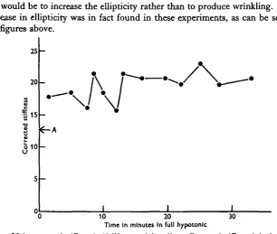
The high stiffness value in hypotonic solutions was maintained for long periods. The behaviour over half an hour is shown in Fig. 2, where the points are the corrected stiffness of individual eggs from one batch. The average diameter of the eggs also remained constant. There can therefore be little or no plastic flow in the membrane when under tension.
4
6
4 J. M. MlTCHISON AND M. M. SWANNAv. egg in/x
Media diam.
- » 121
- > Full hypo. 117
Half hypo. 112 Normal
KM
Half hyper.
101
Full hyper
8 r
V
M / I I
0-2 0-3 0-4 0-5 0 6 0-7 Molarity of medium
0-8 0-9 1-0
Fig. i. Stiffness of eggs in media of different tonicity. Corrected stiffness is in dynes/cm.*/f* deformation for a iooji diameter egg and a 50 ft diameter pipette.
and subsequently measured. Figures are given below for the average of the mean diameters (mean of major and minor axes) of fifty eggs, for the standard deviation of the mean diameters, and for the average ellipticity (major axis/minor axis).
Normal sea water ' Full hypertonic'
Average diameter
(/*) 114 1 0 0
6 0
6-8
Average ellipticity
1-054 1-078
egg would be to increase the ellipticity rather than to produce wrinkling. Such an increase in ellipticity was in fact found in these experiments, as can be seen from the figures above.
20
I 15
e
0 1 0
I I
[image:5.451.62.369.83.342.2]0 10 20 30 Time In minutes In full h/potonlc
Fig. a. Maintenance of stiffness in ' full hypotonic' medium. Corrected stiffness is in dynes/cm.1//1
deformation for a ioo/i diameter egg and a 50 ^ diameter pipette. A is the corrected stiffness value for the same batch of eggs in normal sea water.
DISCUSSION
In the first paper of this series, it was argued that the sea-urchin egg, having a membrane thickness of about 1-5 /x, resists deformation by virtue of its inherent rigidity, rather than by having an internal pressure. It is behaving therefore like a tennis ball, which maintains its shape even when punctured, rather than a rubber balloon or a fluid drop. We must now consider these arguments again, and see how far they are substantiated by the present experiments, and by earlier work.
466 J. M. M I T C H I S O N AND M. M. SWANN
that the cytoplasm of unfertilized sea-urchin eggs has a viscosity of about 3 centi-poises, which is only 3 times that of water. It is unlikely that a fluid of such a low viscosity would show any elastic effects comparable with those of the membrane, so that the measurements of stiffness made with the elastimeter must reflect largely, if not entirely, the mechanical properties of the surface. Within the unfertilized egg there is, of course, the nucleus, and within the fertilized egg the nucleus and at certain stages, the asters. Both these structures almost certainly have a greater rigidity than normal cytoplasm, but the deformations produced by the elastimeter are so slight that these bodies can scarcely affect the issue. It was in fact with the object of overcoming these objections to other methods of measuring the mechanical properties of the cell surface, that the elastimeter was made in the first place.
Turning to the question of the thickness of the cell membrane, it should be said in advance that we are using the term in a wide sense to include both the permeability barrier at the surface, and the cortex. Presumably, however, most of the mechanical properties are due to the cortex or 'structural membrane' (Mitchison, 1952). After the measurements of Cole (1932), and the many microdissection experiments of Chambers, there can be little doubt that the membrane is elastic and that its degree of extensibility is so great that it must have rubber-like properties. Apart from one observation by Chambers (1938) on cleaving eggs, there is general agreement that the thickness of the membrane is about 1-2 p. For the modulus figures in this paper we have used a model with a wall thickness equivalent to i-6/x in the egg. This is near enough the approximate value of 1-5^1 found by Mitchison (1955) from a number of observations on centrifuged eggs. In any case these modulus figures are not very sensitive to small changes in the membrane thickness.
The straight-line pressure-deformation curve given by the egg is further evidence of a thick membrane. It was shown in the first paper that rubber balls with an appreciable wall thickness also give a straight line, whereas solid rubber balls give a concave curve and very thin-walled balls (e.g. rubber balloons) give a convex one. Calculations show that pure surface tension would also give a convex curve.
The most difficult problem to be considered is whether or not the normal egg has a tension at the surface, and therefore an internal pressure. This question cannot be answered from elastimeter experiments alone, since the model experi-ments of the first paper have shown that the same stiffness can be given either by a high Young's modulus in the membrane and no internal pressure, or by a low modulus and high pressure. Nor is it possible to measure the pressure directly since the difficulties of measuring absolute pressures of the order of io~6 atmosphere in a microscopic object appear at the moment to be insuperable. We must therefore turn to the indirect methods which have been described earlier in this paper.
decrease in stiffness caused by the smaller thickness compensates for the in-creased stiffness that would be caused by the lower hydration of the membrane material.
The experiments on the wrinkling point have the advantage that they can be used to set an upper limit on the possible internal pressure. If it is assumed that the membrane of the shrunken egg in 'full hypertonic' is unstretched and that there is no internal pressure in the egg, then the maximum possible stretch of the membrane in the normal egg is 14% (7/x radial displacement). Using the data in text-fig. 16 of the first paper, this gives for the normal egg a maximum internal pressure of 95 dynes/cm.8, with a reduction of Young's modulus from 1 x io4 dynes/cm.2 to 0-54 x io4 dynes/cm.2. We must emphasize that because the radial displacement is a maximum figure (p. 463), therefore this value of the internal pressure is only an upper limit. It is not an average value for the internal pressure of the unfertilized egg, nor does it imply that there is necessarily any internal pressure at all.
A further piece of evidence which indicates the absence of internal pressure is the oval shape of normal unfertilized eggs. It seems unlikely that they would maintain this shape with an internal pressure when it is remembered that they become spherical as soon as a definite internal pressure is produced by hypotonic media. Summing up the observations on the internal pressure of the unfertilized egg, we believe that the balance of the evidence suggests the absence of such a pressure; but, if it exists, it cannot exceed about 95 dynes/cm.2.
There appear in any case to be no good general grounds for believing in the existence of a resting tension in the normal cell. The idea of a tension has been popular in the past because most previous theories of the cell surface have been in terms of a surface tension whose value would be independent of the extension. Surface tension in the strict sense, however, is a phenomenon which only occurs at a liquid/liquid or liquid/gas interface, and there is little reason to suppose that the surface of a cell behaves in this way. It is clear, both from our measurements and from those of earlier workers, that most of the cell surface behaves like an elastic solid, whose tension varies from the extension and which can exist without any resting tension. The only possible place for a true liquid surface tension would be the outer boundary layer between the cell surface and the outside medium. The exact structure of this layer is unknown, but the most plausible suggestions are that it is composed either of the proteins of the vitelline membrane or the antigenic layer, or of close packed lipid molecules lying radially. Neither of these would be expected to behave as a liquid and show a true surface tension, and there is no particular reason why they should show any resting tension.
Turning to other work on the mechanical properties of the cell membrane, the most important paper to be considered is that by Cole (1932). This is an admirable piece of work, which, by a most ingenious method, gave definite proof that the behaviour of the cell surface must be governed by an elastic membrane rather than by an interfacial surface tension. Our only major criticism concerns Cole's deriva-tion of an internal pressure of 40 dynes/cm.2 in the normal unfertilized sea-urchin egg. His method is briefly as follows. Eggs were compressed between a movable
468 J. M . MlTCHISON AND M . M . SWANN
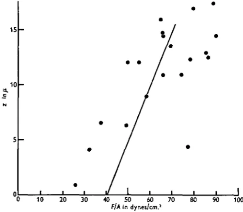
flat gold leaf and a fixed parallel surface. The force (F) exerted on egg was found from the deflexion of the gold leaf, and the compression of the egg (z) was observed down a microscope. The area of contact (A) between the egg and the surfaces was calculated from drawings and photographs, and then plotted against z. F\A was found from the curves of z/F and z/A, and was equated with the internal pressure of the egg (P). Finally P was plotted against z, and, by extrapolating the curve, P was found to be 40 dynes/cm.2 when z = o (no compression). This method seems to us to be inherently unsatisfactory. Since the plot of P (= F/A) against z is not a straight line, and does not follow any known theoretical formula, the extrapolation
15
10
J
10 20 30 40 50 60 70
f/A In dynes/cm.2
[image:8.451.101.352.211.429.2]80 90 100
Fig. 3. Replotting of curve and points from Cole (1932).
than Cole's curve. If the three lowest points (where the largest error is likely) were ignored, it might well be taken to pass through the origin, and to indicate no internal pressure.
Apart from these particular criticisms of Cole's figures, there is a serious objection to the use of this method of finding resting pressures when dealing with comparatively thick-walled and rigid membranes. In a structure like a tennis ball the main resistance to small indentations comes from the bending of the wall near the indentation. Cole's calculations, however, assume a thin elastic membrane where the resistance comes from the increased internal pressure due to the general stretching of the walls. The greatest error will occur in the calculations for small deformations, which are the critical ones for the determination of the resting pressure. In consequence, as Cole himself pointed out in a later paper, the internal pressure determined in this way will not be correct when the measurements are made on a rigid membrane (i.e. an elastic one of appreciable thickness).
For these reasons, we believe that Cole's figure of 40 dynes/cm.2 for the internal pressure (or the figure of 0-08 dyne/cm, for the surface tension which follows from it) is of doubtful value, and we have not felt justified in using a similar method to calculate internal pressures from the elastimeter figures. It is possible that one could get accurate information from Cole's method if it was used together with model experiments similar to those with rubber balls that we have described earlier. However, we think that the elastimeter is a more satisfactory method since it is easier, quicker, and deforms the egg much less. It is worth pointing out that the largest compression of the egg by Cole's method increases the surface area by about 50 %, whereas the maximum increase of surface area with the elastimeter is only about 5 %.
Most of the other experiments on the surface forces in cells have been reviewed by Harvey & Danielli (1938). All of them suffer from the defect that they measure tension at a single point when the cells are deformed or stretched. This is adequate when dealing with a true surface tension which does not vary with extension, but in the case of an elastic body it is not very informative since it only gives one point on the tension/extension curve, and neither the elastic modulus nor the resting tension, if any, can be found from it. One of the most widely used methods has been to measure the force necessary to split cells in two with a centrifuge, and it is satisfactory to find that when applied to sea-urchin eggs the results are comparable with those given in this paper. Harvey (1931) found in unfertilized Arbacia
pustulosa eggs that this method gave a surface tension of 0-2 dynes/cm, when the
470 J. M . MlTCHISON AND M . M . SWANN
reasonable. It must be remembered, however, that neither this, nor any of the comparable experiments, provide evidence that there is any tension and hence internal pressure in the undeformed cell.
In the light of the cell elastimeter experiments, and the discussion above, it is evident the unfertilized sea-urchin egg is bounded by a solid elastic membrane, and filled with fluid or nearly fluid cytoplasm. Under normal conditions there is probably no internal pressure (and therefore no tension in the membrane) but if there is a pressure, it does not exceed 95 dynes/cm.2. The thickness of the membrane is about 1*5 /x (1-2 % of the diameter), and it has a Young's modulus of the order of 1-2 x io4 dynes/cm.2. There is no great difference between the different species of sea urchin, but Arbacia has a rather higher modulus (208x io4) than the other species (0-91-1-37 x io4). For comparison, some other values of Young's modulus are (in dynes/cm.2): steel, 2X10¹²; rubber, about io7; muscle (static), 0 5 x io6 (Buchthal & Kaiser, 1951); Myxomycete threads, 9x10* (Norris, 1940); chick fibroblast cytoplasm, io2 (Crick & Hughes, 1950). A modulus of io4 shows that the egg membrane is not a very rigid structure. It would have a consistency similar to a weak table jelly, but, in an object as small as a sea-urchin egg and only slightly denser than its surrounding medium, this degree of rigidity is sufficient to ensure that the cell maintains its shape even when there is no internal pressure (as in hypertonic media). In everyday terms, the egg resembles a tennis ball or a child's rubber ball, rather than an inflated balloon or an oil drop in water.
NOTE ON THE EFFECT OF TEMPERATURE
The action of various chemical and physical agents on the mechanical properties of the cell membrane will be described in subsequent papers, but it seems appropriate at this point to describe some experiments on the effect of temperature on stiffness. Measure-ments were made on unfertilized eggs {Psammechmus microtuberculatus) at room tempera-ture, and at a temperature of 30 C. in a cold room. Each of the figures below is the average corrected stiffness for five eggs.
21-5° C
9 8
9 - i 8-i
9 3
3°C i6-5 22-0 2O-Q Av. 19-8 Av.
These figures show an average increase in stiffness by a factor of 2-i for a drop in temperature of 18-5° C.
One characteristic phenomenon with the eggs at low temperature is the appearance of the 'yield point' mentioned in the first paper. When the bulge in the pipette is nearly hemispherical, it often appears to give way suddenly and move up the pipette. If the pressure is not released at once the whole egg may be sucked up the pipette; even so it does not cytolyse and recovers its normal shape if released.
Although it is beyond the scope of this paper, it should be mentioned that we have found a similar increase of stiffness on lowering the temperature with fertilized eggs.
more is known about this temperature effect and the physical properties of the membrane material we can only speculate, but it is tempting to suggest that the stiffness is con-trolled by a living or enzyme-determined process which is slowed down at low tempera-tures. If so, this process must be working to keep the stiffness low, and the 'dead' membrane should therefore be stiffer than the 'living' one. There is some evidence that this is the case.
Marsland (1950) has measured the effect of temperature on the 'structural strength' of the cortical gel of unfertilized Arbacia eggs. He takes as a measure of this 'structural strength' the time required with a given centrifugal force to produce a standard stratifica-tion of the pigment granules from the cortex. At first sight his results would seem to be at variance with our own, since he finds a fall in the 'structural strength' with falling temperature, but it is doubtful how far these two sets of results are comparable. In the first place, Marsland's figure is really a measure of the viscosity of a fluid, and not of the elasticity of a solid. When applied to a solid, it is uncertain exactly what physical property would be measured, but it is possible that the yield point would be involved, and we have shown that there is some evidence that this changes with temperature in the opposite way from stiffness. There is a second, and more serious objection to Marsland's experiments which has been pointed out by Wilson (1951). Marsland assumes that the pigment granules in the unfertilized egg are located in the cortex. There are, in fact, a number of these granules in the cortex but there are also a large number in the cytoplasm, and it is only after fertilization that the majority of the granules move into the cortex (Harvey, 1910). This is shown by a count of the granules in the surface made with a high-power water-immersion objective in green light (to render the red granules conspicuous). An un-fertilized Arbacia punctulata egg has about twenty granules per 100 ft* of surface, whereas the fertilized egg has about fifty. There is no evidence that the number of granules changes at fertilization, so the majority of the granules in the unfertilized egg must be in the cytoplasm.
This implies that one of the components of Marsland's figures may be the cytoplasmic viscosity. It seems unlikely, however, that it can be simply this viscosity because Costello (1934), using yolk granule stratification, found that the cytoplasmic viscosity increased about 3 times on a drop of temperature from 20 to 30° C. Costello also pointed out that the pigment granules behaved in a different way from the yolk granules. Whereas the latter gave an apparent viscosity rise, the former would have given a constant viscosity or a slight fall. This has been taken by Marsland to be a confirmation of his views on the cortical strength, but it may well be that one or other of the types of granule has different physical properties (e.g. size or density) at different temperatures. It is worth remembering that Harris (1939) showed that the pigment granules behaved more like vacuoles than solid granules.
It may be noted that Norris (1940) found a temperature effect on the Young's modulus of Myxomycete threads which is very similar to our own results. The modulus increased by a factor of about two for a drop in temperature of 140 C. (from 24 to 10° C ) .
SUMMARY
1. Measurements were made with the cell elastimeter on the stiffness of the cell membrane in the unfertilized eggs of five species of sea urchin. Young's modulus varies in the different species between the values of 091 x io4 and 2-08x io4 dynes/cm.2.
472 J. M . MlTCHISON AND M . M . SWANN
egg. If, however, there is an internal pressure, measurements of the minimum shrinkage of the membrane necessary to produce wrinkling showed that this pressure cannot exceed 95 dynes/cm.2.
3. A drop in temperature of 18-5° C. produced an increase of stiffness by a factor of 2-i.
4. These experiments, together with other evidence, suggest that for mechanical purposes the unfertilized sea-urchin egg can be compared to a hollow sphere filled with fluid and surrounded by a solid elastic wall (the membrane or cortex) about i-5/x thick and with an elastic modulus about 1-2 x io4 dynes/cm.2. This degree of rigidity is sufficient to ensure the maintenance of shape of the egg without the presence of an internal pressure or a tension in the membrane. In everyday terms, the egg therefore resembles a tennis ball or a child's rubber ball, rather than an inflated balloon or an oil drop in water.
It is a pleasure to record our gratitude to Prof. Sir James Gray for his help in these experiments. We should also like to express our thanks to the Director and Staff of the Marine Station, Millport, and of the Stazione Zoologica, Naples, for their kindness and co-operation.
Much of the work in this paper was done while the authors were at the Depart-ment of Zoology, University of Cambridge.
REFERENCES
BUCHTHAL, F. & KAISER, E. (1951). The rheology of the cross-striated muscle fibre. Biol. Medd.,
Kbh., 31, no. 7.
CHAMBERS, R. (1938). Structural aspects of cell division. Arch. Exp. Zelfforsch. 22, 252-6. COLE, K. S. (1932). Surface forces of the Arbada egg. J. Cell. Comp. Physiol. 1, 1-9.
COSTELLO, D. P. (1934). The effects of the temperature on the viscosity of Arbacia egg protoplasm.
J. Cell. Comp. Physiol. 4, 421-33.
CRICK, F. H. C. & HUGHES, A. F. W. (1950). The physical properties of cytoplasm: a study by means of the magnetic particle method. Exp. Cell. Res. 1, 37-80.
HARRIS, D. L. (1939). An experimental study of the pigment granules of the Arbacia egg. Biol.
Bull., Woods Hole, 77, 310.
HARVEY, E. N. (1910). The mechanism of membrane formation and other early changes in developing sea-urchin eggs as bearing on the problem of artificial parthenogenesis. J. Exp. Zool. 8, 355—76. HARVEY, E. N. (1931). The tension at the surface of marine eggs, especially those of the
sea-urchin, Arbacia. Biol. Bull., Woods Hole, 61, 273-9.
HARVEY, E. N. & DANIELLI, J. F. (1938). Properties of the cell surface. Biol. Rev. 13, 319-41. HEILBRUNN, L. V. (1952). An Outline of General Physiology, 3rd ed. p. 84. Philadelphia: Saunders. MARSLAND, D. A. (1950). The mechanism of cell division; temperature-pressure experiments on the
cleaving eggs of Arbacia punctulata. J. Cell. Comp. Physiol. 36, 205-27.
MITCHISON, J. M. (1952). Cell membranes and cell division. Symp. Soc. Exp. Biol. 6, 105-27. MITCHISON, J. M. (1955). In preparation.
MITCHISON, J. M. & SWANN, M. M. (1954). The mechanical properties of the cell surface. I. The cell elastimeter. J. Exp. Biol. 31, 443.
NORMS, C. H. (1940). Elasticity studies on the Myxomycete, Pkysantm polycephalum. J. Cell.
Comp. Physiol. 16, 313-22.