Original Article
Changes of cervical sagittal balance
parameters after anterior cervical corpectomy
and fusion: correlations with clinical outcomes
Hai-Ting Wu1, Yun Wang1, Jiang-Tao Liu1, Lu-Yong Jiang1, Qing-Jiang Pang2
Departments of 1Spinal Surgery, 2Orthopaedics, HwaMei Hospital, University of Chinese Academy of Sciences/
Ningbo No.2 Hospital, Ningbo 315010, China
Received April 2, 2019; Accepted July 9, 2019; Epub August 15, 2019; Published August 30, 2019
Abstract: Objectives: To explore the changes of cervical sagittal balance parameters after anterior cervical corpectomy and fusion and its correlation with clinical outcomes. Methods: A retrospective study of 127 cases of anterior cervical corpectomy and fusion in cervical spondylosis from Jan 2011 to Dec 2016 was performed. The patient’s age, gender, JOA score, VAS score, NDI score and other indicators were measured. The sagittal parameters of the cervical vertebrae (T1-S, C2-7 Cobb, C0-2 Cobb, SVA) were measured by X-ray, and the sagittal position before and after surgery was an-alyzed for parameter changes and correlation with clinical outcomes. Measurement data were expressed as mean ± standard deviation. Single-sample Kolmogorov-smirnov test (K-S test) and paired sample t test were used for analysis.
Spearman sagittal parameters were used for clinical efficacy correlation analysis. Results: A total of 127 patients were
enrolled, including 60 males and 67 females, aged (51.2 ± 10.8) years old. After 1 year follow-up of ACCF, the JOA score of this group increased from 10.03 ± 4.24 points to 14.22 ± 3.99 points, the VAS score decreas- ed from 3.34 ± 2.00 points to 1.40 ± 1.36 points, the NDI index decreased from 41.70% ± 14.87% to 22.09% ±
12.90%, and the difference was statistically significant (P<0.05). The sagittal parameter T1-S was increased
from preoperative 23.54 ± 6.18 to 27.06 ± 7.13, the C2-7 Cobb was increased from preoperative 12.79 ± 5.29 to 15.31 ± 6.44, the SVA was increased from 24.81 ± 8.74 mm to 27.92 ± 8.45 mm, the C0-2 Cobb was decreas-
ed from preoperation 22.13 ± 7.93 to 20.37 ± 7.64, and the difference was statistically significant (P<0.05). Cor -relation analysis between the sagittal parameters and the changes of clinical outcomes index showed that the C2-7 Cobb angle change value was positively correlated with the JOA change value and the T1-S change value (P=0.008/P=0.001). The NDI (Neck disability index, NDI) and C0-2 Cobb change values were negatively correlated (P=0.042/P=0.001). Conclusions: The short-term clinical effect of cervical vertebrae ACCF in the treatment of
cervi-cal spondylosis is significant. The cervicervi-cal vertebra has a certain self-compensation mechanism, which can main -tain the local sagittal balance by changing C2-7, C0-2 and T1-S. The changes of sagittal parameters of the cervical spine before and after surgery have a certain correlation with clinical outcomes.
Keywords: Cervical sagittal parameters, anterior cervical surgery, clinical effects
Introduction
Anterior cervical corpectomy decompression and fusion (ACCF) surgery is one of the com-mon treatments for cervical spondylosis. ACCF can resect the diseased disc and epiphysis, and fully decompress the spinal cord and/or nerve root, thus effectively improving clinical symptoms [1]. At the same time, the cervical curvature can be corrected to some extent, and a part of sagittal sequence can be restored. ACCF may be more effective in improving post-operative symptoms in patients with greater cervical curvature [2, 3].
At present, the study of the spinal sagittal parameters is mainly focused on the thoraco-lumbar and spine-pelvic sagittal deformities while few studies are conducted on the cervical spine. As the best segment of spine mobility
and flexibility, the sagittal parameters of cervi -cal spine are larger than other segments in the normal range [4]. Patients with cervical spondy-losis often face changes in the loss in cervical intervertebral space height, normal physiologi-cal curvature, reduction in the area of nerve root canal, posterior longitudinal ligament and
ligamentum flavum compression spinal cord,
degener-ation of the intervertebral disc, followed by the change in the sagittal parameters of the cervi-cal vertebra. Both cervicervi-cal anterior and poste-rior surgery can restore the sagittal sequence to a certain extent. In addition to the
compre-hensive assessment of physical fitness and
strict control of surgical indications, patients with cervical spondylosis should also fully con-sider the curvature of the cervical vertebrae and its degree of correction in order to effec-tively improve the symptoms of the pain in neck and shoulder and numbness of the limbs, thus to obtain satisfactory clinical effects [2, 5]. Therefore, a full understanding of the cervical spine compensation mechanism is a challenge for spine surgeons.
This paper made a review on the cases of patients treated by ACCF and analyzed the pre- and post-operative one-year sagittal parame-ters and the relevant data about clinical effects. The paper aims to: 1. analyze the clinical effect of ACCF surgery for cervical spondylosis; 2. compare the cervical sagittal parameters pre- and post- operations; 3. discuss the correlation between cervical sagittal parameters and clini-cal effects after ACCF.
Materials and methods
General data
The retrospective analysis was made on the 127 patients who accepted ACCF surgery for cervical spondylosis in HwaMei Hospital from January 2011 to December 2016, including 60 males and 67 females, aged 33-76 years, mean ages 51.2 ± 10.8 year old. The inclusion standards: 1. Patients who were diagnosed as cervical spondylosis with the obvious symp-toms of nerve compression had accepted con-servative treatment, but in vain; 2. Patients who accepted single-phase ACCF surgery; 3. Patients who had the retrospective study with the complete data and follow-up more than 12 months. The exclusion standards: 1. Patients who suffered cervical vertebrae fracture; 2. Spinal infections, tumors or developmental malformations; 3. History of previous neck trauma or surgery; 4. Severe osteoporosis
(BMD T<-2.5 SD).
Surgical process
Patients were under general anesthesia, at the supine position. The shoulder and back were
set on the thin pillow and the neck was slightly extended. After disinfecting the drape, the transverse incision on the right side of the neck was taken, followed by cutting the skin, subcu-taneous and fascia. After separating between the vascular sheath and the visceral sheath, the incision was extended to the front edge of the vertebral body. Then, the anterior vertebral
fascia was incised and the C-arm fluoroscopy
was used to determine the responsible seg-mental intervertebral space. The intervertebral disc and the degenerated nucleus pulposus tis-sue were removed to reach the posterior longi-tudinal ligament. Subtotal resection (groovec-tomy) was performed on vertebral bone between lesions. The posterior longitudinal lig-ament and the posterior margin of the vertebral body were incised, and the nucleus pulposus from the spinal canal were removed to decom-press thoroughly. The Caspar spreader to the appropriate height was moderately open to restore physiological lordosis and interverte-bral height. The end plate was dealt with, and a titanium mesh of a suitable size was selected. The broken bone block of the vertebral body
was filled and then placed in a decompression tank, and pressed to a firm fit. The surface of the titanium mesh is flush with the front edge of
the vertebral body or is 1 to 2 mm lower, and should not be placed too deeply. Fix and lock
the steel plate. After the internal fixation posi
-tion of the fluoroscopy is satisfactory, the nega -tive pressure drainage tube is placed, and then closing the incision.
Follow-up and efficacy evaluation criteria
tangen-tial line on C2 and the tangentangen-tial line of the end-plate of C7 is a protective factor for maintaining cervical stability; 3. C0-2 Cobb: the angle between McGregor’S line and the endplate of the C2 vertebral body; 4. SVA: The horizontal distance from the plumb line of the geometric center of the C2 vertebral body to the upper posterior angle of the endplate of the C7
[image:3.612.90.525.72.364.2]verte-bral body is an effective parameter for measur-ing the sagittal balance of the cervical spine (Figure 1).
Statistical method
SPSS 20.0 software (produced by SPSS Company, the United States) was adopted for statistical analysis. Measurement data were represented by mean ± standard deviation, and analyzed by a single sample Kolmogorov-smirnov test (K-S test) and paired sample t test. Spearman’s rank correlation analysis was used to determine the sagittal parameters of the cer-vical spine and the correlation between clinical outcomes. p values less than 0.05 were
con-sidered statistically significant.
Results
All the operations were performed by the same group. The operation time was 45~191 min, with an average of 97 ± 32 min. The blood loss was 30~200 ml, with an average of 96.0 ±
22.5 ml. The surgical incision healed by the first
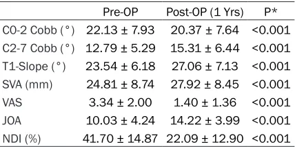
intention. Table 1. Cervical imaging and clinical effects
statistics (n=127)
Pre-OP Post-OP (1 Yrs) P*
C0-2 Cobb (°) 22.13 ± 7.93 20.37 ± 7.64 <0.001
C2-7 Cobb (°) 12.79 ± 5.29 15.31 ± 6.44 <0.001
T1-Slope (°) 23.54 ± 6.18 27.06 ± 7.13 <0.001
SVA (mm) 24.81 ± 8.74 27.92 ± 8.45 <0.001
VAS 3.34 ± 2.00 1.40 ± 1.36 <0.001
JOA 10.03 ± 4.24 14.22 ± 3.99 <0.001
NDI (%) 41.70 ± 14.87 22.09 ± 12.90 <0.001 Note: Values are presented as mean ± standard deviation.
*p<0.05, statistical significance(paired sample t test,). SVA,
[image:3.612.91.305.458.565.2]Sagittal vertical axis; VAS, Visual Analogue Scale; JOA, Japa-nese Orthopaedic Association Scores; NDI, Neck disability index.
Evaluation of the clinical effects of ACCF
After 1-year follow-up, the JOA score of this group was increased from preoperative 10.03 ± 4.24 points to 14.22 ± 3.99 points, and VAS score was decreased from 3.34 ± 2.00 points to 1.40 ± 1.36 points, NDI index was decreased from 41.70% ± 14.87% to 22.09% ± 12.90%.
The differences were statistically significant (P<0.05) (Table 1). During the follow-up period,
all the patients with X-ray films showed that the internal fixation was safe and effective, with no
broken nails, no obvious intervertebral cage displacement. One patient had the symptom of C5 nerve root palsy about 1 week after the sur-gery, and got better after six months of conser-vative treatment. There were no cerebrospinal
fluid leakage, recurrent laryngeal nerve injury,
and complications such as postoperative
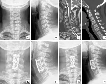
infec-tion, esophageal fistula and death (Typical case
Figure 2).
Changes of sagittal parameters of cervical vertebrae after ACCF
The 1-year follow-up found that the sagittal parameters T1-S of this group were increased from preoperative 23.54 ± 6.18 to 27.06 ± 7.13, the C2-7 Cobb was increased from preop-erative 12.79 ± 5.29 to 15.31 ± 6.44, SVA was increased from preoperative 24.81 ± 8.74 mm to 27.92 ± 8.45 mm, and C0-2 Cobb was decreased from preoperative 22.13 ± 7.93 to 20.37 ± 7.64. The difference was statistically
significant (P<0.05) (Table 2).
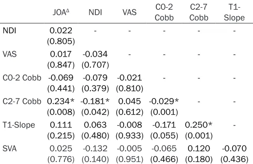
Correlation between the changes of the sagit-tal parameters and the clinical effects
The correlation analysis between the sagittal
parameters and the changes of clinical efficacy
[image:4.612.91.525.72.413.2]variation showed that the change value of C2-7 Cobb angle was positively correlated with the
Figure 2.Typical imaging data. F, 40 y, both upper limbs pain and numbness for 1 year, aggravated for 2 months. A, B. Cervical X-ray indicates the degeneration of the cervical curvature and the formation of the anterior and posterior margin of the vertebrae. C, D. Preoperative MRI showed C5/6 disc herniation with spinal stenosis. E, F. Cervical X-ray
JOA change value and the T1-S change value (P=0.008/P=0.001), and was negatively corre-lated with NDI (Neck disability index) and C0-2 Cobb change value (P=0.042/P=0.001) (Table 3).
Discussion
Analysis of the clinical effects of ACCF for cervical spondylosis
Currently, a variety of surgical methods are widely used to treat cervical spondylosis, including anterior cervical discectomy and bone graft fusion (ACDF), anterior cervical sub-total corpectomy and decompression fusion (ACCF), laminoplasty, laminectomy, and so on [10, 11]. ACDF and ACCF can achieve good postoperative effects such as segmental decompression, recovery of intervertebral
height and cervical spine anterior flexion cor -rection [12]. When compression comes from posterior vertebral body (such as osteophyte, OPLL, etc.), ACCF is widely used because it can directly remove compression behind vertebral
body and fully expose decompression field
of vision. The short-medium-term follow-up research showed that in patients with cervical spondylosis, the symptoms of spinal nerve compression decreased or disappeared 1 year after ACCF, and the quality of life of these
[image:5.612.90.349.97.265.2]patients was significantly improved [13, 14].
Table 2. Correlation between the change of imaging param-eters and clinical effects
JOAΔ NDI VAS C0-2
Cobb CobbC2-7 Slope
T1-NDI 0.022
(0.805) - - - -
-VAS 0.017
(0.847) (0.707)-0.034 - - -
-C0-2 Cobb -0.069
(0.441) (0.379)-0.079 (0.810)-0.021 - -
-C2-7 Cobb 0.234*
(0.008) -0.181* (0.042) (0.612)0.045 -0.029* (0.001) -
-T1-Slope 0.111
(0.215) (0.480)0.063 (0.933)-0.008 (0.055)-0.171 0.250* (0.001)
-SVA 0.025
(0.776) (0.140)-0.132 (0.951)-0.005 (0.466)-0.065 (0.180)0.120 (0.436)-0.070
Note: *When the confidence (on both sides) is 0.01, suggesting signifi -cantly correlated (Spearman’s rank correlation analysis). JOAΔ:
Improve-ment rate of JOA score = (Postoperative score - Preoperative score)/(17 - Preoperative score) × 100%).
Relationship between sagittal parameters of cervical vertebrae after ACCF and its changes
The ACCF treatment can change the sagittal parameters of the cervical spine and affect its part of or even total sagittal sequence. As an effective parameter to measure the sagittal position of the cervical spine, there is a certain correlation between T1-S, C2-7 and SVA param-eters. In the past, T1-S was considered to be a risk factor for cervical instability [15, 16]. An increase in T1-S resulted in a compensatory forward tilt of the cervical spine and an increase in SVA. The lordosis angle (Cobb angle) is the protective factor for maintaining the sagittal balance of the cervical spine. The lordosis angle changes adaptively due to the change in the size and direction of T1-S. One year after the follow-up of this study, T1-S was increased compared with preoperative, and the lordosis angle and SVA were also increased. In other words, any size of T1-S will have a C2-7 Cobb and SVA accordingly. The three parameters are closely related and compensate for each other. The sagittal sequence of the cervical spine remains relatively stable. In addition, this study found that the postoperative C0-2 Cobb was reduced compared with preoperative, which may be related to the compensatory adjust-ment of C2-7 Cobb increased spine to ensure that the patients are able to maintain function-al horizontfunction-al gaze. The preoperative T1-S was
127 patients with ACCF were fol-lowed up for 1 year in this study, which found that most patients felt that the neck pain and shoulder pain were reduced or disappeared after the surgery, the numbness of the
upper limbs and fingers was
decreased or returned to normal, the cotton sensation was disap-peared, and the cervical vertebra movement was normal.
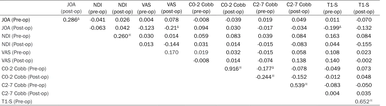
Table 3. Correlation between imaging parameters and clinical effects before and after ACCF JOA
(post-op) (pre-op)NDI (post-op)NDI (pre-op)VAS (post-op)VAS C0-2 Cobb (pre-op) C0-2 Cobb (post-op) C2-7 Cobb (pre-op) C2-7 Cobb (post-op) (pre-op)T1-S (post-op)T1-S JOA (Pre-op) 0.286Δ -0.041 0.026 0.004 0.078 -0.008 -0.039 0.019 0.049 0.011 -0.070
JOA (Post-op) -0.063 0.042 -0.123 -0.21Δ 0.094 0.030 -0.017 -0.034 -0.199Δ -0.132
NDI (Pre-op) 0.260※ 0.030 0.014 0.059 0.083 0.039 0.084 0.163 0.084
NDI (Post-op) 0.013 -0.144 0.031 0.014 -0.015 -0.083 0.044 -0.155
VAS (Pre-op) 0.170 0.019 0.032 -0.015 0.058 0.108 0.023
VAS (Post-op) -0.008 0.014 -0.074 0.138 0.140 -0.002
C0-2 Cobb (Pre-op) 0.916※ -0.177Δ -0.078 -0.049 0.073
C0-2 Cobb (Post-op) -0.244※ -0.152 -0.012 0.048
C2-7 Cobb (Pre-op) 0.539※ -0.083 -0.050
C2-7 Cobb (Post-op) 0.004 0.035
T1-S (Pre-op) 0.652※
Note: ΔWhen the confidence (on both sides) is 0.05, suggesting significantly correlated; ※when the confidence (on both sides) is 0.01, suggesting significantly correlated (Spear
positively correlated with lordosis angle and SVA, and the lordosis angle was negatively cor-related with SVA; the correlation remained the same 1 year after surgery.
Changes of sagittal parameters and clinical effects after cervical fusion
ACCF can effectively treat cervical spondylosis
and significantly improve the quality of life
of patients. Xiao [17] performed cervical anterior/posterior decompression surgery on 55 patients with cervical spondylosis and Parkinson’s disease. The symptoms of post-
operative static tremor were significantly
improved. After 1-year follow-up, the sagittal parameters were correlated to the scoring of the quality of life, and SVA was negatively cor-related with the SF.36 score. When SVA>40
mm, the surgical outcome was significantly
affected. The results of this study showed that after one year, the difference in the value of sagittal parameters of the cervical spine SVA, the clinical effects of VAS Score, and the NDI
index had no statistics significance. Kim [18]
found that 64 patients who underwent posteri-or decompression of the posteriposteri-or longitudinal ligament of the cervical spine were found to have kyphosis when the T1-S>25° preopera-tively, which had a strong impact on the clinical effects. The results of this study indicate that the cervical sagittal parameters T1-S and SVA
have no significant correlation with the clinical
effects of ACDF after cervical spondylosis. The JOA score, VAS score and NDI index were
significantly improved in the first year after operation, indicating that ACCF has significant
clinical effects on the treatment of cervical spondylosis. Cervical sagittal parameters were positively correlated with JOA and NDI before and 1 year after operation. Therefore, we believe that the sagittal parameter C2-7 Cobb is related to clinical effects after ACCF.
In conclusion, the short-term clinical effects of ACCF in the treatment of cervical spondylosis are notable. The cervical vertebra has a certain self-compensatory mechanism, which can maintain the sagittal balance of the spine by changing C2-7, C0-2 and T1-S. After 1-year fol-low-up, it was found that cervical C2-7 Cobb had a certain correlation with the clinical effects. However, the article still has some defects. For example, the number of cases is
limited, the data is slightly biased, and prob-lems of long-term titanium net deposition and loss of intervertebral space height are not considered.
Acknowledgements
This study was supported by the Ningbo natural science foundation (No. 2018A610265). Disclosure of conflict of interest
None.
Address correspondence to: Qing-Jiang Pang, Depar- tment of Orthopaedics, HwaMei Hospital, University of Chinese Academy of Sciences/Ningbo No.2 Hospital, Ningbo 315010, China. E-mail: [email protected]
References
[1] Roussouly P, Labelle H, Rouissi J, Bodin A. Pre- and post-operative sagittal balance in idio-pathic scoliosis: a comparison over the ages of two cohorts of 132 adolescents and 52 adults. Eur Spine J 2013; 22 Suppl 2: s203-215. [2] Ling FP, Chevillotte T, Leglise A, Thompson W,
Bouthors C, Le Huec JC. Which parameters are relevant in sagittal balance analysis of the cer-vical spine? A literature review. Eur Spine J 2018; 27 Suppl 1: 8-15.
[3] Carreon LY, Smith CL, Dimar JR 2nd, Glassman SD. Correlation of cervical sagittal alignment parameters on full-length spine radiographs compared with dedicated cervical radiographs. Scoliosis Spinal Disord 2016; 11: 12.
[4] Le Huec JC1, Demezon H, Aunoble S. Sagittal parameters of global cervical balance using EOS imaging: normative values from a pro-spective cohort of asymptomatic volunteers. Eur Spine J 2015; 24: 63-71.
[5] Koeppen D, Piepenbrock C, Kroppenstedt S,
Čabraja M. The influence of sagittal profile al
-teration and final lordosis on the clinical out -come of cervical spondylotic myelopathy. A Delta-Omega-analysis. PLoS One 2017; 12: e0174527.
[6] Bakhsheshian J, Mehta VA, Liu JC. Current di-agnosis and management of cervical spondy-lotic myelopathy. Global Spine J 2017; 7: 572-586.
[7] Kim M, Rhim SC, Roh SW, Jeon SR. Analysis of the risk factors associated with prolonged intubation or reintubation after anterior cervi-cal spine surgery. J Korean Med Sci 2018; 33: e77.
in healthy cervical spine adults and patients with cervical disc degeneration. BMC Musculo-skelet Disord 2018; 19: 37.
[9] Lee SH, Son DW, Lee JS, Kim DH, Sung SK, Lee SW, Song GS. Differences in cervical sagittal alignment changes in patients undergoing laminoplasty and anterior cervical discectomy and fusion. Neurospine 2018; 15: 91-100. [10] Ilharreborde B, Vidal C, Skalli W, Mazda K.
Sag-ittal alignment of the cervical spine in adoles-cent idiopathic scoliosis treated by posterome-dial translation. Eur Spine J 2013; 22: 330-337.
[11] Oe S, Togawa D, Yoshida G, Hasegawa T, Yam-ato Y, Kobayashi S, Yasuda T, Banno T, Mihara Y, Matsuyama Y. Difference in spinal sagittal alignment and health-related quality of life be-tween males and females with cervical defor-mity. Asian Spine J 2017; 11: 959-967. [12] Vidal C, Ilharreborde B, Azoulay R, Sebag G,
Mazda K. Reliability of cervical lordosis and global sagittal spinal balance measurements in adolescent idiopathic scoliosis. Eur Spine J 2013; 22: 1362-1367.
[13] Quinn JC, Kiely PD, Lebl DR, Hughes AP. Ante-rior surgical treatment of cervical spondylotic myelopathy: review article. HSS J 2014; 11: 15-25.
[14] Oni P, Schultheiß R, Scheufler KM, Roberg J,
Harati A. Radiological and clinical outcome af-ter multilevel anaf-terior cervical discectomy
and/or corpectomy and fixation. J Clin Med
2018; 7: E469.
[15] Brasil AVB, Fruett da Costa PR, Vial ADM, Bar-cellos GDC, Zauk EB, Worm PV, Ferreira MP,
Ferreira NP. Cervicothoracic lordosis can influ -ence outcome after posterior cervical spine surgery. Open Orthop J 2018; 12: 91-98. [16] Siasios I, Winograd E, Khan A, Vakharia K,
Di-mopoulos VG, Pollina J. Cervical sagittal bal-ance parameters after single-level anterior cervical discectomy and fusion: correlations with clinical and functional outcomes. J Cranio-vertebr Junction Spine 2018; 9: 56-62. [17] Xiao R, Miller JA, Lubelski D, Alberts JL, Mroz
TE, Benzel EC, Krishnaney AA, Machado AG. Quality of life outcomes following cervical de-compression for coexisting parkinson’s dis-ease and cervical spondylotic myelopathy. Spine J 2016; 16: 1358-1366.
[18] Kim B, Yoon DH, Ha Y, Yi S, Shin DA, Lee CK, Lee N, Kim KN. Relationship between T1 slope and loss of lordosis after laminoplasty in
pa-tients with cervical ossification of the posterior