Mathematical Modelling of the Interaction of
Chlamydia Trachomatis
with the Immune
System
Masoumeh Bagher Oskouei
∗†, Dann G. Mallet
∗‡, Ashkan Amirshahi
‡, Graeme J. Pettet
∗‡,
Abstract—In this paper, we present a two dimen-sional mathematical model capable of describing the process of the ascension of infection to the upper gen-ital tract in females and investigating the number of free chlamydial particles and infected cells. This model includes diffusion and chemotaxis terms corre-sponding to the motion of free chlamydial particles and the migration of immune cells toward the infec-tion site in response to diffusible chemical cues such asIF N−γ, respectively. The qualitative results of the model reflect experimentally observed phenomena.
Keywords: mathematical model,Chlamydia trachoma-tis, partial differential equation, immune
1
Introduction
Chlamydia trachomatis is an obligate intracellular bac-terial pathogen that infects the genital and ocular mu-cosa of humans causing sexually transmitted disease and trachoma. It is estimated that 70-75% of endocervi-cal infections in women caused by C. trachomatis are asymptomatic and may persist for months to years [4]. The sequelae of C. trachomatis genital tract infections in women, namely chronic pain, pelvic inflammatory dis-ease (PID), infertility and ectopic pregnancy are the most costly outcomes of any sexually transmitted infection ex-cept human immunodeficiency virus (HIV/AIDS), result-ing in an estimated $US4 billion in health care costs per annum only United States alone [5].
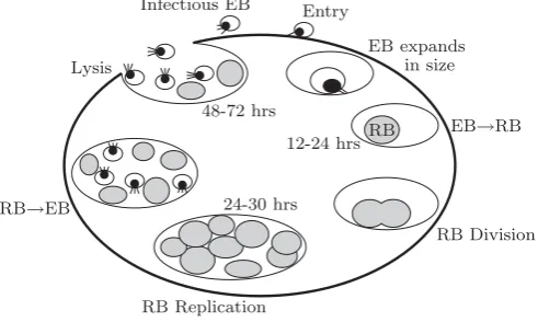
C. trachomatis is an intracellular, complex and multi-functional process pathogen, unique among prokaryotes because of a biphasic developmental cycle of replication in which the organism exists in two distinctive forms; the Elementary Body (EB), which is the infectious form, and the Reticulate Body (RB), which is the replicating struc-ture. In this developmental cycle (see Fig. 1), the infec-tious but metabolically inactive elementary body, 200-300 nm in diameter, is endocytosed by eukaryotic cells and resides within a cytoplasmic inclusion. Within the
∗Mathematical Sciences Discipline, Queensland University of
Technology, Brisbane, Australia
†Email:[email protected]
‡Institute of Health and Biomedical Innovation, Queensland
University of Technology, Brisbane, Australia
Infectious EB
12-24 hrs
24-30 hrs 48-72 hrs
Entry
EB expands in size
EB→RB RB
RB Division
RB Replication RB→EB
[image:1.595.300.546.254.401.2]Lysis
Figure 1: Schematic of the Chlamydia trachomatis
Serovar D Developmental Cycle.
inclusion, the EBs transform into the non-infectious but metabolically active reticulate body which is larger at 1000-1500 nm in diameter. The RBs divide within the cell by binary fission and transform back to the infectious form before being released to the cell exterior [1, 3].
Wilson [16] indicates that 200-500 new EBs are released from each infected cell following the replication process. This is an initial indicator of how theC. trachomatis in-fection can progress and subsequently reach the upper genital tract. It is also important though to note that the host immune system has a significant role in the clear-ance of Chlamydia throughout the infection period. C. trachomatisis subjected to both innate and adaptive im-mune responses of the host. Wilson for example, has shown that CD4+ T cells play a significant role in adap-tive immunity toChlamydia trachomatis infection of the genital tract [14]. Moreover the recent studies on human clinical samples and also animal models clearly show that it normally takes 4 to 7 days for the immune response to be induced. The next section will focus more closely on how chlamydial particles interact with immune cells.
t
Follicular Ovulatory Luteal
[image:2.595.43.278.136.252.2]7 14 21 28
Figure 2: Schematic of hormone levels during the normal menstrual cycle (progesterone – dotted line, estrogen – dashed line).
hormone (LH), follicle-stimulating hormone (FSH), es-trogen, and progesterone. A sharp drop in the levels of estrogen and progesterone results in a shedding of the en-dometrial lining of the uterus (see Fig.3). Estrogen and progesterone are produced in the ovaries and are regu-lated by other hormones produced in the brain. Of im-portance to this study is the fact that a simple vaginal infection, such as a chlamydial infection, will not affect the production or regulation of these reproductive hor-mones. However, the hormones have a significant effect on Chlamydia growth. Recent studies [7, 8] show that susceptibility and inflammation are induced in high pro-gesterone environments and also estrogen decreases flammation and protection from infection. The rate of in-fection increases when the level of progesterone is higher [17].
Figure 3: Endometrial changes during the normal men-strual cycle.
In vitro, providing all the factors mentioned above is not straightforward. Moreover, there are the several differ-ences in the Chlamydia infection of laboratory animals such as mice, compared with the infection in humans such as length of infection and the immune-evasion mech-anism. Therefore a mathematical model describing the interaction betweenC. trachomatis and the immune sys-tem is an extremely useful way to obtain further insights into the dynamics of the infection process, and the sub-sequent effects and possible effective control strategies.
In this paper we develop a mathematical model to investi-gate the interaction betweenC. trachomatisand the host immune system. We present preliminary results regard-ing infection dynamics and progression of infection in the genital tract and to track the motion of free chlamydial particles toward the upper genital level.
The remainder of this paper is organised as follows. A brief description of C. trachomatis and immune system interactions is provided in Section 2. Section 3 is a detailed description of our partial differential equation based mathematical model of the immune response re-lated to Chlamydia trachomatis. Then we present pre-liminary numerical results from simulations of the math-ematical model. Finally, a discussion and conclusions re-garding the model are made in Section 5.
2
The host immune system and
C.
tra-chomatis
The innate and adaptive immunity are two essential bar-riers against Chlamydia. The innate immune system is a non-specific system that provides immediate defence against infection. Macrophages, neutrophils and den-dritic cells are the key elements of the innate immune system. Pattern recognition receptors (PRRs) are pro-teins on the surface of both epithelial cells and circulating cells of the innate immune system. The toll-like receptors (TLRs) and NOD-like receptors (NLRs) are the two most important families of PRRs and are able to recognise and bind pathogen-associated molecular patterns (PAMPs). The adaptive immune system, on the other hand, is a specific system which develops after first contact with a pathogen. Humoral immunity (B lymphocytes) and cell mediated immunity (T lymphocytes) are two arms of the adaptive immune response (see Fig. 4).The B lympho-cytes are triggered by antigen-presenting cells (APCs), cells of the innate response or T cells. APCs stimulate B cells to produce antibodies which bind to antigens on the surface of the invading pathogen to flag them for neutral-isation. Similarly, T cells are activated by APCs, cells of the innate response or B lymphocytes. T lympho-cytes consist of T helper cells (Th cells) and cytotoxic T cells (CD8+ cells or killer cells). Th cells produce pro-inflammatory cytokines such as IL-4, IL-5, IL-6, and
IFN-γwhich both support the the humoral system and inhibit pathogen growth. Cytotoxic T cells directly attack and eliminate a pathogen. Although humoral immunity is the principal immunological response effective against extra-cellular bacteria, the major defensive and protective re-sponse against intracellular bacteria is performed by T cells [11, 10]. BecauseChlamydia is present both extra-and intracellularly, both of these elements of the immune system are important when modelling chlamydial infec-tions.
[image:2.595.40.287.524.617.2]A
Humoral immune response Cellular immune response
+
B lymphocyte Antigen
ASPC
Eliminating antigen
Dendritic cells
Act’d T helper Act’d cytotoxic
T helper eff Cytotoxic T eff
[image:3.595.118.457.94.262.2]Cytokines Killing infected cell
Figure 4: Schematic showing components of the immune system.
particle enters the body, epithelial cells are the first line of defence against the pathogen. PRRs on the surface of the epithelial cells bind to the antigens expressed on the surface ofC. trachomatis. This connection stimulates the epithelial cells to secrete chemokines and other pro-inflammatory cytokines which initiate circulating cells of the innate immune response. Once the innate immune cells such as macrophages, dendritic cells and neutrophils reach the infection site, they are activated and their PRRs bind to chlamydial antigens leading to pathogen destruc-tion. Activated macrophages and dendritic cells are able to express pathogen antigens bound to major histocom-patibility complex (MHC) proteins on the surface and to serve as APCs which can activate the adaptive immune cells. Under stimulation of activated dendritic cells, T cells move toward the site of infection and differentiate into either cytotoxic T cells (CD8+ T cells orTc cells) or helper T cells (CD4+ T cells). Cytotoxic T cells (CD8+) are activated when their receptors strongly interact with the MHC class I molecule presented at the site of infec-tion and perform their funcinfec-tion by releasing lymphotoxins that form pores in the target cell’s plasma membrane and cause it to burst or lyse. On the other hand, T helper cells (CD4+) interact with MHC class II molecules and produce pro-inflammatory cytokines such as interferon-gamma (IFN-γ) that inhibit chlamydial growth, and acti-vate macrophages and humoral cells in order to accelerate clearance of infection [12, 13, 9].
3
Mathematical model
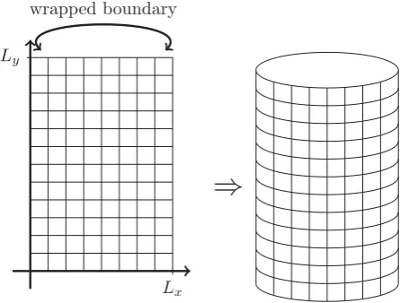
To model chlamydial infection in the genital tract, we use a rectangular domain Ω = [0, Lx]×[0, Ly], where the side boundaries are subject to a wrapping condition. This results in a domain shaped like the exterior surface of a cylinder – an oversimplified picture of the surface of the genital tract (see Fig. 5).
Lx Ly
wrapped boundary
⇒
Figure 5: Computationally, the genital tract is modelled using a rectangular grid that is then subjected to wrapped boundary conditions forx= 0 andx=Lx.
In order to simulate the infection process and the ef-fectiveness of immune responses during three different phases of the female menstrual cycle, we will solve the model equations over a time range of 28 days, the aver-age length of the female menstrual cycle.
[image:3.595.310.533.328.497.2]byM the components of the immune system that attack the intracellular Chlamydia. The free chlamydial par-ticles, uninfected epithelial cells, and infected cells are represented byC, EandI respectively.
To model the events described in the previous section, we assume that initially there are some free chlamydial par-ticles at the lower region of the genital tract and represent this using the initial condition
C(x, y,0) = 10e−(y−1)2.
Initially there are no infected cells (all chlamydial parti-cles are initially outside of the host cells) and epithelial cells are at their normal concentration levels through-out the domain. Further, there is initially an absence of chemokines/cytokines and of components of the im-mune system that attackChlamydiawhen it is inside host cells. Immune cells that clear freeChlamydiaparticles are initially circulating in the system at some ‘normal’ level given. Hence, initially we have
E(x, y,0) =Emax,
I(x, y,0) =K(x, y,0) =M(x, y,0) = 0,
H(x, y,0) =H0.
We make the assumption that each of the species of in-terest in our model is subject to a no-flux condition at the upper and lower ends of the domain; in the other words, they do not leave or enter the system via those end regions. Therefore, we have boundary conditions
∂C ∂y =
∂E ∂y =
∂I ∂y =
∂H ∂y =
∂M ∂y =
∂K
∂y = 0 (1)
at the boundariesy= 0 andy=Ly.
For free chlamydial particles, we assume that the parti-cles diffuse in the Fickian sense, with diffusion coefficient
DC. When infected cells burst at rateκ,P new EBs are released from each infected cell. The internalisation of EBs into the epithelial cells during the infection process occurs at rateg(t) and varies depending on the current phase of the female menstrual cycle. Chlamydial par-ticles are cleared due to the humoral immune response, however as previously mentioned, the immune response to Chlamydia is delayed and this is modelled using the expressionh(t). Combining these elements, we obtain the free particle conservation equation
∂C
∂t =DC∇2C+P κI−g(t)CE−h(t)CH. (2)
By day 5 to 7 of the menstrual cycle, shedding of the endometrial lining of the uterus results in a drop in the rate of EB internalisation. Vaginal epithelial thickness is minimal during the first phase, and increases in thickness
throughout the reproductive phase [18]. Therefore, the rate of EB internalisation gradually increases right after follicular phase until reaching a steady state level in the high progestrone environment. Thus we model the rate of EB internalisation using
g(t) =
⎧ ⎪ ⎨ ⎪ ⎩
2.7×10⁻⁶(exp (t)−1) + 0.001 0≤t≤7 1.1×10⁻⁴t2−0.0013 7< t≤14 0.02 otherwise.
The delay of the innate and humoral immune response is imposed using the function
h(t) =
7.3×10⁻⁶(exp(t)−1) + 0.001 0≤t≤7 0.1 otherwise.
The population of uninfected epithelial cells is also al-lowed to undergo Fickian diffusion, with coefficientDE. Epithelial cells are also assumed to be produced accord-ing to a logistic growth law to the normal/maximum cell level ofEmax and to die naturally at rateμ1. The cells are also lost to the infected population when they come in contact with free Chlamydia particles at rateg(t). Com-bining these we obtain the conservation equation
∂E
∂t =DE∇
2E
+E(Emax−E)−g(t)CE−μ1E. (3)
Infected cells also diffuse randomly with coefficient DI. The internalisation of EBs into the epithelial cells causes an increase in the number of infected cells at rate g(t). The infected cell population decreases due to the lysis of cells when the intracellular developmental cycle is com-plete, with a lysis rate ofκ. Finally, the cell mediated im-mune system, with a delayed response modelled bys(t), decreases the number of infected cells. This gives
∂I
∂t =DI∇2I+g(t)CE−κI−s(t)IM (4)
for the population of infected cells.
Studies [6] show that CD4+ and CD8+ cells have a signif-icant role in the clearance ofChlamydiacompared to the innate immune response and that the rate of clearance by cell mediated immunity is higher than humoral clear-ance rate. Similarly, it takes 4 to 7 days for cell mediated response to be induced. The delay of the cell mediated immune response is thus modelled by the function
s(t) =
7.3×10⁻⁶(exp(t)−1) + 0.001 0≤t≤7 0.3 otherwise
coefficientsDH andDM respectively. The second term in equations (5) and (6) represents the activation of humoral immunity in response to free chlamydial particles and cell mediated immunity in response to infected cells respec-tively. Natural death for elements of the humoral and cell mediated immune response has been characterised by the third terms in the corresponding equations, at rates of μ2 and μ3 respectively. The final terms in equations (5) and (6), model chemotactic responses. We propose that humoral and cell mediated immune cells direct their movement toward higher concentrations of chemokines and cytokines produced in response to either free chlamy-dial particles outside the host cell or infected cells, with chemotactic coefficients ofχH andχM respectively. To-gether, these components give the conservation equations
∂H
∂t =DH∇
2H
+γ1CH−μ2H− ∇(χH(H∇K)), (5)
∂M
∂t =DM∇
2M
+γ2IM−μ3M− ∇(χM(M∇K)). (6)
Cytokines and chemokines are also assumed to un-dergo random Fickian diffusion with coefficient DK. Chemokines produced by epithelial cells and some cy-tokines such as IFN-γsecreted by macrophages stimulate humoral immune cells to defend against free extracellular chlamydial particles at rateβ1. Additionally, chemokines secreted by infected cells at the RB stage of chlamydial developmental cycle, trigger T cells to produce cytokines in order to inhibit chlamydial growth or clear/control in-fection. In short, once chlamydial particles are inside the host cells, cell mediated immunity may prevent infec-tion. These components combine to give the conservation equation
∂K
∂t =DK∇
2K
+β1CH+β2IM. (7)
4
Numerical results
In this preliminary study, we simulate chlamydial parti-cles growing on a square domain Ω = [0,5]×[0,5]. An ex-plicit finite difference method was used to solve the model equations (2–7) subject to the initial and boundary con-ditions given in Section 3. In this paper, we assume the first day ofChlamydia infection corresponds to the first day of menstruation and then continue to simulate the following 28 days of infection.
Wilson [15] provides a number of estimates for rate con-stants and other parameters, including P = 350, κ = 0.45, μ1 = 0.3. We estimate that μ2 = μ3 = 0.01,
β1 = 0.4 and β2 = 0.1. Without access to further experimentally-informed parameters, we assume that the diffusion coefficients for cell species are an order of mag-nitude smaller than the coefficients for particles (simply due to relative sizes). Hence, we set DC = 10−4 and
DE =DI =DH =DM =DK = 10−5. Similarly, we set the chemotactic coefficients toχH =χM = 10−5.
t
2×10⁵
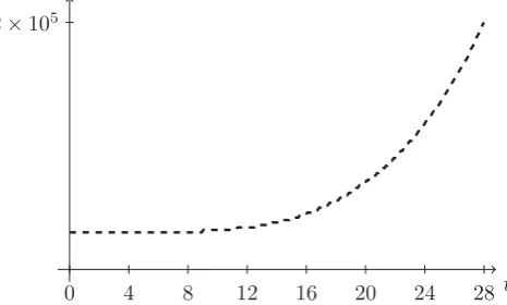
[image:5.595.304.537.136.276.2]0 4 8 12 16 20 24 28
Figure 6: Number of free particlesCover time (days).
t
1.5×10⁴
[image:5.595.309.545.320.461.2]0 4 8 12 16 20 24 28
Figure 7: Number of infected cellsIover time (days).
t
3.8×10⁴
[image:6.595.42.286.96.244.2]0 4 8 12 16 20 24 28
Figure 8: Humoral immunity measure H over time (days).
5
Conclusion
In this research we have extended the ordinary differential equation mathematical model of Wilson [15] to a partial differential equation model that allows for the consider-ation of spatio-temporal variconsider-ation in the relevant biolog-ical species. This includes the incorporation of random motion of particles and cells, as well as the chemotac-tic response of components of the immune system. We have also introduced more explicit tracking of factors such hormones and the delayed immune response that have a significant role in the growth of Chlamydia. The model presented here has successfully reproduced experimental results observed in the laboratory by Amirshahi and oth-ers and as such this new model provides a framework for further investigation of Chlamydial infection through ex-tension of the mathematical description. Such exex-tensions are a topic of current research of the authors.
References
[1] Abdelrahman, Y.M., Belland, R.J., The chlamydial developmental cycle, FEMS Microbiology Reviews, V29, N5, 949–959, 2005.
[2] Amirshahi, A., Effect of female sex hormones on Chlamydia trachomatis growth and gene expres-sion, Master of Applied Science (Research) Thesis, Queensland University of Technology, 2009.
[3] Belland, R.J., et al., Genomic transcriptional pro-filing of the developmental cycle of Chlamydia tra-chomatis,PNAS, V100, N14, pp. 8478–8483, 2003. [4] Dixon, R.E., et al., Chlamydia infection causes loss
of oviduct pacemaker cells and inhibits oocyte trans-port in the mouse oviduct, Biol Reprod., V80, N4, pp. 665–673, 2009.
[5] Hickey, D.K., Aldwell, F.E., Beagley, K.W., Tran-scutaneous immunization with a novel lipid-based
adjuvant protects againstChlamydiagenital and res-piratory infections, Vaccine, V27, N44, pp. 6217– 6225, 2009.
[6] Su, H., Caldwell, H.D., CD4+ T Cells Play a Sig-nificant Role in Adoptive Immunity to Chlamydia trachomatis Infection of the Mouse Genital Tract,
Infect. Immun., V63, N9, pp. 3302–3308, 1995. [7] Kaushic, C., Zhou, F., Murdin, A.D., Wira, C.R.,
Effects of Estradiol and Progesterone on Suscep-tibility and Early Immune Responses to Chlamy-dia trachomatis Infection in the Female Reproduc-tive Tract,Infect. Immun., V68, N7, pp. 4207–4216, 2000.
[8] Kaushic, C., Ashkar, A.A., Reid, L.A., Rosenthal, K.L., Progesterone Increases Susceptibility and De-creases Immune Responses to Genital Herpes Infec-tion,J Virol., V77, N8, pp. 4558–4565, 2003. [9] Morrison, S.G., et al., Immunity to Murine
Chlamy-dia trachomatis Genital Tract Reinfection Involves B Cells and CD4+ T Cells but Not CD8+ T Cells,
Infec. Immun., V68, N12, pp. 6979–6987, 2000. [10] Paul, W.E., ed. Fundamental immunology,
Lippin-cott Williams & Wilkins, Philadelphia, 2008.
[11] Revillard, J.P., ed.Cell-mediated immunity: in vitro correlates, Karger Basel, 1971.
[12] Starnbach, M.N., et al., An inclusion membrane pro-tein from Chlamydia trachomatis enters the MHC class I pathway and stimulates a CD8+ T cell re-sponse,J Immunol., V171, N9, pp. 4742–4749, 2003. [13] Stephens, R.S., ed. Chlamydia: intracellular biology, pathogenesis, and immunity, ASM Press: Washing-ton, DC, 1999.
[14] Wilson, D.P., Timms, P., McElwain, D.L.S., A mathematical model for the investigation of the Th1 immune response to Chlamydia trachomatis, Math Biosci., V182, N1, pp. 27–44, 2003.
[15] Wilson, D.P., Mathematical modelling of Chlamy-dia,ANZIAM J, V45(E), pp. C201-C214, 2004. [16] Wilson, D.P, et al., Type III Secretion,
Contact-dependent Model for the Intracellular Development of Chlamydia, Bull Math Biol., V68, N1, pp. 161– 178, 2006.